A case of an unruptured duplicated middle cerebral artery aneurysm–An unusual presentation of the distal internal carotid artery aneurysm
- Affiliations
-
- 1Department of Neurosurgery, Pusan National University Yangsan Hospital, Yangsan, Korea
- KMID: 2520883
- DOI: http://doi.org/10.7461/jcen.2021.E2020.10.004
Abstract
- The duplicated middle cerebral artery (DMCA) is an anatomic variation that arises from the distal internal carotid artery (ICA) and supplies blood to the middle cerebral artery (MCA) territory. Aneurysms of the DMCA have been reported in 36 cases in 2020. We also report a case of a 3.7 mm saccular aneurysm originating from the DMCA. A 52-year-old woman visited our hospital with worsening headache. She had no neurological abnormalities. Magnetic resonance imaging (MRI) and magnetic resonance angiography (MRA) revealed a right distal ICA aneurysm at the anterior choroidal artery. Cerebral angiography was performed to confirm the shape and the size of the aneurysm. Cerebral angiography revealed that the vessel that was originally identified as the anterior choroidal artery by the MRA was actually the duplicated MCA that was originating from the aneurysm neck and was supplying the MCA territory. The patient’s aneurysm was clipped using a transsylvian approach and she recovered without any neurological symptoms. DMCAs are rare and often associated with aneurysms and require preoperative evaluation to confirm the vascular status, aneurysm characteristics, and the shape of the parent artery.
Keyword
Figure
-
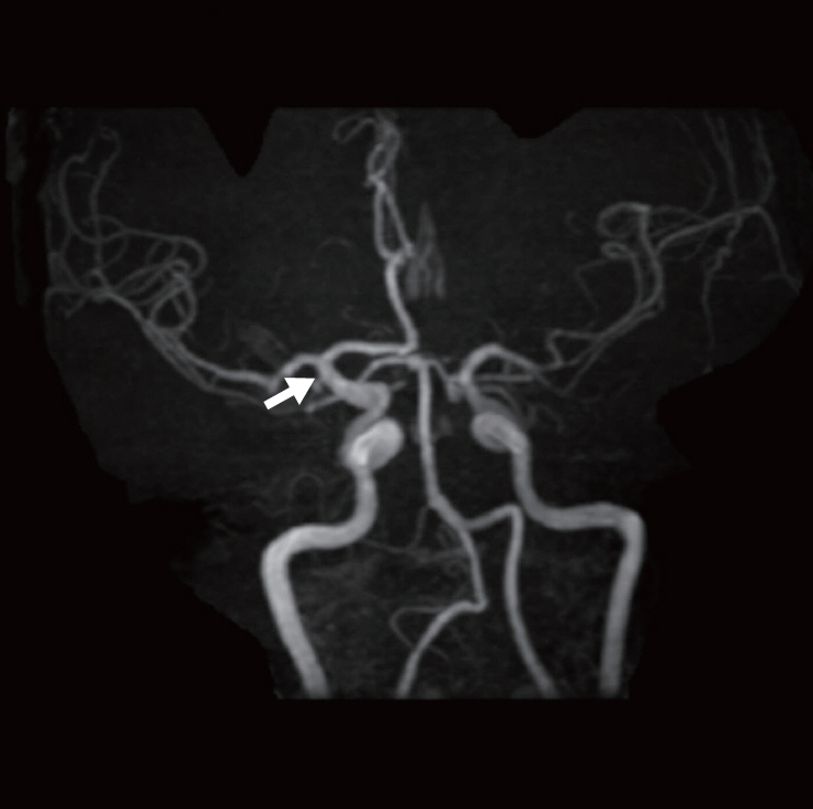
Fig. 1. Magnetic resonance angiography showing a right distal ICA aneurysm (arrow) with lateral projection, suggestive of a typical AChA aneurysm. ICA, internal carotid artery; AChA, anterior choroidal artery.
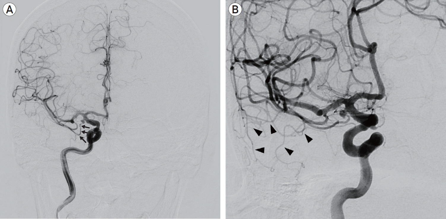
Fig. 2. Conventional cerebral angiogram of the right ICA (A) AP view of the right ICA angiogram demonstrating type B DMCA. Note the sharp separation angle from the ICA. (B) Oblique view of the right ICA angiogram showing the territories supplied by the DMCA (arrow heads). ICA, internal carotid artery; DMCA, duplicated middle cerebral artery.

Fig. 3. Intraoperative findings (A) Intraoperative microscopic view showing the DMCA (arrows) and related aneurysm. Note the DMCA running laterally to supply the anterior pole of the temporal lobe. (B) Microscopic view showing well clipped aneurysm. (C) ICGVA demonstrating the patent DMCA flow. DMCA, duplicated middle cerebral artery; ICG-VA, indocyanine green video angiography.
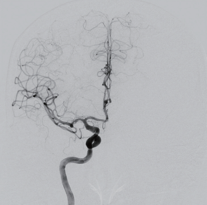
Fig. 4. Postoperative cerebral angiography revealing the complete occlusion of the aneurysm and the patent DMCA. DMCA, duplicated middle cerebral artery.
Reference
-
1. Alpers BJ, Berry RG, Paddison RM. Anatomical studies of the circle of Willis in normal brain. AMA Arch Neurol Psychiatry. 1959; Apr. 81(4):409–18.
Article2. Chang HY, Kim MS. Middle cerebral artery duplication: classification and clinical implications. J Korean Neurosurg Soc. 2011; Feb. 49(2):102–6.3. Crompton MR. The pathology of ruptured middle-cerebral aneurysms with special reference to the differences between the sexes. Lancet. 1962; Sep. 2(7253):421–5.
Article4. Hori E, Kurosaki K, Matsumura N, Yamatani K, Kusunose M, Kuwayama N, et al. Multiple aneurysms arising from the origin of a duplication of the middle cerebral artery. J Clin Neurosci. 2005; Sep. 12(7):812–5.
Article5. Iwata M, Kawaguchi S, Manaka H. A case of unruptured cerebral aneurysm arising from duplicate origin of the middle cerebral artery. No Shinkei Geka. 2020; Jun. 48(6):515–20.6. Kai Y, Hamada J, Morioka M, Yano S, Kudo M, Kuratsu J. Treatment of unruptured duplicated middle cerebral artery aneurysm: case report. Surg Neurol. 2006; Feb. 65(2):190–3. discussion 193.
Article7. Kim JS, Lee CH, Park H, Han JW. An unruptured cerebral aneurysm at the origin of the duplicated middle cerebral artery. J Cerebrovasc Endovasc Neurosurg. 2015; Sep. 17(3):223–6.
Article8. Komiyama M, Nakajima H, Nishikawa M, Yasui T. Middle cerebral artery variations: duplicated and accessory arteries. AJNR Am J Neuroradiol. 1998; Jan. 19(1):45–9.9. Mori K, Tamase A, Seki S, Iida Y, Kawabata Y, Nakano T, et al. Duplicated middle cerebral artery associated with aneurysm at M1/M2 bifurcation: a case report. J Med Case Rep. 2018; Oct. 12(1):283.
Article10. Pico F, Labreuche J, Touboul PJ, Leys D, Amarenco P. Intracranial arterial dolichoectasia and small-vessel disease in stroke patients. Ann Neurol. 2005; Apr. 57(4):472–9.
Article11. Ren H, Ma L, Wei M, Li J, Yu M, Yin L. Duplicated middle cerebral artery origin with an aneurysm. Medicine (Baltimore). 2018; Mar. 97(9):e9947.
Article12. Stojanović NN, Kostić A, Mitić R, Berilažić L. Correlation between multiple cerebral aneurysms and a rare type of segmental duplication of the middle cerebral artery. BMC Neurol. 2020; Jan. 20(1):3.
Article13. Teal JS, Rumbaugh CL, Bergeron RT, Segall HD. Anomalies of the middle cerebral artery: accessory artery, duplication, and early bifurcation. Am J Roentgenol Radium Ther Nucl Med. 1973; Jul. 118(3):567–75.
Article14. Tsang COA, Smith L, Klostranec J, Orru E, Pereira VM. Ruptured duplicated middle cerebral artery aneurysm successfully treated by coil embolization with balloon remodeling. World Neurosurg. 2018; Dec. 120:509–10.
Article15. Umansky F, Dujovny M, Ausman JI, Diaz FG, Mirchandani HG. Anomalies and variations of the middle cerebral artery: a microanatomical study. Neurosurg. 1988; Jun. 22(6 Pt 1):1023–7.
- Full Text Links
-
- Actions
-
Cited
- CITED
-
- Close
- Share
-
- Similar articles
-
- Cerebral Aneurysm in the Long Fenestration at the Middle Portion of M1 Segment
- Postoperative Vasospasm in Unruptured Intracranial Aneurysm
- An Unruptured Cerebral Aneurysm at the Origin of the Duplicated Middle Cerebral Artery
- Middle Cerebral Artery Variations Associated with Intracranial Aneurysmal Rupture
- Intracranial Aneurysm Associated with Aplasia of the Internal Cartoid Artery
