J Korean Assoc Oral Maxillofac Surg.
2021 Aug;47(4):321-326. 10.5125/jkaoms.2021.47.4.321.
Diagnostic challenge and management of intraosseous mandibular hemangiomas: a case report and literature review
- Affiliations
-
- 1Department of Oral Surgery and Oral Implantology, Faculty of Dentistry, University Alfonso X el Sabio, Spain
- 2Department of Dental Clinical Specialties, Faculty of Dentistry, Complutense University of Madrid, Spain
- 3Department of Oral and Maxillofacial Surgery, Hospital Clínico San Carlos, Madrid, Spain
- KMID: 2519829
- DOI: http://doi.org/10.5125/jkaoms.2021.47.4.321
Abstract
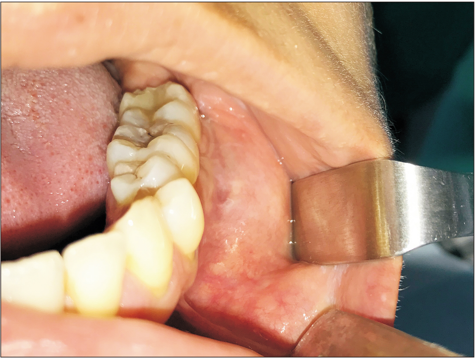
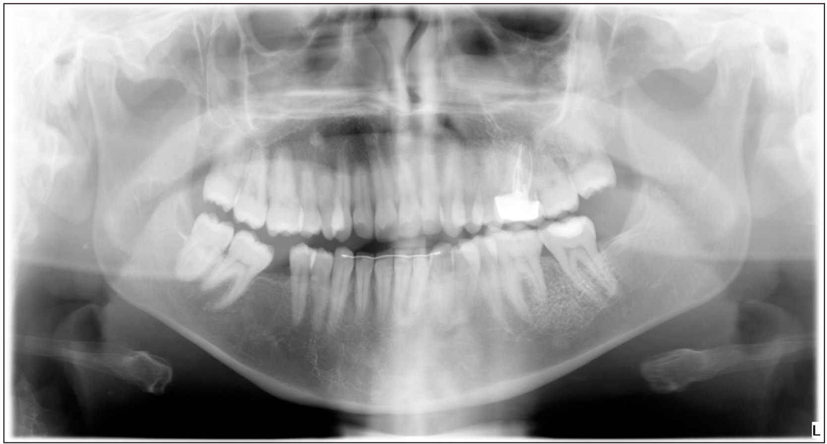
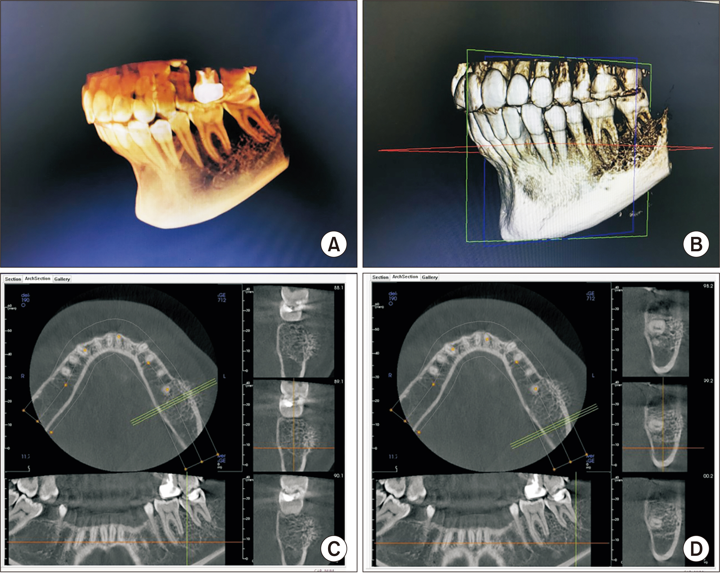
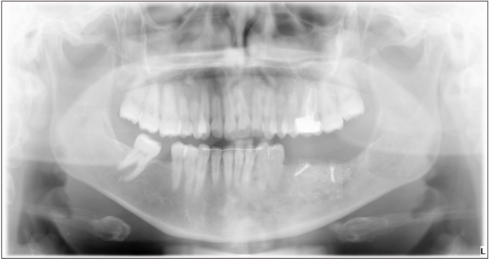
- Hemangioma is a benign tumor characterized by the proliferation of blood vessels. Although it often appears in soft tissues, its occurrence in bone tissue, particularly the mandible, is extremely rare. A 32-year-old female sought attention at the dental clinic complaining of a painless swelling in the posterior region of the left side of the mandible. A panoramic radiograph and computed axial tomography scan were taken, showing honeycomb and sunburst images, respectively, in the affected area. The patient underwent a biopsy, which led to the diagnosis of intraosseous hemangioma. Having assessed the characteristics of the lesion, it was decided to perform complete excision including safety margins, followed by an iliac crest bone graft to reconstruct the mandible. Awareness of the possible clinical and radiographic presentations of intraosseous hemangioma is considered important, as non-diagnosis could have severe consequences given its possible relation to dental structures.
Keyword
Figure
Reference
-
References
1. Donohue CA, de la Torre MA, de la Torre MG, Sánchez AJG, López MJA, Guzmán GDA, et al. 2016; Hemangioma intraóseo: reto diagnóstico. Presentación de un caso y revisión de la literatura. Rev ADM. 73:39–43. Spanish.2. Elif B, Derya Y, Gulperi K, Sevgi B. 2017; Intraosseous cavernous hemangioma in the mandible: a case report. J Clin Exp Dent. 9:e153–6. https://doi.org/10.4317/jced.52864 . DOI: 10.4317/jced.52864. PMID: 28149481. PMCID: PMC5268109.
Article3. Treviño AMG, Valdés MJ, Martínez MHR, Moreno TMG, Rivera SG. 2016; Hemangioma intraóseo de la mandíbula. Reporte de un caso clínico. Rev ADM. 73:96–8. Spanish.4. Sepulveda I, Spencer ML, Platin E, Trujillo I, Novoa S, Ulloa D. 2013; Intraosseous hemangioma of the mandible: case report and review of the literature. Int J Odontostomat. 7:395–400. https://doi.org/10.4067/S0718-381X2013000300010 .
Article5. Gómez Oliveira G, García-Rozado A, Luaces Rey R. 2008; Intraosseous mandibular hemangioma. A case report and review of the literature. Med Oral Patol Oral Cir Bucal. 13:E496–8. PMID: 18667983.6. Luaces Rey R, García-Rozado González A, López-Cedrún Cembranos L, Ferreras Granado J, Charro Huerga E. 2006; Intraosseous hemangioma of the mandible. An intraoral approach. Rev Esp Cirug Oral y Maxilofa. 28:195–9.7. Zlotogorski A, Buchner A, Kaffe I, Schwartz-Arad D. 2005; Radiological features of central haemangioma of the jaws. Dentomaxillofac Radiol. 34:292–6. https://doi.org/10.1259/dmfr/37705042 . DOI: 10.1259/dmfr/37705042. PMID: 16120879.
Article8. Fernández LR, Luberti RF, Domínguez FV. 2003; Radiographic features of osseous hemangioma in the maxillo-facial region. Bibliographic review and case report. Med Oral. 8:166–77. PMID: 12730651.9. Chandra SR, Chen E, Cousin T, Oda D. 2017; A case series of intraosseous hemangioma of the jaws: various presentations of a rare entity. J Clin Exp Dent. 9:e1366–70. https://doi.org/10.4317/jced.54285 . DOI: 10.4317/jced.54285. PMID: 29302291. PMCID: PMC5741852.
Article10. Dhiman NK, Jaiswara C, Kumar N, Patne SC, Pandey A, Verma V. 2015; Central cavernous hemangioma of mandible: case report and review of literature. Natl J Maxillofac Surg. 6:209–13. https://doi.org/10.4103/0975-5950.183866 . DOI: 10.4103/0975-5950.183866. PMID: 27390499. PMCID: PMC4922235.
Article11. Dereci O, Acikalin MF, Ay S. 2015; Unusual intraosseous capillary hemangioma of the mandible. Eur J Dent. 9:438–41. https://doi.org/10.4103/1305-7456.163236 . DOI: 10.4103/1305-7456.163236. PMID: 26430377. PMCID: PMC4570000.
Article12. Chetan BI, Shruthi DK, Karthik B. Sharmila. 2015; Diagnostic and surgical aspects of central hemangioma of mandible: a surgical approach for the reconstruction of mandible. J Int Oral Health. 7:56–8. PMID: 25709370. PMCID: PMC4336663.13. Eliot CA, Castle JT. 2010; Intraosseous hemangioma of the anterior mandible. Head Neck Pathol. 4:123–5. https://doi.org/10.1007/s12105-010-0170-x . DOI: 10.1007/s12105-010-0170-x. PMID: 20512636. PMCID: PMC2878621.
Article14. Kalsi H, Scannell J. 2013; Unusual presentation of an intraosseous hemangioma of the maxilla and displaced canine. Int J Clin Pediatr Dent. 6:124–6. https://doi.org/10.5005/jp-journals-10005-1203 . DOI: 10.5005/jp-journals-10005-1203. PMID: 25206206. PMCID: PMC4086593.
Article15. Kaya B, Işılgan SE, Cerkez C, Otrakçı V, Serel S. 2014; Intraosseous cavernous hemangioma: a rare presentation in maxilla. Eplasty. 14:e35. PMID: 25328568. PMCID: PMC4194598.16. Handa H, Naidu GS, Dara BG, Deshpande A, Raghavendra R. 2014; Diverse imaging characteristics of a mandibular intraosseous vascular lesion. Imaging Sci Dent. 44:67–73. https://doi.org/10.5624/isd.2014.44.1.67 . DOI: 10.5624/isd.2014.44.1.67. PMID: 24701461. PMCID: PMC3972408.
Article17. Omeje K, Efunkoya A, Amole I, Akhiwu B, Osunde D. 2014; A two-year audit of non-vascularized iliac crest bone graft for mandibular reconstruction: technique, experience and challenges. J Korean Assoc Oral Maxillofac Surg. 40:272–7. https://doi.org/10.5125/jkaoms.2014.40.6.272 . DOI: 10.5125/jkaoms.2014.40.6.272. PMID: 25551091. PMCID: PMC4279977.
Article

