Preoperative Radiological Parameters to Predict Clinical and Radiological Outcomes after Laminoplasty
- Affiliations
-
- 1Department of Neurosurgery, Research Institute for Convergence of Biomedical Science and Technology, Pusan National University Yangsan Hospital, Yangsan, Korea
- 2Department of Neurosurgery, School of Medicine, Pusan National University, Busan, Korea
- 3Department of Neurosurgery, Yongin Severance Hospital, Yonsei University College of Medicine, Yongin, Korea
- 4Department of Neurosurgery, Spine and Spinal Cord Institute, Severance Hospital, Yonsei University College of Medicine, Seoul, Korea
- KMID: 2519695
- DOI: http://doi.org/10.3340/jkns.2020.0294
Abstract
- Many studies have focused on pre-operative sagittal alignment parameters which could predict poor clinical or radiological outcomes after laminoplasty. However, the influx of too many new factors causes confusion. This study reviewed sagittal alignment parameters, predictive of clinical or radiological outcomes, in the literature. Preoperative kyphotic alignment was initially proposed as a predictor of clinical outcomes. The clinical significance of the K-line and K-line variants also has been studied. Sagittal vertical axis, T1 slope (T1s), T1s-cervical lordosis (CL), anterolisthesis, local kyphosis, the longitudinal distance index, and range of motion were proposed to have relationships with clinical outcomes. The relationship between loss of cervical lordosis (LCL) and T1s has been widely studied, but controversy remains. Extension function, the ratio of CL to T1s (CL/T1s), and Sharma classification were recently proposed as LCL predictors. In predicting postoperative kyphosis, T1s cannot predict postoperative kyphosis, but a low CL/T1s ratio was associated with postoperative kyphosis.
Keyword
Figure
-

Fig. 1. Flow chart diagram of our search mechanism in accordance to the PRISMA. PRISMA : preferred reporting items for systematic reviews and meta-analyses, ACDF : anterior cervical discectomy and fusion.
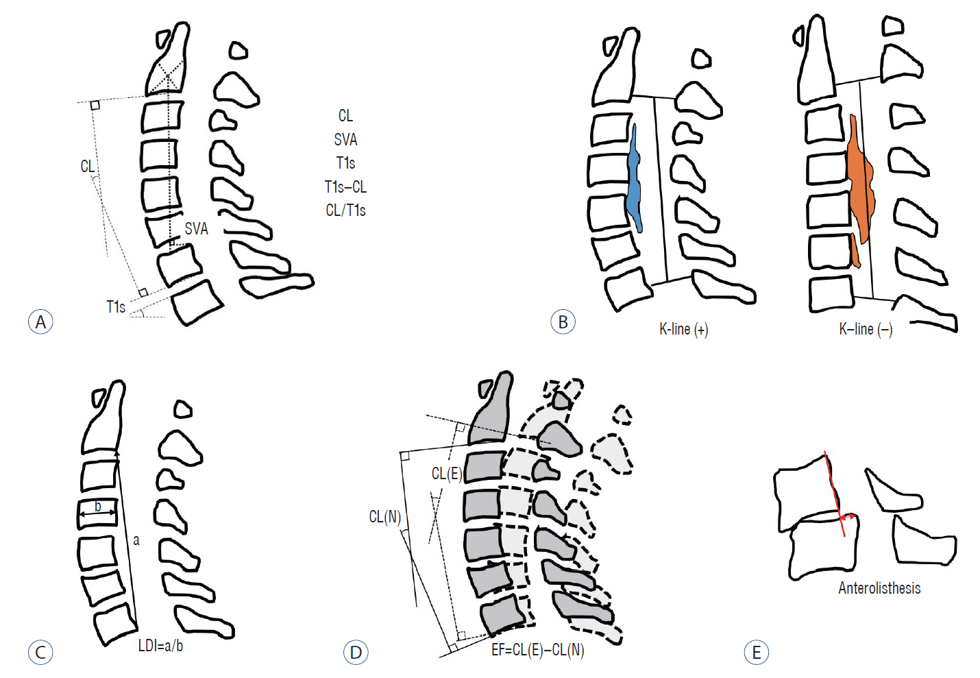
Fig. 2. Schematic images of sagittal radiological parameters. A : Routine sagittal parameters. B : K-line. C : a means length of a vertical line drawn between the postero-inferior edges of C2 and C7, b means the antero-posterior diameter of C4, and LDI of the cervical spine. D : Extension function (EF). E : Anterolisthesis in flexion position. CL : cervical lordosis, SVA : sagittal vertical axis, T1s : T1 slope, LDI : longitudinal distance index, CL(N) : cervical lordosis in neutral position, CL(E) : cervical lordosis in extension position.

Fig. 3. Schema of the indication of laminoplasty according to preoperative alignment, diagnosis, K-line, and loss of cervical lordosis. Preop : preoperative, OPLL : ossified posterior longitudinal ligament, CSM : cervical spondylotic myelopathy, LCL : loss of cervical lordosis.
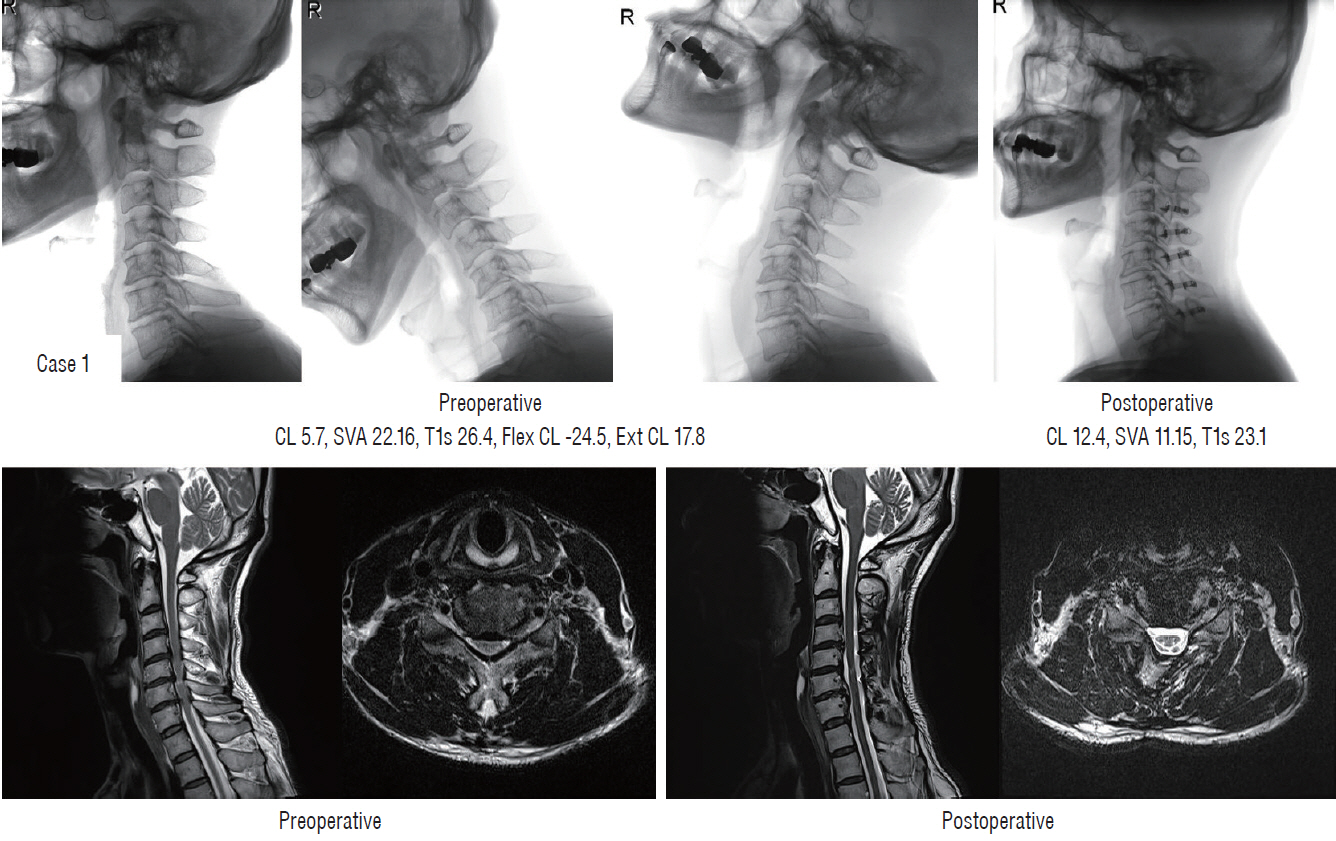
Fig. 4. Sufficient indirect decompression (case 1). CL : cervical lordosis, SVA : sagittal vertical axis, T1s : T1 slope, Flex CL : cervical lordosis in flexion position, Ext CL : cervical lordosis in extension position.

Fig. 5. Different postoperative alignment and clinical outcomes in two patients with preoperative kyphotic alignment (case 2 [upper] and case 3 [lower]). CL : cervical lordosis, SVA : sagittal vertical axis, T1s : T1 slope, Flex CL : cervical lordosis in flexion position, Ext CL : cervical lordosis in extension position, EF : extension function.

Fig. 6. Failure of indirect decompression in patients with preoperative kyphotic alignment (case 3). CL : cervical lordosis, SVA : sagittal vertical axis, T1s : T1 slope.

Fig. 7. Failure of indirect decompression following loss of cervical lordosis in preoperative straight alignment patients (case 4). CL : cervical lordosis, SVA : sagittal vertical axis, T1s : T1 slope, Flex CL : cervical lordosis in flexion position, Ext CL : cervical lordosis in extension position, EF : extension function.
Reference
-
References
1. Aita I, Wadano Y, Yabuki T. Curvature and range of motion of the cervical spine after laminaplasty. J Bone Joint Surg Am. 82:1743–1748. 2000.
Article2. Baba H, Uchida K, Maezawa Y, Furusawa N, Azuchi M, Imura S. Lordotic alignment and posterior migration of the spinal cord following en bloc open-door laminoplasty for cervical myelopathy: a magnetic resonance imaging study. J Neurol. 243:626–632. 1996.
Article3. Bajamal AH, Kim SH, Arifianto MR, Faris M, Subagio EA, Roitberg B, et al. Posterior surgical techniques for cervical spondylotic myelopathy: WFNS Spine Committee recommendations. Neurospine. 16:421–434. 2019.
Article4. Batzdorf U, Batzdorff A. Analysis of cervical spine curvature in patients with cervical spondylosis. Neurosurgery. 22:827–836. 1988.
Article5. Batzdorf U, Flannigan BD. Surgical decompressive procedures for cervical spondylotic myelopathy. A study using magnetic resonance imaging. Spine (Phila Pa 1976). 16:123–127. 1991.
Article6. Cao J, Zhang J, Yang D, Yang L, Shen Y. Multivariate analysis of factors associated with kyphotic deformity after laminoplasty in cervical spondylotic myelopathy patients without preoperative kyphotic alignment. Sci Rep. 7:43443. 2017.
Article7. Chen HY, Yang MH, Lin YP, Lin FH, Chen PQ, Hu MH, et al. Impact of cervical sagittal parameters and spinal cord morphology in cervical spondylotic myelopathy status post spinous process-splitting laminoplasty. Eur Spine J. 29:1052–1060. 2020.
Article8. Chiba K, Toyama Y, Watanabe M, Maruiwa H, Matsumoto M, Hirabayashi K. Impact of longitudinal distance of the cervical spine on the results of expansive open-door laminoplasty. Spine (Phila Pa 1976). 25:2893–2898. 2000.
Article9. Chiba K, Ogawa Y, Ishii K, Takaishi H, Nakamura M, Maruiwa H, et al. Long-term results of expansive open-door laminoplasty for cervical myelopathy--average 14-year follow-up study. Spine (Phila Pa 1976). 31:2998–3005. 2006.
Article10. Cho JH, Ha JK, Kim DG, Song KY, Kim YT, Hwang CJ, et al. Does preoperative T1 slope affect radiological and functional outcomes after cervical laminoplasty? Spine (Phila Pa 1976). 39:E1575–E1581. 2014.
Article11. Deora H, Kim SH, Behari S, Rudrappa S, Rajshekhar V, Zileli M, et al. Anterior surgical techniques for cervical spondylotic myelopathy: WFNS Spine Committee recommendations. Neurospine. 16:408–420. 2019.
Article12. Fujiwara H, Oda T, Makino T, Moriguchi Y, Yonenobu K, Kaito T. Impact of cervical sagittal alignment on axial neck pain and health-related quality of life after cervical laminoplasty in patients with cervical spondylotic myelopathy or ossification of the posterior longitudinal ligament: a prospective comparative study. Clin Spine Surg. 31:E245–E251. 2018.13. Fujiyoshi T, Yamazaki M, Kawabe J, Endo T, Furuya T, Koda M, et al. A new concept for making decisions regarding the surgical approach for cervical ossification of the posterior longitudinal ligament: the K-line. Spine (Phila Pa 1976). 33:E990–E993. 2008.14. Head J, Rymarczuk G, Stricsek G, Velagapudi L, Maulucci C, Hoelscher C, et al. Ossification of the posterior longitudinal ligament: surgical approaches and associated complications. Neurospine. 16:517–529. 2019.
Article15. Hirabayashi K, Miyakawa J, Satomi K, Maruyama T, Wakano K. Operative results and postoperative progression of ossification among patients with ossification of cervical posterior longitudinal ligament. Spine (Phila Pa 1976). 6:354–364. 1981.
Article16. Hirabayashi K, Watanabe K. A review of my invention of expansive laminoplasty. Neurospine. 16:379–382. 2019.
Article17. Ijima Y, Furuya T, Ota M, Maki S, Saito J, Kitamura M, et al. The K-line in the cervical ossification of the posterior longitudinal ligament is different on plain radiographs and CT images. J Spine Surg. 4:403–407. 2018.
Article18. Ishibashi K. Expansive laminoplasty by sagittal splitting of the spinous process for cervical myelopathy: correlation of clinical results with morphological changes in the cervical spine. Kurume Med J. 47:135–145. 2000.
Article19. Ito K, Yukawa Y, Ito K, Machino M, Kanbara S, Nakashima H, et al. Dynamic changes in the spinal cord cross-sectional area in patients with myelopathy due to cervical ossification of posterior longitudinal ligament. Spine J. 15:461–466. 2015.
Article20. Itoki K, Kurokawa R, Shingo T, Kim P. Effect of myoarchitectonic spinolaminoplasty on concurrent hypertension in patients with cervical spondylotic myelopathy. Neurospine. 15:77–85. 2018.
Article21. Iwasaki M, Kawaguchi Y, Kimura T, Yonenobu K. Long-term results of expansive laminoplasty for ossification of the posterior longitudinal ligament of the cervical spine: more than 10 years follow up. J Neurosurg. 96(2 Suppl):180–189. 2002.
Article22. Joshi RS, Haddad AF, Lau D, Ames CP. Artificial intelligence for adult spinal deformity. Neurospine. 16:686–694. 2019.
Article23. Kato M, Namikawa T, Matsumura A, Konishi S, Nakamura H. Effect of cervical sagittal balance on laminoplasty in patients with cervical myelopathy. Global Spine J. 7:154–161. 2017.
Article24. Kawakami M, Tamaki T, Ando M, Yamada H, Yoshida M. Relationships between sagittal alignment of the cervical spine and morphology of the spinal cord and clinical outcomes in patients with cervical spondylotic myelopathy treated with expansive laminoplasty. J Spinal Disord Tech. 15:391–397. 2002.
Article25. Kawanabe Y, Fujimoto M, Sato T. Cervical open-door laminoplasty by hydroxyapatite implant insertion without suturing. Neurospine. 15:362–367. 2018.
Article26. Khan O, Badhiwala JH, Wilson JRF, Jiang F, Martin AR, Fehlings MG. Predictive modeling of outcomes after traumatic and nontraumatic spinal cord injury using machine learning: review of current progress and future directions. Neurospine. 16:678–685. 2019.
Article27. Kim B, Yoon DH, Ha Y, Yi S, Shin DA, Lee CK, et al. Relationship between T1 slope and loss of lordosis after laminoplasty in patients with cervical ossification of the posterior longitudinal ligament. Spine J. 16:219–225. 2016.
Article28. Kim DH, Lee CH, Ko YS, Yang SH, Kim CH, Park SB, et al. The clinical implications and complications of anterior versus posterior surgery for multilevel cervical ossification of the posterior longitudinal ligament; an updated systematic review and meta-analysis. Neurospine. 16:530–541. 2019.
Article29. Kim M, Yun J, Cho Y, Shin K, Jang R, Bae HJ, et al. Deep learning in medical imaging. Neurospine. 16:657–668. 2019.
Article30. Kim SW, Hai DM, Sundaram S, Kim YC, Park MS, Paik SH, et al. Is cervical lordosis relevant in laminoplasty? Spine J. 13:914–921. 2013.
Article31. Kim TH, Lee SY, Kim YC, Park MS, Kim SW. T1 slope as a predictor of kyphotic alignment change after laminoplasty in patients with cervical myelopathy. Spine (Phila Pa 1976). 38:E992–E997. 2013.
Article32. Kwon SY, Shin JJ, Lee JH, Cho WH. Prognostic factors for surgical outcome in spinal cord injury associated with ossification of the posterior longitudinal ligament (OPLL). J Orthop Surg Res. 10:94. 2015.
Article33. Lee BJ, Park JH, Jeon SR, Rhim SC, Roh SW. Importance of the preoperative cross-sectional area of the semispinalis cervicis as a risk factor for loss of lordosis after laminoplasty in patients with cervical spondylotic myelopathy. Eur Spine J. 27:2720–2728. 2018.
Article34. Lee CK, Shin DA, Yi S, Kim KN, Shin HC, Yoon DH, et al. Correlation between cervical spine sagittal alignment and clinical outcome after cervical laminoplasty for ossification of the posterior longitudinal ligament. J Neurosurg Spine. 24:100–107. 2016.
Article35. Lee DH, Kim H, Lee HS, Noh H, Kim NH, Hwang C, et al. The Kappa line - a predictor of neurologic outcome after cervical laminoplasty. In : ISASS13 Oral Podium and Oral Poster Presentation; 2013 Apr 3-5; Vancouver, Canada. Rosemont (IL): ISASS;2013.36. Lee JS, Son DW, Lee SH, Kim DH, Lee SW, Song GS. The predictable factors of the postoperative kyphotic change of sagittal alignment of the cervical spine after the laminoplasty. J Korean Neurosurg Soc. 60:577–583. 2017.
Article37. Lee SH, Kim KT, Lee JH, Kang KC. Presentation #32: posterior cervical spinal cord shift following posterior decompression and prediction of persistent anterior spinal cord compression using K‐plane: a three dimensional modification of K‐line on MRI. Spine Journal Meeting Abstracts. 2016:155–157. 2016.38. Lee SH, Son DW, Lee JS, Kim DH, Sung SK, Lee SW, et al. Differences in cervical sagittal alignment changes in patients undergoing laminoplasty and anterior cervical discectomy and fusion. Neurospine. 15:91–100. 2018.
Article39. Lee SH, Son DW, Lee JS, Sung SK, Lee SW, Song GS. Does extension dysfunction affect postoperative loss of cervical lordosis in patients who undergo laminoplasty? Spine (Phila Pa 1976). 44:E456–E464. 2019.
Article40. Li J, Zhang Y, Zhang N, Xv ZK, Li H, Chen G, et al. Clinical outcome of laminoplasty for cervical ossification of the posterior longitudinal ligament with K-line (-) in the neck neutral position but K-line (+) in the neck extension position: a retrospective observational study. Medicine (Baltimore). 96:e6964. 2017.41. Li XY, Kong C, Sun XY, Guo MC, Ding JZ, Yang YM, et al. Influence of the ratio of C2-C7 Cobb angle to T1 slope on cervical alignment after laminoplasty. World Neurosurg. 124:e659–e666. 2019.
Article42. Lin BJ, Hong KT, Lin C, Chung TT, Tang CT, Hueng DY, et al. Impact of global spine balance and cervical regional alignment on determination of postoperative cervical alignment after laminoplasty. Medicine (Baltimore). 97:e13111. 2018.
Article43. Maruo K, Moriyama T, Tachibana T, Inoue S, Arizumi F, Daimon T, et al. The impact of dynamic factors on surgical outcomes after double-door laminoplasty for ossification of the posterior longitudinal ligament of the cervical spine. J Neurosurg Spine. 21:938–943. 2014.
Article44. Masaki Y, Yamazaki M, Okawa A, Aramomi M, Hashimoto M, Koda M, et al. An analysis of factors causing poor surgical outcome in patients with cervical myelopathy due to ossification of the posterior longitudinal ligament: anterior decompression with spinal fusion versus laminoplasty. J Spinal Disord Tech. 20:7–13. 2007.
Article45. Matsuoka Y, Suzuki H, Endo K, Sawaji Y, Murata K, Nishimura H, et al. Small sagittal vertical axis accompanied with lumbar hyperlordosis as a risk factor for developing postoperative cervical kyphosis after expansive open-door laminoplasty. J Neurosurg Spine. 29:176–181. 2018.
Article46. Matsuyama Y, Kawakami N, Yanase M, Yoshihara H, Ishiguro N, Kameyama T, et al. Cervical myelopathy due to OPLL: clinical evaluation by MRI and intraoperative spinal sonography. J Spinal Disord Tech. 17:401–404. 2004.47. Miyazaki M, Ishihara T, Notani N, Kanezaki S, Tsumura H. Relationship of T1 slope with loss of lordosis and surgical outcomes after laminoplasty for cervical ossification of the posterior longitudinal ligament. Clin Neurol Neurosurg. 164:19–24. 2018.
Article48. Ogawa Y, Toyama Y, Chiba K, Matsumoto M, Nakamura M, Takaishi H, et al. Long-term results of expansive open-door laminoplasty for ossification of the posterior longitudinal ligament of the cervical spine. J Neurosurg Spine. 1:168–174. 2004.
Article49. Oh JK, Hong JT, Kang DH, Kim SW, Kim SW, Kim YJ, et al. Epidemiology of C5 palsy after cervical spine surgery: a 21-center study. Neurospine. 16:558–562. 2019.
Article50. Oichi T, Oshima Y, Taniguchi Y, Matsubayashi Y, Chikuda H, Takeshita K, et al. Cervical anterolisthesis: a predictor of poor neurological outcomes in cervical spondylotic myelopathy patients after cervical laminoplasty. Spine (Phila Pa 1976). 41:E467–E473. 2016.51. Oshima Y, Takeshita K, Taniguchi Y, Matsubayashi Y, Doi T, Ohya J, et al. Effect of preoperative sagittal balance on cervical laminoplasty outcomes. Spine (Phila Pa 1976). 41:E1265–E1270. 2016.
Article52. Papavero L, Schmeiser G, Kothe R, Boszczyk B, Heese O, Kawaguchi Y, et al. Degenerative cervical myelopathy: a 7-letter coding system that supports decision-making for the surgical approach. Neurospine. 17:164–171. 2020.
Article53. Parthiban J, Alves OL, Chandrachari KP, Ramani P, Zileli M. Value of surgery and nonsurgical approaches for cervical spondylotic myelopathy: WFNS Spine Committee recommendations. Neurospine. 16:403–407. 2019.
Article54. Rao H, Huang Y, Lan Z, Xu Z, Li G, Xu W. Does preoperative T1 slope and cervical lordosis mismatching affect surgical outcomes after laminoplasty in patients with cervical spondylotic myelopathy? World Neurosurg. 130:e687–e693. 2019.
Article55. Sakai K, Yoshii T, Hirai T, Arai Y, Matsukura Y, Okawa A. The K-line tilt, a novel radiographic parameter of cervical sagittal balance, is a predictor of postoperative kyphotic deformity after laminoplasty for cervical myelopathy caused by ossification of the posterior longitudinal ligament. In : Proceedings of the 44th Annual Meeting of the Cervical Spine Research Society; 2016 Dec 1-3; Toronto, Canada. Brookfield (WI): Milwaukee;2017.56. Sakai K, Yoshii T, Hirai T, Arai Y, Torigoe I, Tomori M, et al. Cervical sagittal imbalance is a predictor of kyphotic deformity after laminoplasty in cervical spondylotic myelopathy patients without preoperative kyphotic alignment. Spine (Phila Pa 1976). 41:299–305. 2016.
Article57. Sakaura H, Ohnishi A, Yamagishi A, Ohwada T. Differences in postoperative changes of cervical sagittal alignment and balance after laminoplasty between cervical spondylotic myelopathy and cervical ossification of the posterior longitudinal ligament. Global Spine J. 9:266–271. 2019.
Article58. Satomi K, Nishu Y, Kohno T, Hirabayashi K. Long-term follow-up studies of open-door expansive laminoplasty for cervical stenotic myelopathy. Spine (Phila Pa 1976). 19:507–510. 1994.
Article59. Schwartz JT, Gao M, Geng EA, Mody KS, Mikhail CM, Cho SK. Applications of machine learning using electronic medical records in spine surgery. Neurospine. 16:643–653. 2019.
Article60. Seichi A, Takeshita K, Ohishi I, Kawaguchi H, Akune T, Anamizu Y, et al. Long-term results of double-door laminoplasty for cervical stenotic myelopathy. Spine (Phila Pa 1976). 26:479–487. 2001.
Article61. Sharma R, Borkar SA, Goda R, Kale SS. Which factors predict the loss of cervical lordosis following cervical laminoplasty? A review of various indices and their clinical implications. Surg Neurol Int. 10:147. 2019.
Article62. Sharma R, Borkar S, Katiyar V, Goda R, Phalak M, Joseph L, et al. Interplay of dynamic extension reserve and T1 slope in determining the loss of cervical lordosis following laminoplasty: a novel classification system. World Neurosurg. 136:e33–e40. 2020.
Article63. Shimokawa N, Sato H, Matsumoto H, Takami T. Review of radiological parameters, imaging characteristics, and their effect on optimal treatment approaches and surgical outcomes for cervical ossification of the posterior longitudinal ligament. Neurospine. 16:506–516. 2019.
Article64. Suda K, Abumi K, Ito M, Shono Y, Kaneda K, Fujiya M. Local kyphosis reduces surgical outcomes of expansive open-door laminoplasty for cervical spondylotic myelopathy. Spine (Phila Pa 1976). 28:1258–1262. 2003.
Article65. Suk KS, Kim KT, Lee JH, Lee SH, Lim YJ, Kim JS. Sagittal alignment of the cervical spine after the laminoplasty. Spine (Phila Pa 1976). 32:E656–E660. 2007.
Article66. Sun LQ, Li M, Li YM. Prediction of incomplete decompression after cervical laminoplasty on magnetic resonance imaging: the modified K-line. Clin Neurol Neurosurg. 146:12–17. 2016.
Article67. Tamai K, Suzuki A, Yabu A, Terai H, Hoshino M, Toyoda H, et al. Clinical impact of cervical imbalance on surgical outcomes of laminoplasty: a propensity score-matching analysis. Clin Spine Surg. 33:E1–E7. 2020.68. Taniyama T, Hirai T, Yamada T, Yuasa M, Enomoto M, Yoshii T, et al. Modified K-line in magnetic resonance imaging predicts insufficient decompression of cervical laminoplasty. Spine (Phila Pa 1976). 38:496–501. 2013.
Article69. Taniyama T, Hirai T, Yoshii T, Yamada T, Yasuda H, Saito M, et al. Modified K-line in magnetic resonance imaging predicts clinical outcome in patients with nonlordotic alignment after laminoplasty for cervical spondylotic myelopathy. Spine (Phila Pa 1976). 39:E1261–E1268. 2014.
Article70. Vaziri S, Lockney DT, Dru AB, Polifka AJ, Fox WC, Hoh DJ. Does ossification of the posterior longitudinal ligament progress after fusion? Neurospine. 16:483–491. 2019.
Article71. Wilson JRF, Badhiwala JH, Moghaddamjou A, Martin AR, Fehlings MG. Degenerative cervical myelopathy; a review of the latest advances and future directions in management. Neurospine. 16:494–505. 2019.
Article72. Yamazaki A, Homma T, Uchiyama S, Katsumi Y, Okumura H. Morphologic limitations of posterior decompression by midsagittal splitting method for myelopathy caused by ossification of the posterior longitudinal ligament in the cervical spine. Spine (Phila Pa 1976). 24:32–34. 1999.
Article73. Yonenobu K, Abumi K, Nagata K, Taketomi E, Ueyama K. Interobserver and intraobserver reliability of the japanese orthopaedic association scoring system for evaluation of cervical compression myelopathy. Spine (Phila Pa 1976). 26:1890–1894. discussion 1895. 2001.
Article74. Zhang JT, Li JQ, Niu RJ, Liu Z, Tong T, Shen Y. Predictors of cervical lordosis loss after laminoplasty in patients with cervical spondylotic myelopathy. Eur Spine J. 26:1205–1210. 2017.
Article75. Zileli M. Recommendations of WFNS Spine Committee. Neurospine. 16:383–385. 2019.
Article76. Zileli M, Borkar SA, Sinha S, Reinas R, Alves ÓL, Kim SH, et al. Cervical spondylotic myelopathy: natural course and the value of diagnostic techniques -WFNS Spine Committee recommendations. Neurospine. 16:386–402. 2019.
Article77. Zileli M, Maheshwari S, Kale SS, Garg K, Menon SK, Parthiban J. Outcome measures and variables affecting prognosis of cervical spondylotic myelopathy: WFNS Spine Committee recommendations. Neurospine. 16:435–447. 2019.
Article
- Full Text Links
-
- Actions
-
Cited
- CITED
-
- Close
- Share
-
- Similar articles
-
- Long-term Clinical and Radiological Outcomes after Central Decompressive Laminoplasty for Lumbar Spinal Stenosis
- Effectiveness of the Laminoplasty in the Elderly Patients with Cervical Spondylotic Myelopathy
- A Comparison of Implants Used in Double Door Laminoplasty : Allogeneic Bone Spacer versus Hydroxyapatite Spacer
- Technical Modification and Comparison of Results with Hirabayashi's Open-door Laminoplasty
- Comparison of Early Surgical Outcome between Unilateral Open-Door Laminoplasty and Midline Splitting Laminoplasty
