Korean J Gastroenterol.
2021 Aug;78(2):138-143. 10.4166/kjg.2021.044.
Pancreatic Acinar Cell Cystadenoma Mimicking Pancreatic Serous Cystadenoma
- Affiliations
-
- 1Department of Internal Medicine, Green Hospital, Seoul, Korea
- 2Departments of Pathology , Busan Paik Hospital, Inje University College of Medicine, Busan, Korea
- 3Departments of Surgery, Busan Paik Hospital, Inje University College of Medicine, Busan, Korea
- 4Departments of Internal Medicine, Busan Paik Hospital, Inje University College of Medicine, Busan, Korea
- KMID: 2519383
- DOI: http://doi.org/10.4166/kjg.2021.044
Abstract
- Acinar cell cystadenoma, also known as an acinar cystic transformation of the pancreas, is an exceedingly rare but benign pancreatic lesion. A 51-year-old woman was transferred to Inje University Busan Paik Hospital because of an 8 cm-sized calcified, multiseptated, and multilocular cystic mass in the pancreatic tail observed during abdominal CT performed at another hospital. The patient did not complain of abdominal pain or other symptoms, and her laboratory findings were normal. MRI showed that the cyst was not connected to the main pancreatic duct. A pancreatic serous cystadenoma was suspected, and a laparoscopic distal pancreatectomy was performed. The resected mass was composed of variable sized multilocular cysts with incomplete septa and focally lined by epithelium with acinar differentiation. The patient was diagnosed with acinar cell cystadenoma and is currently being followed up regularly. No complications or recurrences have been observed.
Keyword
Figure
-
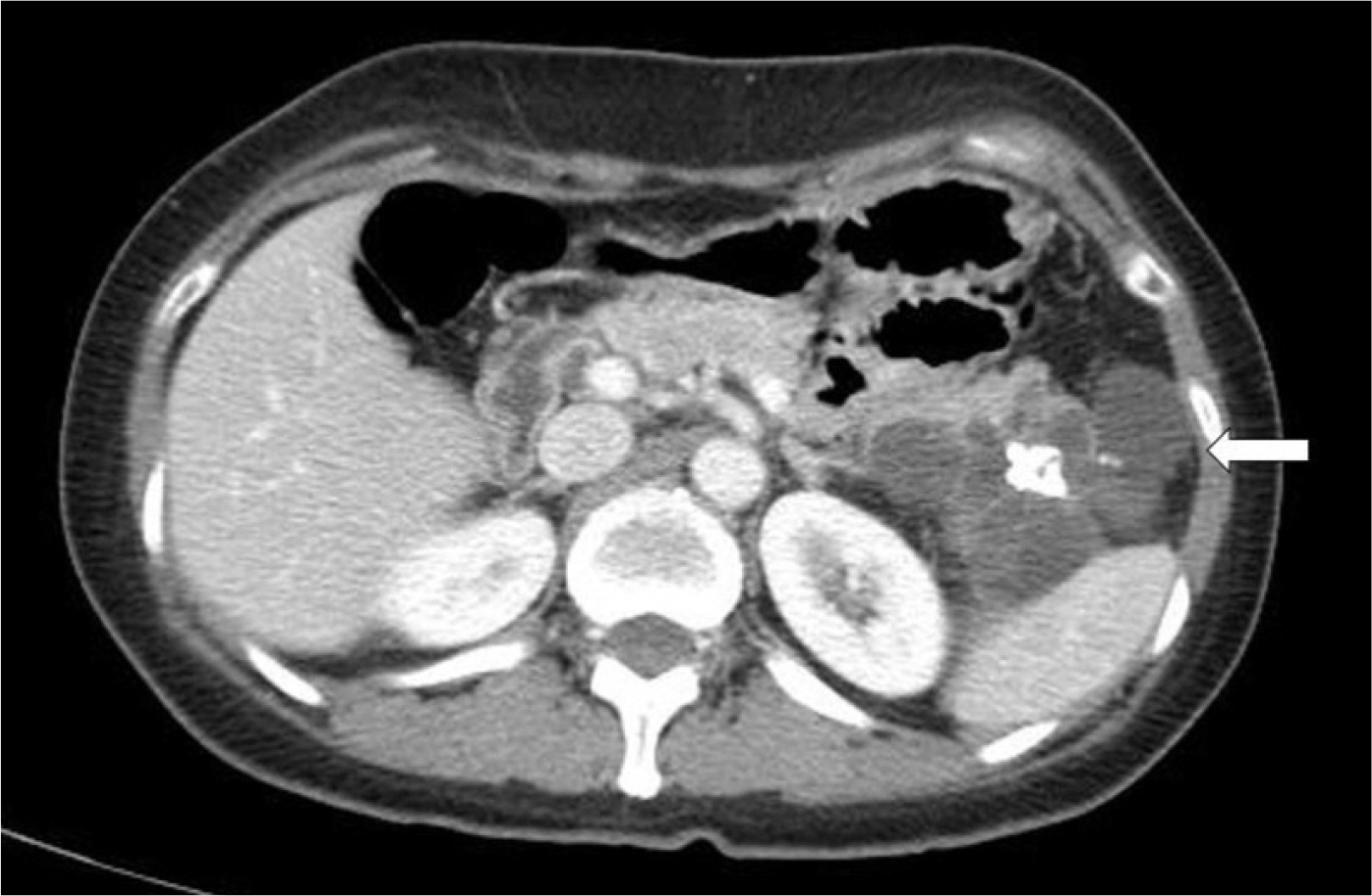
Fig. 1 Abdominal computed tomography revealed an 8 cm-sized calcified, multiseptated, and multilocular cystic mass in the pancreatic tail (arrow).

Fig. 2 Pancreatic magnetic resonance imaging (T2-weighted axial image) revealed an 8 cm-sized lobulated multilocular cyst in the pancreatic tail (arrow).

Fig. 3 Distal pancreatectomy specimen showed well delineated mutilocular cystic mass measuring 7.0×4.0×3.0 cm (arrows indicate mass boundaries).

Fig. 4 (A) Microscopic findings revealed variable sized multilocular cysts with incomplete septa (H&E,×100), and (B) focally linned by epithelium with acinar differentiation (arrows) (H&E,×100).
Reference
-
1. Gumus M, Ugras S, Algin O, Gundogdu H. 2011; Acinar cell cystadenoma (acinar cystic transformation) of the pancreas: the radiologic-pathologic features. Korean J Radiol. 12:129–134. DOI: 10.3348/kjr.2011.12.1.129. PMID: 21228949. PMCID: PMC3017877.
Article2. Kim BH, Park SY, Kim B, Kang GH. 2007; Acinar cell cystadenoma of the pancreas: report of a case with metaplastic ossification. J Pathol Transl Med. 41:203–206.3. Wang G, Ji L, Qu FZ, et al. 2016; Acinar cell cystadenoma of the pancreas: a retrospective analysis of ten-year experience from a single academic institution. Pancreatology. 16:625–631. DOI: 10.1016/j.pan.2016.03.020. PMID: 27086062.
Article4. Klöppel G. 2000; Pseudocysts and other non-neoplastic cysts of the pancreas. Semin Diagn Pathol. 17:7–15. PMID: 10721803.5. Albores-Saavedra J. 2002; Acinar cystadenoma of the pancreas: a previously undescribed tumor. Ann Diagn Pathol. 6:113–115. DOI: 10.1053/adpa.2002.32379. PMID: 12004359.
Article6. Singhi AD, Norwood S, Liu TC, et al. 2013; Acinar cell cystadenoma of the pancreas: a benign neoplasm or non-neoplastic ballooning of acinar and ductal epithelium? Am J Surg Pathol. 37:1329–1335. DOI: 10.1097/PAS.0b013e3182a1ad72. PMID: 24076773.7. Tanaka H, Hatsuno T, Kinoshita M, et al. 2016; A resected case of symptomatic acinar cell cystadenoma of the pancreas displacing the main pancreatic duct. Surg Case Rep. 2:39. DOI: 10.1186/s40792-016-0166-1. PMID: 27108123. PMCID: PMC4842199.
Article8. Wolf AM, Shirley LA, Winter JM, et al. 2013; Acinar cell cystadenoma of the pancreas: report of three cases and literature review. J Gastrointest Surg. 17:1322–1326. DOI: 10.1007/s11605-013-2199-0. PMID: 23605178.
Article9. Delavaud C, Cros J, et al. d'Assignies G. 2014; CT and MR imaging of multilocular acinar cell cystadenoma: comparison with branch duct intraductal papillary mucinous neoplasia (IPMNs). Eur Radiol. 24:2128–2136. DOI: 10.1007/s00330-014-3248-0. PMID: 24895037.
Article10. Sopha SC, Terhune JH, Hoover L, Uradomo L, Boutros CN. 2018; Acinar cell cystadenoma of the pancreas: a multidisciplinary and contemporary approach. J Gastrointest Surg. 22:1797–1798. DOI: 10.1007/s11605-018-3698-9. PMID: 29380117.
Article11. Volkan Adsay N. 2007; Cystic lesions of the pancreas. Mod Pathol. 20 Suppl 1:S71–S93. DOI: 10.1038/modpathol.3800706. PMID: 17486054.
Article12. Chen AL, Misdraji J, Brugge WR, Ferrone CR, Pitman MB. 2017; Acinar cell cystadenoma: a challenging cytology diagnosis, facilitated by moray® micro-forceps biopsy. Diagn Cytopathol. 45:557–560. DOI: 10.1002/dc.23693. PMID: 28236434.
Article13. Sigel CS, Klimstra DS. 2013; Cytomorphologic and immunophenotypical features of acinar cell neoplasms of the pancreas. Cancer Cytopathol. 121:459–470. DOI: 10.1002/cncy.21279. PMID: 23408736.
Article14. Orr J, Lockwood R, Roberts J, Shi C, Yachimski P. 2018; EUS and confocal endomicroscopic diagnosis of pancreatic acinar cell cystadenoma. Gastrointest Endosc. 88:769–770. DOI: 10.1016/j.gie.2018.06.003. PMID: 29906415.
Article15. Bergmann F, Aulmann S, Welsch T, et al. 2014; Molecular analysis of pancreatic acinar cell cystadenomas: evidence of a non-neoplastic nature. Oncol Lett. 8:852–858. DOI: 10.3892/ol.2014.2163. PMID: 25009661. PMCID: PMC4081433.
Article16. Zhang X, Zhu H, Yang X, Adsay VN, Jain D. 2017; Post-obstructive cyst formation in pancreas and cystic acinar transformation: Are they same? Pathol Res Pract. 213:997–1001. DOI: 10.1016/j.prp.2017.03.013. PMID: 28599853.
Article17. Alkhateeb MA, Boqari D, Mansi NK. 2020; Pancreatic acinar cystadenoma in a background of diffuse multifocal pancreatic cystic lesions: a case report. Int J Surg Case Rep. 73:223–227. DOI: 10.1016/j.ijscr.2020.07.026. PMID: 32712551. PMCID: PMC7390793.
Article18. Fahlbusch T, Tannapfel A, Uhl W, Braumann C. 2018; Acinar cell cystadenoma - a rarity in advanced von Hippel-Lindau disease: a case report. Visc Med. 34:73–75. DOI: 10.1159/000480372. PMID: 29594173. PMCID: PMC5869601.
Article19. Cosgrove N, DiPalma J, Katz D, Kowalski T. 2016; A rare case of acinar cell cystadenoma in a 14-year-old adolescent: a case report. Case Rep Pancreat Cancer. 2:3–5. DOI: 10.1089/crpc.2015.29009.nco. PMID: 30631807. PMCID: PMC6319691.
Article20. Khor TS, Badizadegan K, Ferrone C, et al. 2012; Acinar cystadenoma of the pancreas: a clinicopathologic study of 10 cases including multilocular lesions with mural nodules. Am J Surg Pathol. 36:1579–1591. DOI: 10.1097/PAS.0b013e318265fa4b. PMID: 23060352.21. Rift CV, Hasselby JP, Hansen CP, Federspiel B. 2020; Acinar cystic transformation of the pancreas: report of a case and a review of the literature. Pathol Res Pract. 216:152928. DOI: 10.1016/j.prp.2020.152928. PMID: 32204924.
Article22. Scott BB, Price TP, Callahan ZM, Poling JS, Lavu H. 2016; Intraductal papillary mucinous neoplasm of the pancreas arising in the setting of an intermixed acinar cell cystadenoma of the pancreas: report of a rare case. Case Rep Pancreat Cancer. 2:75–78. DOI: 10.1089/crpc.2016.0018. PMID: 30631822. PMCID: PMC6319694.
Article23. Wen X, Bandovic J. 2020; Fifteen-year follow-up of a patient with acinar cystic transformation of the pancreas and literature review. Case Rep Pathol. 2020:8847550. DOI: 10.1155/2020/8847550. PMID: 33425418. PMCID: PMC7781703.
Article
- Full Text Links
-
- Actions
-
Cited
- CITED
-
- Close
- Share
-
- Similar articles
-
- A Case of Serous Cystadenoma of the Pancreas Communicating with the Pancreatic Duct
- Pancreatic serous cystadenoma mimicking pseudocyst
- A Case of Pancreatic Serous Cystadenoma Associated with Papillary Thyroid Cancer
- Acinar Cell Cystadenoma of the Pancreas: Report of a Case with Metaplastic Ossification
- Acinar Cell Cystadenoma (Acinar Cystic Transformation) of the Pancreas: the Radiologic-Pathologic Features
