Primary pleomorphic liver liposarcoma: A case series and literature review
- Affiliations
-
- 1Department of General Surgery, Pusat Perubatan Universiti Kebangsaan Malaysia, Kuala Lumpur, Malaysia
- 2Department of General Surgery, Hospital Sultanah Aminah, Ministry of Health Malaysia, Johor Bahru, Malaysia
- 3Department of General Surgery, Sarawak General Hospital, Ministry of Health Malaysia, Kuching, Malaysia
- 4Department of Pathology, Sarawak General Hospital, Ministry of Health Malaysia, Kuching, Malaysia
- KMID: 2519298
- DOI: http://doi.org/10.14701/ahbps.2021.25.3.395
Abstract
- Primary hepatic liposarcoma is an extremely rare mesenchymal tumor that accounts for only 0.1% to 2% of primary malignant liver tumors. Due to its rarity, there is a lack of knowledge about its clinical course, management, and prognosis. Only 15 cases of primary liposarcoma of the liver have been reported since 1973. Among these 15 cases, only two involved primary liver liposarcoma with a pleomorphic subtype. Here we report the third and fourth cases of primary pleomorphic liver liposarcoma. A 57-year-old female presented with abdominal discomfort and progressive abdominal distension for two weeks. Computed tomography (CT) of her abdomen revealed a large well-defined solid nodule mass with an area of necrosis and hemorrhage occupying segment IV-B of the liver. Wide local excision was performed. She had an uneventful recovery and remained well at six months post-treatment. A 65-year-old male presented with an abdominal mass for two-month. CT demonstrated a mass in the left lobe of the liver with mixed soft tissues and fat attenuation. He underwent wide local excision. He was discharged on day three postoperatively. Histological analysis for both cases revealed liposarcoma of the liver with a pleomorphic subtype.
Keyword
Figure
-
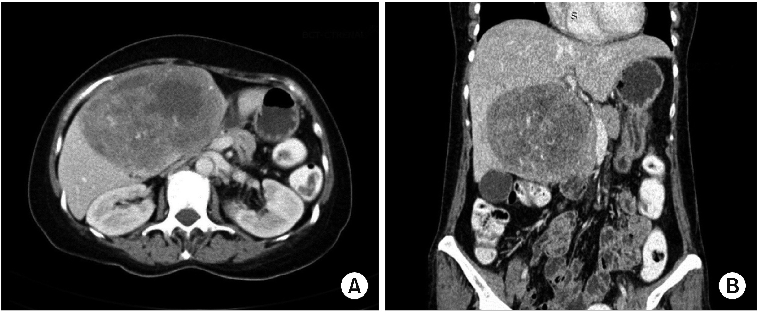
Fig. 1 Axial (A) and coronal (B) computed tomographyof the abdomen showing a well-defined predominantly solid mass with area of necrosis and hemorrhage at segment IV of liver.
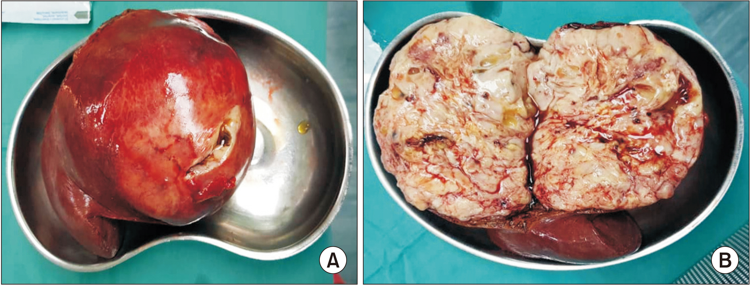
Fig. 2 (A) Well circumscribed and encapsulated fleshy pale yellowish tumour. (B) Bivalved section of specimen appearing brownish gelatinous with haemorrhagic foci.

Fig. 3 (A) Sheets of pleomorphic tumour cells (H&E, ×40). (B) Multiple bizarre tumour giant cells admixed with lipoblasts (blue arrows) are seen (H&E, ×200). (C) Tumour cells are diffusely positive for Vimentin (×200). (D) S100 stain highlighting lipoblasts (×400).
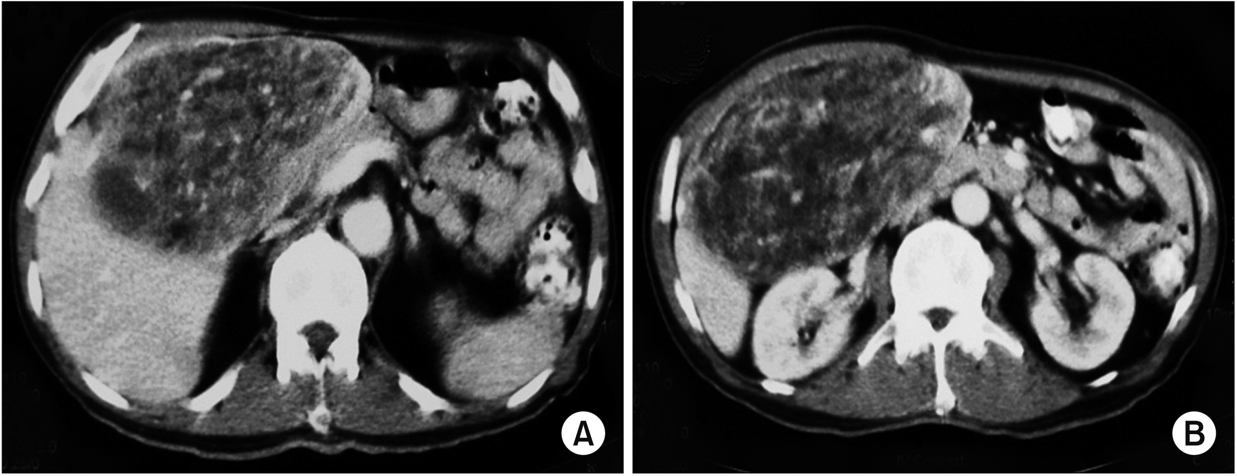
Fig. 4 Abdomen computed tomography (A, B) showing a large mass at the left lobe liver with mixed soft tissues and fat attenuation.
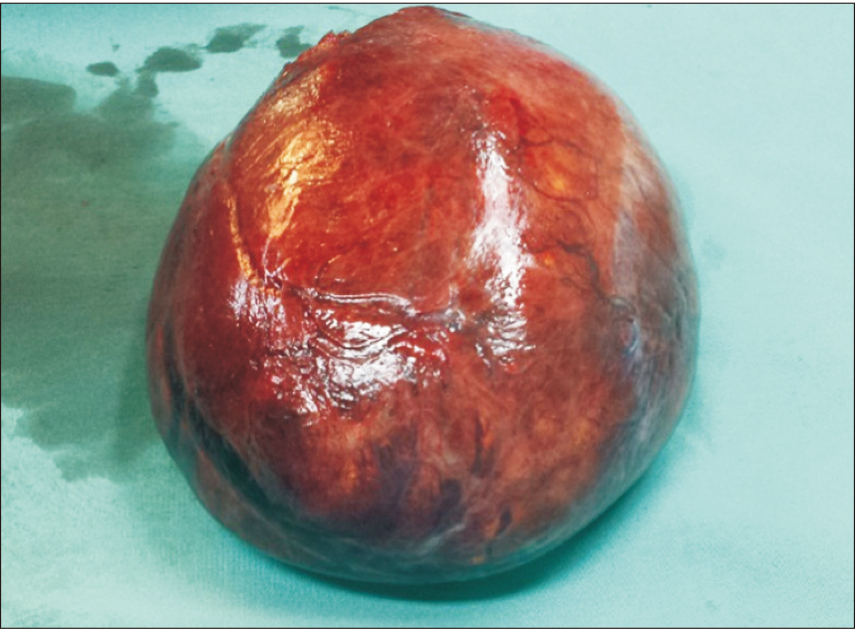
Fig. 5 Gross specimen of the tumour.
Reference
-
1. Liver Cancer Study Group of Japan. 1990; Primary liver cancer in Japan. Clinicopathologic features and results of surgical treatment. Ann Surg. 211:277–287. PMID: 2155591. PMCID: PMC1358432.2. Teas S, Ronan SG, Ghosh L. 1978; Solitary metastatic liposarcoma of the liver. Arch Pathol Lab Med. 102:605. PMID: 581455.3. WHO Classification of Tumours Editorial Board. 2020. Soft tissue and bone tumours. 5th ed. International Agency for Research on Cancer;Lyon:4. Kuo LM, Chou HS, Chan KM, Yu MC, Lee WC. 2006; A case of huge primary liposarcoma in the liver. World J Gastroenterol. 12:1157–1159. DOI: 10.3748/wjg.v12.i7.1157. PMID: 16534865. PMCID: PMC4087916.
Article5. Naik PR, Kumar P, Kumar PV. 2013; Primary pleomorphic liposarcoma of liver: a case report and review of the literature. Case Reports Hepatol. 2013:398910. DOI: 10.1155/2013/398910. PMID: 25374715. PMCID: PMC4208437.
Article6. Murphey MD, Arcara LK, Fanburg-Smith J. 2005; From the archives of the AFIP: imaging of musculoskeletal liposarcoma with radiologic-pathologic correlation. Radiographics. 25:1371–1395. DOI: 10.1148/rg.255055106. PMID: 16160117.7. Nassif NA, Tseng W, Borges C, Chen P, Eisenberg B. 2016; Recent advances in the management of liposarcoma. F1000Res. 5:2907. DOI: 10.12688/f1000research.10050.1. PMID: 28105325. PMCID: PMC5224678.
Article8. Wolloch Y, Dintsman M, Garti I. 1973; Primary malignant tumors of the liver. Isr J Med Sci. 9:6–11. PMID: 4352939.9. Kim TW, Reyes CV. 1985; Myxoid liposarcoma mimicking fluid density. J Surg Oncol. 30:80–82. DOI: 10.1002/jso.2930300204. PMID: 4079429.
Article10. Kim YI, Yu ES, Lee KW, Park EU, Song HG. 1987; Dedifferentiated liposarcoma of the liver. Cancer. 60:2785–2790. DOI: 10.1002/1097-0142(19871201)60:11<2785::AID-CNCR2820601131>3.0.CO;2-O. PMID: 3677011.
Article11. Soares FA, Landell GA, Peres LC, Oliveira MA, Vicente YA, Tone LG. 1989; Liposarcoma of hepatic hilum in childhood: report of a case and review of the literature. Med Pediatr Oncol. 17:239–243. DOI: 10.1002/mpo.2950170314. PMID: 2664442.
Article12. Wright NB, Skinner R, Lee RE, Craft AW. 1993; Myxoid liposarcoma of the porta hepatis in childhood. Pediatr Radiol. 23:620–621. DOI: 10.1007/BF02014984. PMID: 8152881.
Article13. Aribal E, Berberoglu L. 1993; Primary liposarcoma of the liver. AJR Am J Roentgenol. 161:1331–1332. DOI: 10.2214/ajr.161.6.8249754. PMID: 8249754.
Article14. Nelson V, Fernandes NF, Woolf GM, Geller SA, Petrovic LM. 2001; Primary liposarcoma of the liver: a case report and review of literature. Arch Pathol Lab Med. 125:410–412. DOI: 10.5858/2001-125-0410-PLOTL. PMID: 11231494.15. Kim JL, Woo JY, Lee MJ, Kim KR, Jung JP, Lee NJ, et al. 2007; Imaging findings of primary well-differentiated liposarcoma of the liver: a case report. Acta Radiol. 48:1061–1065. DOI: 10.1080/02841850701630334. PMID: 18038349.
Article16. Nakhai B, Motabar AR. 2007; Primary liposarcoma of the liver: a case report and review of the literature. Med J Islam Repub Iran. 21:167–172.17. Lin YC, Tsai WC, Liu YC, Yu CP. 2011; Primary liposarcoma of liver: a case report and literature review. J Med Sci. 31:81–84.18. Binesh F, Akhavan A, Kargar S, Navabii H. 2012; Primary liposarcoma of liver: a rare case and literature review. BMJ Case Rep. 2012:bcr0120125711. DOI: 10.1136/bcr-01-2012-5711. PMID: 22751420. PMCID: PMC4543104.
Article19. Chen L, Luo J, Wen Q, Chu S, Wang W, Fan S. 2016; Primary pleomorphic liposarcoma of liver: a case report and review of the literature. Int J Clin Exp Pathol. 9:7629–7633. DOI: 10.1155/2013/398910,. PMID: 25374715. PMCID: PMC4208437.20. Rayya F. 2017; Resection of a rare hepatic myxoid liposarcoma: case report. Clin Surg. 2:1310.
- Full Text Links
-
- Actions
-
Cited
- CITED
-
- Close
- Share
-
- Similar articles
-
- Recurrent Primary Pleomorphic Liposarcoma of the Breast: A Case Report with Imaging Findings
- A Case of Pleomorphic Lipoma in the Scalp
- Pleural Liposarcoma: Case Report
- Retroperitoneal Pleomorphic Liposarcoma Mimicking Adrenal Cancer in F-18 FDG PET/CT
- Primary Round Cell Liposarcoma of the Omentum: A case report
