Gastric Oxyntic Mucosa Pseudopolyps
- Affiliations
-
- 1Department of Internal Medicine, Pusan National University School of Medicine, and Biomedical Research Institute, Pusan National University Hospital, Busan, Korea
- KMID: 2518869
- DOI: http://doi.org/10.5946/ce.2020.157
Figure
-
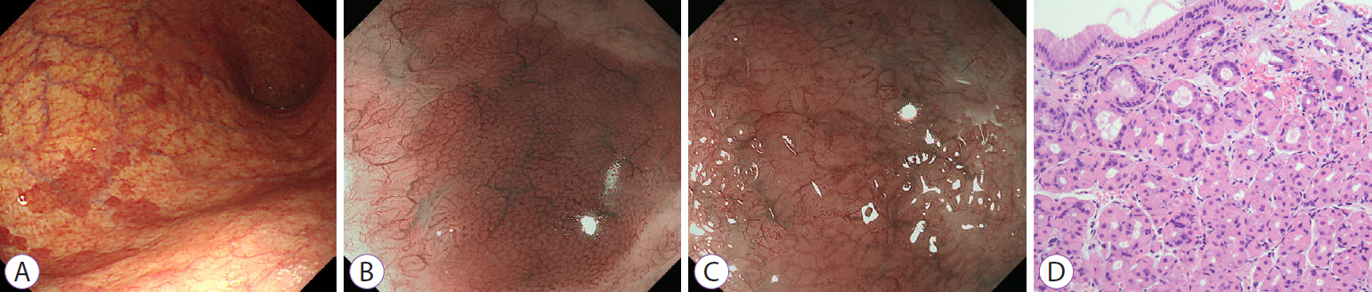
Fig. 1. (A) On conventional endoscopy, multiple, reddish, nodular lesions with variable sizes are seen in the background of atrophic gastritis in the gastric body and fundus. (B) Magnifying endoscopy with narrow-band imaging (ME-NBI) of the reddish nodular lesions reveals small, round pits surrounded by honeycomb-type subepithelial capillary networks (SECNs) with a regular arrangement of collecting venules. (C) ME-NBI of the surrounding atrophic mucosa reveals loss of the normal SECNs and round pits, with an irregular arrangement of the collecting venules. (D) Endoscopic biopsy of the reddish nodular lesions reveals intact oxyntic mucosa without evidence of atrophy or intestinal metaplasia, and pseudohypertrophy of parietal cells with protrusion into the gland lumen (hematoxylin and eosin stain, ×200).
Reference
- Full Text Links
-
- Actions
-
Cited
- CITED
-
- Close
- Share
-
- Similar articles
-
- Oxyntic Gland Area and Density of Parietal Cells and Its Relationship to Acid Output in Helicobacter pylori Infected Duodenal Ulcer Patients
- Topographic Difference of Inflammatory Reactions in Gastric Mucosa in Various Helicobacter pylori-Associated Diseases
- Oxyntic Gland Adenoma Treated by Endoscopic Mucosal Resection
- Prolapsed gastric mucosa through gastrojejunostomy (report of 3 cases with review of the literature)
- Heterotopic Gastric Mucosa in the Umbilicus
