A Case of Small Cell Carcinoma Originated from Sphenoid Sinus in Patient with Recurrent Pituitary Tumor
- Affiliations
-
- 1Department of Otorhinolaryngology-Head and Neck Surgery, Dong-A University, College of Medicine, Busan, Korea
- 2Department of Pathology, Dong-A University, College of Medicine, Busan, Korea
- KMID: 2518677
- DOI: http://doi.org/10.18787/jr.2021.00351
Abstract
- Small cell carcinoma (SmCC) is a type of neuroendocrine tumor commonly originating in the lung, with only about 2-4% of cases arising at extrapulmonary sites. Extrapulmonary SmCC of the head and neck has a poor prognosis and a high rate of distant metastasis. The paranasal sinus is a rare location for extrapulmonary SmCC and only a few related papers have been published to date. We report a rare case of SmCC originating from the sphenoid sinus in a patient with a recurrent pituitary tumor with a literature review.
Keyword
Figure
-
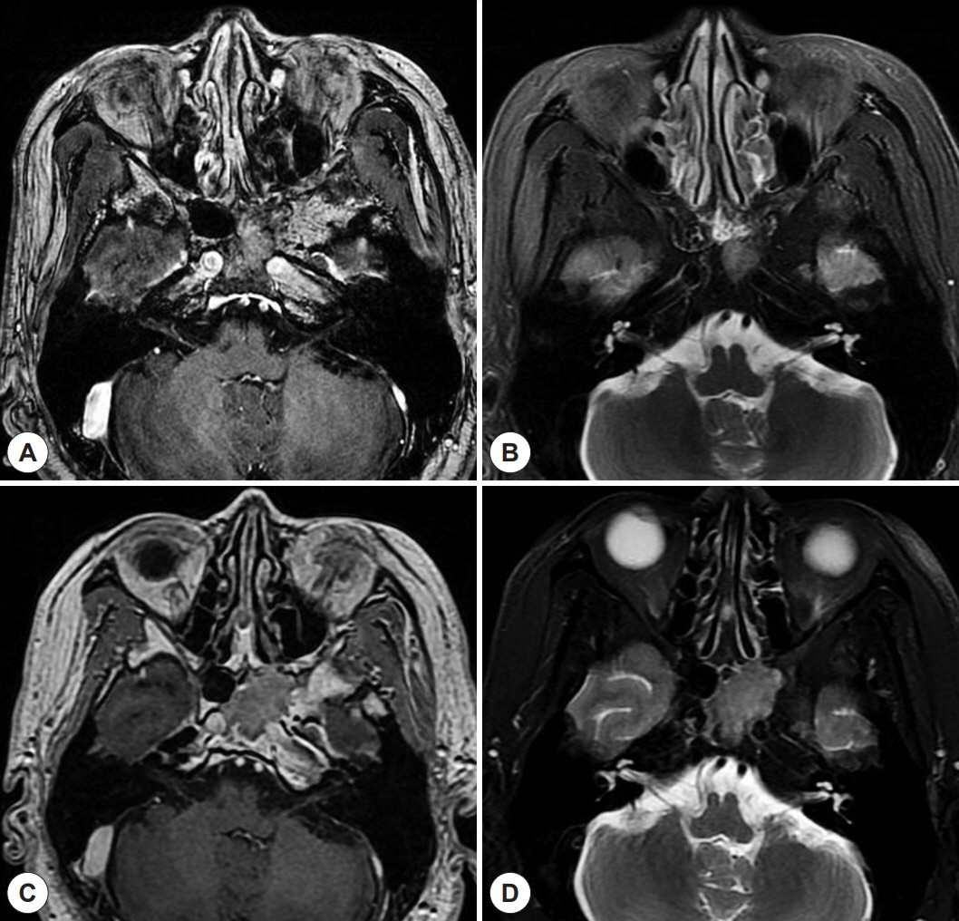
Fig. 1. Post-operative brain magnetic resonance images (MRI). A: Post-operative 5 years Axial view of T1 enhanced brain MRI shows that intermediate signal intensity in left sphenoid sinus with homogenous enhancement, after that, patient had received radiotherapy for recurrent pituitary adenoma. B: Axial T2-weighted imaged. C: Post-operative 11 years Axial view of T1 enhanced brain MRI shows that intermediate signal intensity in left sphenoid sinus and increasing in size compared to previous findings. D: Axial T2-weighted imaged.
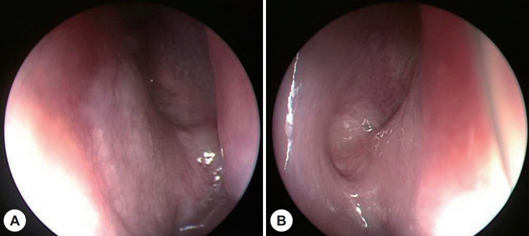
Fig. 2. Endoscopic examination. There is no abnormal finding on sphenoid sinus opening (A: right side, B: left side).

Fig. 3. Initial and follow-up paranasal sinus computed tomography (PNS CT) (A) there is no abnormal finding in initial view of PNS CT. (B) After trans-sphenoidal operation, PNS CT shows that soft tissue density in left sphenoid. (C) Post-operative 5 years PNS CT shows that an increase in size compared to the previous CT image without bony erosion. (D) Post-operative 11 years PNS CT shows that inhomogeneous density mass in left sphenoid sinus has found markedly size increase with internal carotid artery area infiltration and bony destruction was found to lateral sphenoid bone and partially bony erosion to sella turcica. (E) Coronal view of PNS CT (F) Sagittal view of PNS CT.
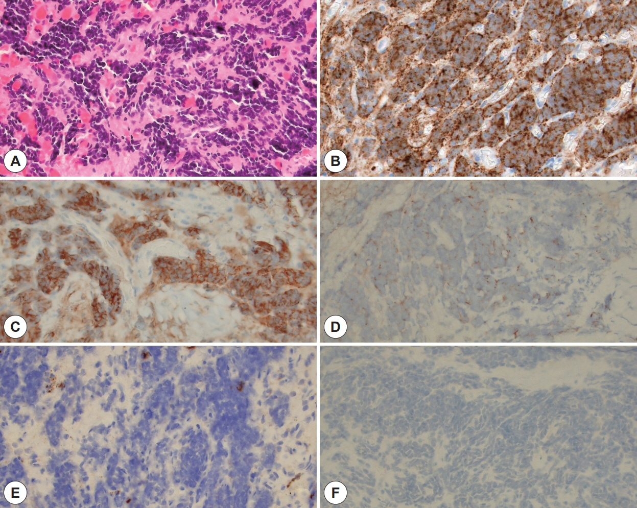
Fig. 4. Pathological findings. A: No pituitary gland tissue is observed. Round to oval cells with scant cytoplasm are arranged in a sheet-like pattern. These cells show finely dispersed chromatin and no distinct nucleoli (H&E, ×400 magnification). B-D: Tumor cells show diffuse membranous positive staining for CD56 and synaptophysin and paranuclear dot-like immunoreactivity for panCK (B: CD56, C: synaptophysin, D: panCK, ×400 magnification). E-F: Only tumor infiltrating lymphocytes demonstrate positive immunoreactivity for CD45 and tumor cells are negative for CD45 and GFAP (E: CD45, F: GFAP, ×400 magnification).
Reference
-
References
1. Pointer KB, Ko HC, Brower JV, Witek ME, Kimple RJ, Lloyd RV, et al. Small cell carcinoma of the head and neck: An analysis of the National Cancer Database. Oral Oncol. 2017; 69:92–8.2. Kim YS, Bae CH, Song SY, Kim YD. A case of a small cell neuroendocrine carcinoma of the nasal septum. Korean J Otorhinolaryngol-Head Neck Surg. 2009; 52(6):529–32.3. Wakasaki T, Yasumatsu R, Masuda M, Matsuo M, Tamae A, Kubo K, et al. Small Cell Carcinoma in the Head and Neck. Ann Otol Rhinol Laryngol. 2019; 128(11):1006–12.4. Raychowdhuri RN. Oat-cell carcinoma and paranasal sinuses. J Laryngol Otol. 1965; 79:253–5.5. Yeo SC, Cho HJ, Kim SW, Jeon SY. A case of small cell carcinoma of the maxillary sinus coexisting with fungus ball. J Rhinol. 2016; 23(2):110–4.6. Ha SY, An HJ, Cho KW, Yoo CY, Cho JH, Kim SH, et al. A case of small cell carcinoma of the maxillary sinus with distant metastasis. Korean J Med. 2015; 88(6):719–23.7. Koo BM, Kim HG, Cho H, Park P. A Case of Delayed Diagnosis of Small Cell Carcinoma Originated from the Maxillary Sinus with Bilateral Fungal Sinusitis. Korean J Otorhinolaryngol-Head Neck Surg. 2020; 63(6):276–81.8. Nagaishi M, Suzuki K, Sugiura Y, Takano I, Tanaka Y, Hyodo A. Undifferentiated sarcoma of the sphenoid sinus. Auris Nasus Larynx. 2018; 45(2):388–91.9. Ma ATW, Lei KIK. Small cell neuroendocrine carcinoma of the ethmoid sinuses presenting with generalized seizure and syndrome of inappropriate antidiuretic hormone secretion: a case report and review of literature. Am J Otolaryngol. 2009; 30(1):54–7.10. Kang JM, Lee HY, Lee KS, Ko SY. Small Cell Neuroendocrine Carcinoma of the Nasal Cavity: a case report. Korean J Otorhinolaryngol-Head Neck Surg. 2003; 46(2):164–7.11. Bishop JA, Guo TW, Smith DF, Wang H, Ogawa T, Pail SI, et al. Human papillomavirus-related carcinomas of the sinonasal tract. Am J Surg Pathol. 2013; 37(2):185–92.12. Nour YA, Al-Madani A, El-Daly A, Gaafar A. Isolated sphenoid sinus pathology: spectrum of diagnostic and treatment modalities. Auris Nasus Larynx. 2008; 35(4):500–8.13. Qingqiang Zhu, Wenrong Zhu, Jingtao Wu, Hongying Zhang. The CT and MRI observations of small cell neuroendocrine carcinoma in paranasal sinuses. World J Surg Oncol. 2015; 13:54.14. Naier Lin, Meng Qi, Zhengyue Wang, Siqi Luo, Yucheng Pan, Fang Zhang, et al. Small Cell Neuroendocrine Carcinoma of Paranasal Sinuses: Radiologic Features in 14 Cases. J Comput Assist Tomogr. 2021; 45(1):135–41.15. Babin E, Rouleau V, Vedrine PO, Toussaint B, de Raucourt D, Malard O, et al. Small cell neuroendocrine carcinoma of the nasal cavity and paranasal sinuses. J Laryngol Otol. 2006; 120(4):289–97.
- Full Text Links
-
- Actions
-
Cited
- CITED
-
- Close
- Share
-
- Similar articles
-
- Sphenoid Sinus Mucocele Complicated With Spontaneous CSF Rhinorrhea: Case Report
- Sphenoid Sinus Mucocele(Case Report)
- Ectopic Pituitary Adenoma within the Sphenoid Sinus
- Isolated Inverted Papilloma of the Sphenoid Sinus Presenting as Ptosis
- A Case of Compressive Optic Neuropathy Caused by Sphenoid Sinus Mucocele
