J Korean Neurosurg Soc.
2021 Jul;64(4):585-591. 10.3340/jkns.2020.0223.
Features of the Filum Terminale in Tethered Cord Syndrome with Focus on Pathology
- Affiliations
-
- 1Department of Neurosurgery, Seoul National University Hospital, Seoul, Korea
- 2Division of Pediatric Neurosurgery, Seoul National University Children’s Hospital, Seoul, Korea
- 3Department of Anatomy and Cell Biology, Seoul National University College of Medicine, Seoul, Korea
- KMID: 2517691
- DOI: http://doi.org/10.3340/jkns.2020.0223
Abstract
Objective
: Filum transection is one of the most commonly performed operative procedure in pediatric neurosurgery. However, the clinical and pathological features as well as the surgical indication are not well-established. This study aimed to analyze the characteristics of patients who underwent transection of the filum during the last 10 years in a single institute.
Methods
: A total of 82 patients underwent transection of the filum during the period. As a general rule, we performed the transection in patients who are symptomatic or have abnormality in the urologic or neuromuscular evaluations. There were exceptions as asymptomatic patients who only fit the definition of thickened filum (width greater than 2.0 mm or conus level below L3 vertebral body) were operated by parent’s wish or surgeon’s preference according to radiological findings, etc.
Results
: Seventy-six out of 82 patients had fibrous tissue in the pathologic specimen of filum. Interestingly, patients who had glial cells were more correlated with no preoperative syrinx, and no progression of syrinx even for those who did have syrinx initially. Also, larger percentage of symptomatic patients had peripheral nerve twigs than asymptomatic patients. No difference in conus level or thickness of filum was found between patients with or without preoperative syrinx. Significantly more patients with syrinx (56%) were chosen to be operated without any symptom or abnormality in study i.e., solely based on radiological findings than those without syrinx (21%). The surgical outcome for syrinx was favorable, as all but one patient had either improved or static syrinx. The exceptional case had increase in size due to the upward displacement of the proximal end of the cut filum.
Conclusion
: This study evaluated the pathological, clinical, radiological features of patients who underwent transection of the filum. Interesting correlations between pathological findings and clinical features were found. Excellent outcome regarding preoperative syrinx was also shown.
Figure
-
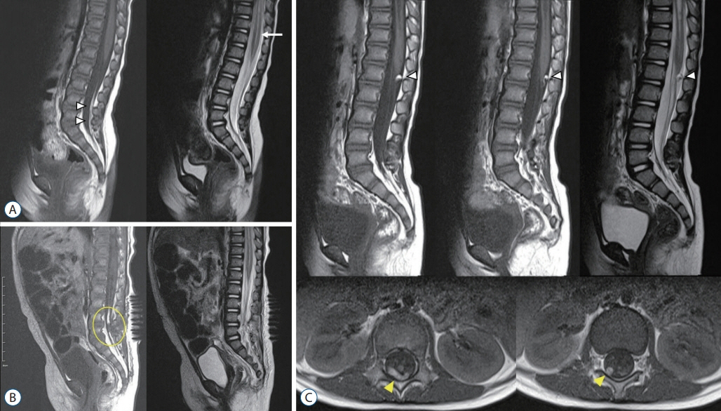
Fig. 1. A : Preoperative MRI showing fatty filum (arrowheads) and syrinx (arrow). B : Immediate postoperative MRI showing cut filum (circle). C : MRI taken 1 year after the operation showing a mass at the dorsal surface of the conus (white arrowheads, high signal intensity on both T2 and T1 images). The axial images reveal that the mass is actually elongated, linear shape (yellow arrowheads) suggesting it is the curled up proximal end of the cut filum. MRI : magnetic resonance imaging.
Reference
-
References
1. Blount JP, Elton S. Spinal lipomas. Neurosurg Focus. 10:1–13. 2001.
Article2. Chong S, Lee JY, Kim KH, Shin HI, Kim K, Park K, et al. Radical excision of lumbosacral lipoma: an early experience of “followers”. Childs Nerv Syst. 35:1591–1597. 2019.
Article3. Cools MJ, Al-Holou WN, Stetler WR Jr, Wilson TJ, Muraszko KM, Ibrahim M, et al. Filum terminale lipomas: imaging prevalence, natural history, and conus position. J Neurosurg Pediatr. 13:559–567. 2014.
Article4. Cornips EM, Vereijken IM, Beuls EA, Weber JW, Soudant DL, van Rhijn LW, et al. Clinical characteristics and surgical outcome in 25 cases of childhood tight filum syndrome. Eur J Paediatr Neurol. 16:103–117. 2012.
Article5. George TM, Cummings TJ. The immunohistochemical profile of the myelomeningocele placode: is the placode normal? Pediatr Neurosurg. 39:234–239. 2003.
Article6. Guerra LA, Pike J, Milks J, Barrowman N, Leonard M. Outcome in patients who underwent tethered cord release for occult spinal dysraphism. J Urol. 176(4 Pt 2):1729–1732. 2006.
Article7. Kashlan ON, Wilkinson DA, Morgenstern H, Khalsa SS, Maher CO. Predictors of surgical treatment in children with tethered fibrofatty filum terminale. J Neurosurg Pediatr. 25:196–203. 2020.
Article8. Metcalfe PD, Luerssen TG, King SJ, Kaefer M, Meldrum KK, Cain MP, et al. Treatment of the occult tethered spinal cord for neuropathic bladder: results of sectioning the filum terminale. J Urol. 176(4 Pt 2):1826–1829. discussion 1830. 2006.
Article9. Selçuki M, Vatansever S, Inan S, Erdemli E, Bağdatoğlu C, Polat A. Is a filum terminale with a normal appearance really normal? Childs Nerv Syst. 19:3–10. 2003.
Article10. Selden NR, Nixon RR, Skoog SR, Lashley DB. Minimal tethered cord syndrome associated with thickening of the terminal filum. J Neurosurg. 105(3 Suppl):214–218. 2006.
Article11. Thompson EM, Strong MJ, Warren G, Woltjer RL, Selden NR. Clinical significance of imaging and histological characteristics of filum terminale in tethered cord syndrome. J Neurosurg Pediatr. 13:255–259. 2014.
Article12. Tsitouras V, Sgouros S. Syringomyelia and tethered cord in children. Childs Nerv Syst. 29:1625–1634. 2013.
Article13. Yundt KD, Park TS, Kaufman BA. Normal diameter of filum terminale in children: in vivo measurement. Pediatr Neurosurg. 27:257–259. 1997.
Article
- Full Text Links
-
- Actions
-
Cited
- CITED
-
- Close
- Share
-
- Similar articles
-
- Case Report of Fibrolipomatous Filum Terminale in Korean Cadaver
- Neurogenic Bladder in the Patients with Tethered Cord Syndrome: A case report
- Fully Endoscopic Interlaminar Detethering of Spinal Cord in Tethered Cord Syndrome: A Case Report and Technical Description
- Isolated Hemangioblastoma of the Filum Terminale
- A Case of Spinal Cord Ependymoma
