J Korean Assoc Oral Maxillofac Surg.
2021 Jun;47(3):233-236. 10.5125/jkaoms.2021.47.3.233.
Pre-contoured reconstruction plate fabricated via three-dimensional printed bending support
- Affiliations
-
- 1Department of Dentistry, Korea University Anam Hospital, Seoul, Korea
- 2Department of Oral and Maxillofacial Surgery, Chung-Ang University School of Medicine, Seoul, Korea
- 3Department of Oral and Maxillofacial Surgery, Dental Center, Chung-Ang University Hospital, Seoul, Korea
- KMID: 2517275
- DOI: http://doi.org/10.5125/jkaoms.2021.47.3.233
Abstract
- A mandibular continuity defect can be repaired using either a prosthetic device or autogenous bone. A titanium reconstruction plate can be used with a localized or vascularized flap over the defect of the mandible. Unfortunately, the plate may fail due to plate exposure, screw loosening, fracture, or infection, and will need to be removed. Plate exposure though the skin or mucosa is one of the main reasons for failure. In the present work, the authors introduced a lingually positioned reconstruction plate fabricated via three-dimensional printed bending support. This custom reconstruction plate can avoid plate re-exposure as well as reduce surgical errors and operation time.
Keyword
Figure
-

Fig. 1 Exposed reconstruction plate.

Fig. 2 Modeling of a reconstruction plate located as medially as possible according to the contours of the lingual cortex. The virtual plate and holes were three-dimensionally simulated along the cortex of the mandible. Fabrication of the lower bending support according to the mandible model. The rod on the bending support was fitted to the hole of the plate, which holds the plate during the bending procedure. Pre-contouring of the plate using a combination of upper and lower bending halves.

Fig. 3 Computed tomography image of the lingual pre-contoured reconstruction plate affixed to the mandibular continuity defect.
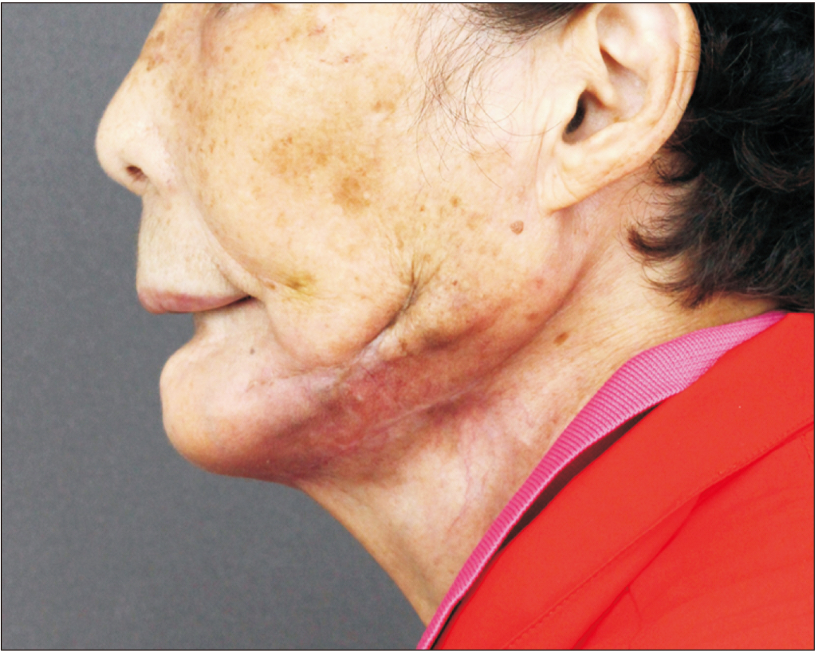
Fig. 4 Postoperative healing of the skin after 6 months.
Reference
-
References
1. Spencer KR, Sizeland A, Taylor GI, Wiesenfeld D. 1999; The use of titanium mandibular reconstruction plates in patients with oral cancer. Int J Oral Maxillofac Surg. 28:288–90. PMID: 10416897.
Article2. Lindqvist C, Söderholm AL, Salo A, Subasinghe J, Ylijoki S, Skutnabb K, et al. 2001; A comparative study on four screw-plate locking systems in sheep: a clinical and radiological study. Int J Oral Maxillofac Surg. 30:160–6. https://doi.org/10.1054/ijom.2000.0037. DOI: 10.1054/ijom.2000.0037. PMID: 11405453.
Article3. Shibahara T, Noma H, Furuya Y, Takaki R. 2002; Fracture of mandibular reconstruction plates used after tumor resection. J Oral Maxillofac Surg. 60:182–5. https://doi.org/10.1053/joms.2002.29817. DOI: 10.1053/joms.2002.29817. PMID: 11815918.
Article4. Katakura A, Shibahara T, Noma H, Yoshinari M. 2004; Material analysis of AO plate fracture cases. J Oral Maxillofac Surg. 62:348–52. https://doi.org/10.1016/j.joms.2003.05.009. DOI: 10.1016/j.joms.2003.05.009. PMID: 15015169.
Article5. Schöning H, Emshoff R. 1998; Primary temporary AO plate reconstruction of the mandible. Oral Surg Oral Med Oral Pathol Oral Radiol Endod. 86:667–72. https://doi.org/10.1016/s1079-2104(98)90201-3. DOI: 10.1016/s1079-2104(98)90201-3. PMID: 9868722.
Article6. Ettl T, Driemel O, Dresp BV, Reichert TE, Reuther J, Pistner H. 2010; Feasibility of alloplastic mandibular reconstruction in patients following removal of oral squamous cell carcinoma. J Craniomaxillofac Surg. 38:350–4. https://doi.org/10.1016/j.jcms.2009.04.011. DOI: 10.1016/j.jcms.2009.04.011. PMID: 19501515.
Article7. Kämmerer PW, Klein MO, Moergel M, Gemmel M, Draenert GF. 2014; Local and systemic risk factors influencing the long-term success of angular stable alloplastic reconstruction plates of the mandible. J Craniomaxillofac Surg. 42:e271–6. https://doi.org/10.1016/j.jcms.2013.10.004. DOI: 10.1016/j.jcms.2013.10.004. PMID: 24296118.
Article8. Liu SP, Cai ZG, Zhang J, Zhang JG, Zhang Y. 2013; [Plate related complication after mandibular reconstruction]. Zhonghua Kou Qiang Yi Xue Za Zhi. 48:586–90. Chinese. PMID: 24438564.9. Nicholson RE, Schuller DE, Forrest LA, Mountain RE, Ali T, Young D. 1997; Factors involved in long- and short-term mandibular plate exposure. Arch Otolaryngol Head Neck Surg. 123:217–22. https://doi.org/10.1001/archotol.1997.01900020107016. DOI: 10.1001/archotol.1997.01900020107016. PMID: 9046293.
Article10. Maurer P, Eckert AW, Kriwalsky MS, Schubert J. 2010; Scope and limitations of methods of mandibular reconstruction: a long-term follow-up. Br J Oral Maxillofac Surg. 48:100–4. https://doi.org/10.1016/j.bjoms.2009.07.005. DOI: 10.1016/j.bjoms.2009.07.005. PMID: 19647911.
Article11. Wei FC, Celik N, Yang WG, Chen IH, Chang YM, Chen HC. 2003; Complications after reconstruction by plate and soft-tissue free flap in composite mandibular defects and secondary salvage reconstruction with osteocutaneous flap. Plast Reconstr Surg. 112:37–42. https://doi.org/10.1097/01.PRS.0000065911.00623.BD. DOI: 10.1097/01.PRS.0000065911.00623.BD. PMID: 12832874.
Article12. Martola M, Lindqvist C, Hänninen H, Al-Sukhun J. 2007; Fracture of titanium plates used for mandibular reconstruction following ablative tumor surgery. J Biomed Mater Res B Appl Biomater. 80:345–52. https://doi.org/10.1002/jbm.b.30603. DOI: 10.1002/jbm.b.30603. PMID: 16850467.
Article13. Azuma M, Yanagawa T, Ishibashi-Kanno N, Uchida F, Ito T, Yamagata K, et al. 2014; Mandibular reconstruction using plates prebent to fit rapid prototyping 3-dimensional printing models ameliorates contour deformity. Head Face Med. 10:45. https://doi.org/10.1186/1746-160X-10-45. DOI: 10.1186/1746-160X-10-45. PMID: 25338640. PMCID: PMC4213462.
Article14. Salgueiro MI, Stevens MR. 2010; Experience with the use of prebent plates for the reconstruction of mandibular defects. Craniomaxillofac Trauma Reconstr. 3:201–8. https://doi.org/10.1055/s-0030-1268520. DOI: 10.1055/s-0030-1268520. PMID: 22132258. PMCID: PMC3052711.
Article15. Cunningham LL Jr, Madsen MJ, Peterson G. 2005; Stereolithographic modeling technology applied to tumor resection. J Oral Maxillofac Surg. 63:873–8. https://doi.org/10.1016/j.joms.2005.02.027. DOI: 10.1016/j.joms.2005.02.027. PMID: 15944992.
Article16. Coopman R, Aerden T, De Temmerman G, Politis C. 2017; Mandibular wing osteotomy: technical modification. Br J Oral Maxillofac Surg. 55:635–6. https://doi.org/10.1016/j.bjoms.2017.04.005. DOI: 10.1016/j.bjoms.2017.07.013. PMID: 28844570.
Article17. Chan HH, Siewerdsen JH, Vescan A, Daly MJ, Prisman E, Irish JC. 2015; 3D rapid prototyping for otolaryngology-head and neck surgery: applications in image-guidance, surgical simulation and patient-specific modeling. PLoS One. 10:e0136370. https://doi.org/10.1371/journal.pone.0136370. DOI: 10.1371/journal.pone.0136370. PMID: 26331717. PMCID: PMC4557980.
Article18. Toro C, Robiony M, Costa F, Zerman N, Politi M. 2007; Feasibility of preoperative planning using anatomical facsimile models for mandibular reconstruction. Head Face Med. 3:5. https://doi.org/10.1186/1746-160X-3-5. DOI: 10.1186/1746-160X-3-5. PMID: 17224060. PMCID: PMC1783647.
Article19. Zweifel DF, Simon C, Hoarau R, Pasche P, Broome M. 2015; Are virtual planning and guided surgery for head and neck reconstruction economically viable? J Oral Maxillofac Surg. 73:170–5. https://doi.org/10.1016/j.joms.2014.07.038. DOI: 10.1016/j.joms.2014.07.038. PMID: 25443385.
Article
- Full Text Links
-
- Actions
-
Cited
- CITED
-
- Close
- Share
-
- Similar articles
-
- Plate prebending using a three-dimensional–printed model affords effective anatomical reduction in clavicular shaft fractures
- Reconstruction of the Cervical Lateral Mass Using 3-Dimensional-Printed Prostheses
- 3D-printed titanium implant with pre-mounted dental implants for mandible reconstruction: a case report
- Anatomical Analysis of Clavicles in Korean Adults and Compatibility of Pre-Contoured Anatomical Plates
- Accuracy of dies fabricated by various three dimensional printing systems: a comparative study
