Unusual Magnetic Resonance Imaging Findings Contrast-induced Encephalopathy following Cerebral Angiography
- Affiliations
-
- 1Department of Neurosurgery, Medical Research Institute, Pusan National University Hospital, Busan, Korea
- 2Department of Diagnostic Radiology, Medical Research Institute, Pusan National University Hospital, Busan, Korea
- KMID: 2517226
- DOI: http://doi.org/10.7180/kmj.2021.36.1.51
Abstract
- Contrast-induced encephalopathy (CIE) following cerebral angiography has similar clinical presentations to ischemic complications of cerebral angiography. Neurologic deficits in CIE are mostly transient, but those caused by acute cerebral infarction (ACI) as ischemic complications of cerebral angiography may be permanent. Therefore, distinguishing CIE from ACI is important. Diffusion restriction on magnetic resonance imaging (MRI) implies ACI, while hyperintensity on diffusion weighted imaging (DWI) without correlation on the apparent diffusion coefficient (ADC) map implies CIE. We reported a rare case of CIE with diffusion restriction on MRI following cerebral angiography that mimicked MRI findings of ACI. The mechanism of this phenomenon remains unknown and requires further investigation.
Figure
-
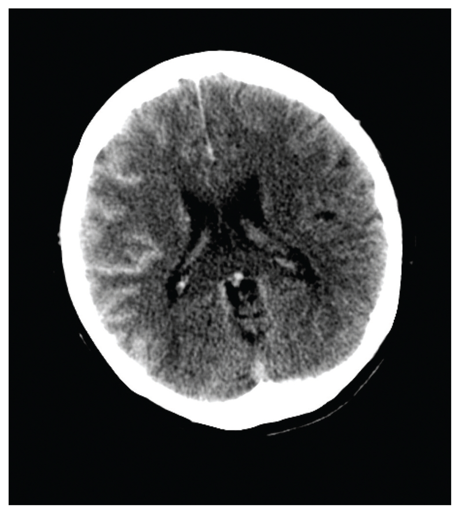
Fig. 1 Brain computed tomography following cerebral angiography shows marked enhancement throughout the right cerebral cortex with concordant diffuse swelling, indicating of contrast-induced encephalopathy.
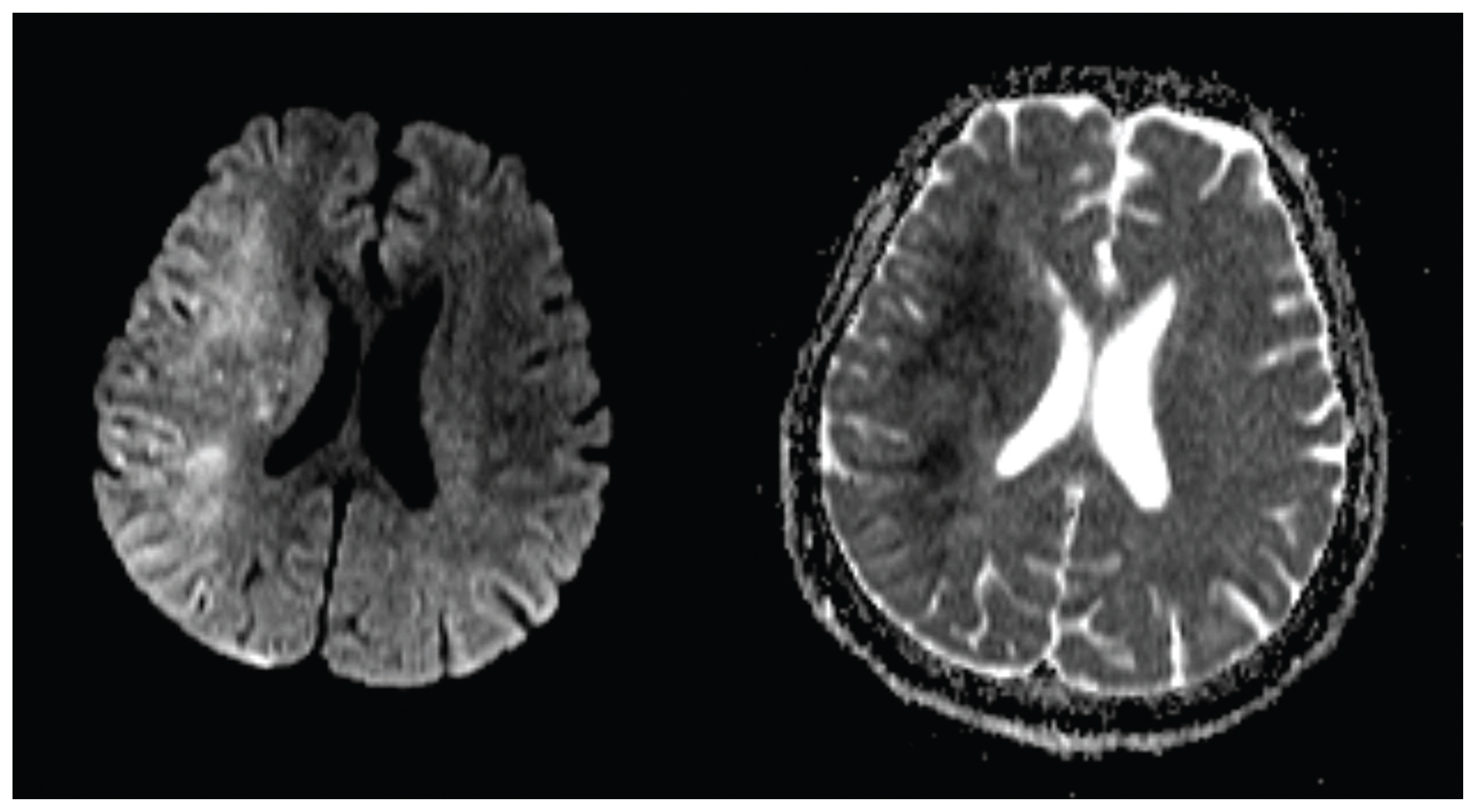
Fig. 2 Magnetic resonance imaging following cerebral angiography shows extensive cortical and striatal hyperintensity within the right hemisphere on diffusion weighted image (left) and corresponding hypointense signal alterations on the apparent diffusion coefficient map (right) nearly matching with the findings of computed tomography.
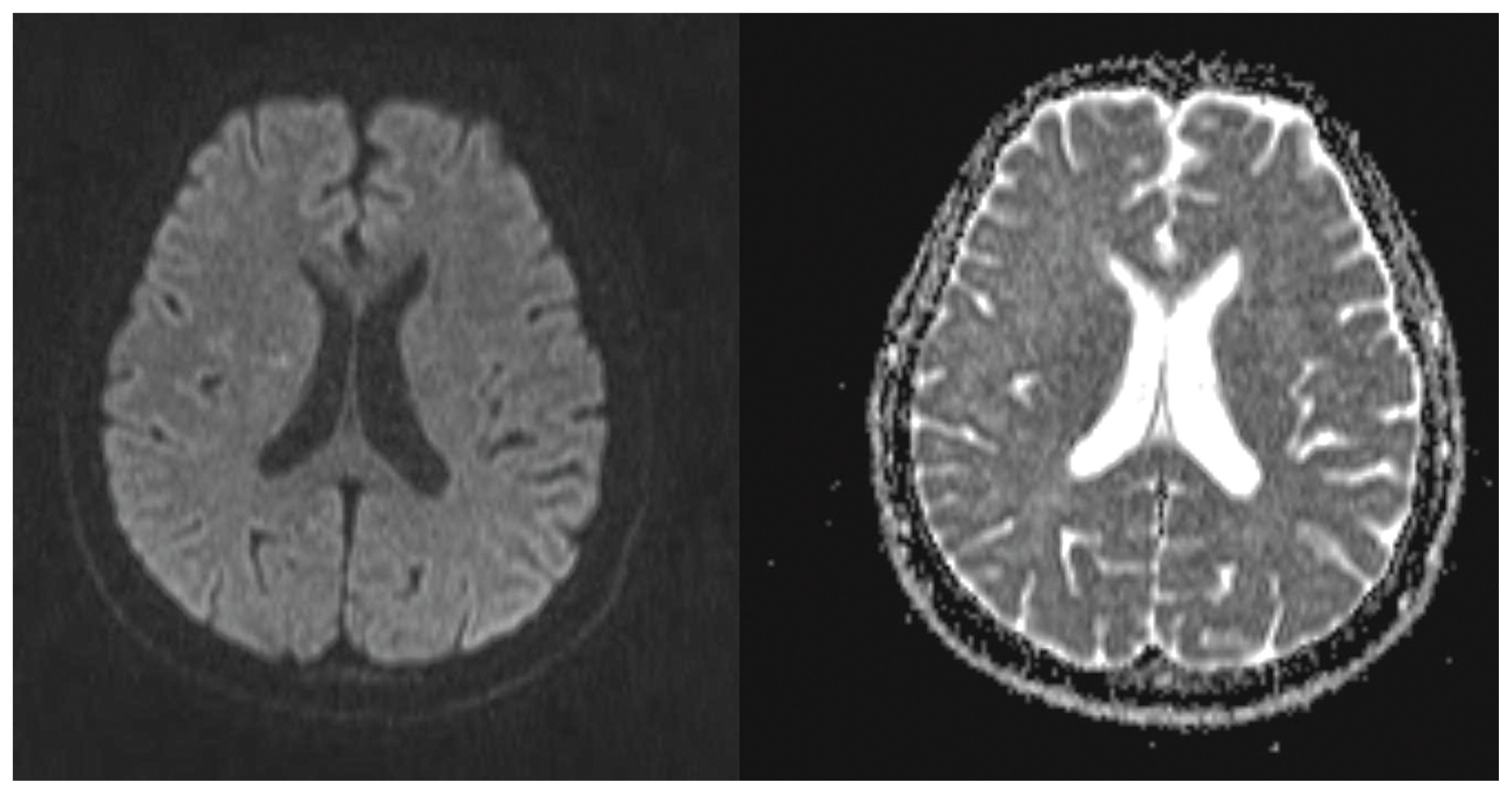
Fig. 3 Follow-up magnetic resonance imaging performed 1 week after cerebral angiography reveals complete reversal of the previously areas on diffusion weighted image (left) and the apparent diffusion coefficient map (right).
Reference
-
1. Uchiyama Y, Abe T, Hirohata M, Tanaka N, Kojima K, Nishimura H, et al. Blood brain-barrier disruption of nonionic iodinated contrast medium following coil embolization of a ruptured intracerebral aneurysm. AJNR Am J Neuroradiol. 2004; 25:1783–6.2. Niimi Y, Kupersmith MJ, Ahmad S, Song J, Berenstein A. Cortical blindness, transient and otherwise, associated with detachable coil embolization of intracranial aneurysms. AJNR Am J Neuroradiol. 2008; 29:603–7.
Article3. Saigal G, Bhatia R, Bhatia S, Wakhloo AK. MR findings of cortical blindness following cerebral angiography: is this entity related to posterior reversible leukoencephalopathy? AJNR Am J Neuroradiol. 2004; 25:252–6.4. Leong S, Fanning NF. Persistent neurological deficit from iodinated contrast encephalopathy following intracranial aneurysm coiling. A case report and review of the literature. Interv Neuroradiol. 2012; 18:33–41.5. Park JC, Ahn JH, Chang IB, Oh JK, Kim JH, Song JH. A Case of Unusual Presentation of Contrast-induced Encephalopathy after Cerebral Angiography Using Iodixanol. J Cerebrovasc Endovasc Neurosurg. 2017; 19(1):84–8.
Article6. Muccio CF, De Simone M, Esposito G, De Blasio E, Vittori C, Cerase A. Reversible post-traumatic bilateral extensive restricted diffusion of the brain. A case study and review of the literature. Brain Inj. 2009; 23:466–72.
Article7. Al Brashdi YH, Albayram MS. Reversible restricted-diffusion lesion representing transient intramyelinic cytotoxic edema in a patient with traumatic brain injury. Neuroradiol J. 2015; 28:409–12.
Article
- Full Text Links
-
- Actions
-
Cited
- CITED
-
- Close
- Share
-
- Similar articles
-
- A Case of Unusual Presentation of Contrast-induced Encephalopathy after Cerebral Angiography Using Iodixanol
- Hyperintense Acute Reperfusion Marker on FLAIR in Patient with Possible Contrast-Induced Encephalopathy Following Cerebral Angiography
- A Case of Congenital Cytomegalovirus Encephalopathy with Patchy, Nodular Lesion of Periventricular area on Brain Magnetic Resonance Imaging
- Corrigendum to: Unusual Magnetic Resonance Imaging Findings Contrast-induced Encephalopathy following Cerebral Angiography
- Wernicke's Encephalopathy With Reversible Cortical Involvement
