Delayed cerebral infarction due to cerebral venous air emboli after cardiac arrest
- Affiliations
-
- 1Department of Neurology, Stroke Center, Dong-A University Hospital, Busan, Republic of Korea
- 2Department of Intensive Care Medicine, Dong-A University Hospital, Busan, Republic of Korea
- KMID: 2517135
- DOI: http://doi.org/10.18700/jnc.210012
Figure
-
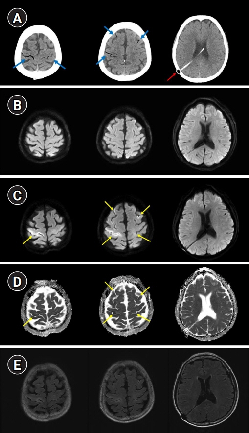
Fig. 1. Brain computed tomography and magnetic resonance imaging (MRI). (A) Non-contrast computed tomography of the head performed immediately after the return of spontaneous circulation (ROSC) showing air bubbles in the frontoparietal sulci (blue arrow). A ventriculoperitoneal shunt catheter was inserted in the right lateral ventricle (red arrow). (B) Diffusion-weighted brain MRI 5 hours after ROSC showing no acute lesions. (C-E) A follow-up brain MRI was performed on the 6th day after ROSC. Diffusion-weighted images and apparent diffusion coefficient images showed focal gyriform infarction in both frontoparietal cortices, especially in the right motor cortex (yellow arrows). Fluid-attenuated inversion recovery images showed high signal intensities in the same area.
Reference
-
1. Jeon SB, Kim JS, Lee DK, Kang DW, Kwon SU. Clinicoradiological characteristics of cerebral air embolism. Cerebrovasc Dis. 2007; 23:459–62.
Article2. Kim YJ, Jeon SB. Cerebral air embolism treated using hyperbaric oxygen therapy. J Neurocrit Care. 2019; 12:64–65.
Article3. Lai D, Jovin TG, Jadhav AP. Cortical vein air emboli with gyriform infarcts. JAMA Neurol. 2013; 70:939–40.
Article
- Full Text Links
-
- Actions
-
Cited
- CITED
-
- Close
- Share
-
- Similar articles
-
- Stroke Caused by Cerebral Air Embolism after Central Venous Catheter Removal: A Case Report
- A Case of Cerebral Infarction Caused by Tumor Emboli from the Site of
- Cerebral Infarction Caused by Direct Cardiac Tumor Emboli Mixed with Thrombus
- A Case of Cerebral Venous Thromobosis Associated with Postsplenectomy Thrombocytosis
- Spontaneous Absorption of Cerebral Air Emboli
