In-House Developed Surface-Guided Repositioning and Monitoring System to Complement In-Room Patient Positioning System for Spine Radiosurgery
- Affiliations
-
- 1Department of Neurosurgery, Neuroscience & Radiosurgery Hybrid Research Center, Inje University Ilsan Paik Hospital, Inje University College of Medicine, Goyang, Korea
- 2Department of Biomedical Engineering, U-Health Research Center, Inje University, Gimhae, Korea
- KMID: 2516963
- DOI: http://doi.org/10.14316/pmp.2021.32.2.40
Abstract
- Purpose
This study aimed to develop a surface-guided radiosurgery system customized for a neurosurgery clinic that could be used as an auxiliary system for improving the accuracy, monitoring the movements of patients while performing hypofractionated radiosurgery, and minimizing the geometric misses.
Methods
RGB-D cameras were installed in the treatment room and a monitoring system was constructed to perform a three-dimensional (3D) scan of the body surface of the patient and to express it as a point cloud. This could be used to confirm the exact position of the body of the patient and monitor their movements during radiosurgery. The image from the system was matched with the computed tomography (CT) image, and the positional accuracy was compared and analyzed in relation to the existing system to evaluate the accuracy of the setup.
Results
The user interface was configured to register the patient and display the setup image to position the setup location by matching the 3D points on the body of the patient with the CT image. The error rate for the position difference was within 1-mm distance (min, 一0.21 mm; max, 0.63 mm). Compared with the existing system, the differences were found to be as follows: x=0.08 mm, y=0.13 mm, and z=0.26 mm.
Conclusions
We developed a surface-guided repositioning and monitoring system that can be customized and applied in a radiation surgery environment with an existing linear accelerator. It was confirmed that this system could be easily applied for accurate patient repositioning and intertreatment motion monitoring.
Keyword
Figure
-

Fig. 1 Three-dimensional surface modeling system architecture and surface-guided radiosurgery (SGRS) in the treatment room: (a) the architecture of the image acquisition using the depth camera, (b) the surface imaging profile in sagittal plane, and (c) the installed SGRS in the treatment room. IR, infrared; LINAC, linear accelerator .

Fig. 2 Acquired three-dimensional images using RGB-D cameras: (a) left, (b) center, (c) right direction, and (d) integrated images through the cameras.
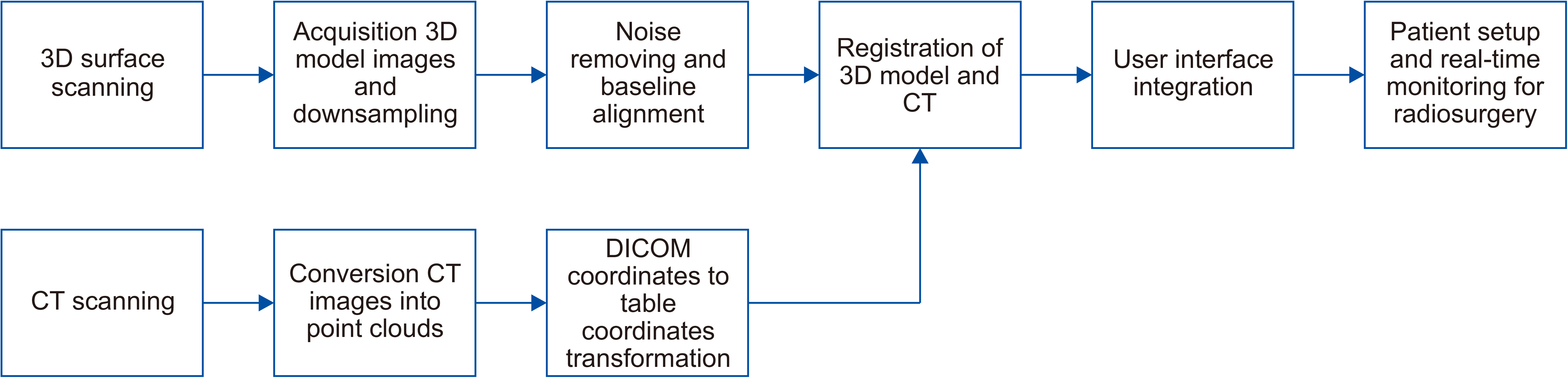
Fig. 3 Image registration process and user interface integration. 3D, three-dimensional; CT, computed tomography.

Fig. 4 Surface-guided radiosurgery user interface in the treatment room. (a) The user interface for the patient setup matching. (b) The phantom experiment using our surface-guided reposition and monitoring system. CT, computed tomography.

Fig. 5 Image registration results of surface-guided image to computed tomography (CT) for a phantom and clinical case. (a) The 3D image registration result for a phantom CT and point cloud. (b) The image registration process in the same coordinate plane. (c) The final image in which the point cloud and CT images are registered.
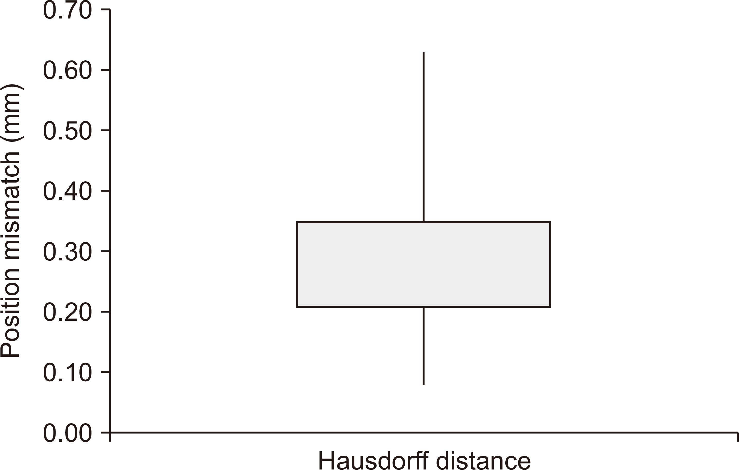
Fig. 6 Multi-fractional setup trials using Hausdorff distance in difference plots.
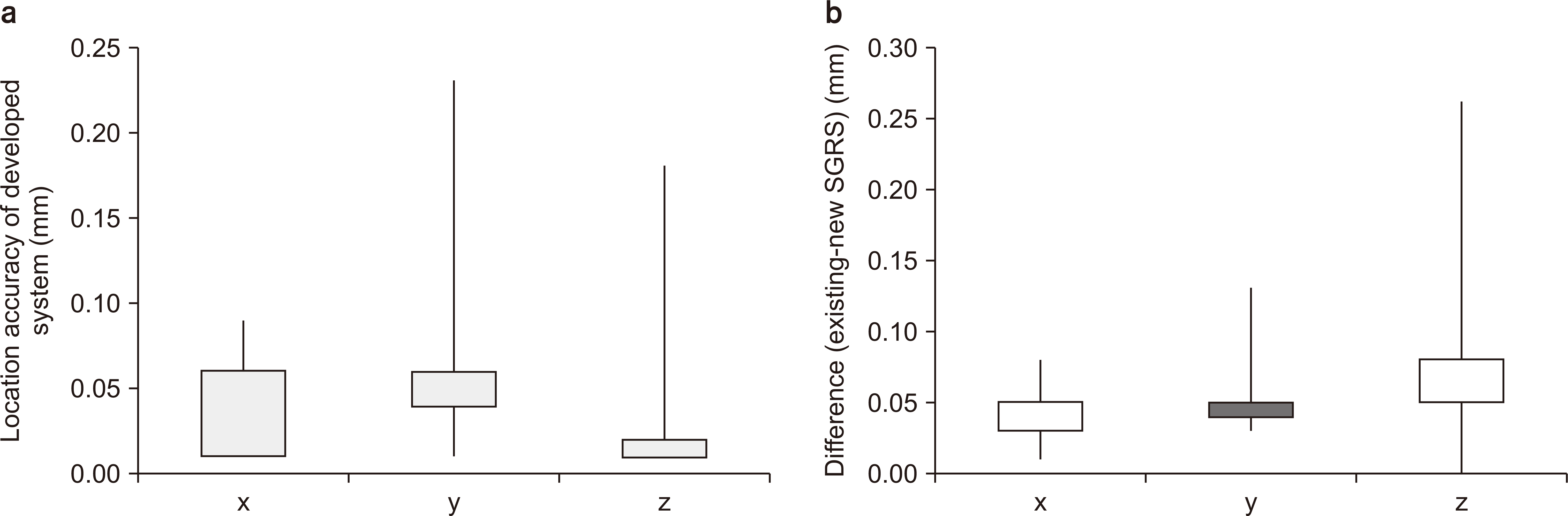
Fig. 7 Multi-fractional setup trials involving existing system (ExacTrac, BrainLab, Munich, Germany) and the developed surface-guided radiosurgery (SGRS) in difference plots. (a) The location accuracy of the developed system for the multi-fractional setup trials. (b) The difference for the x, y, and z axis.
Reference
-
References
1. Li G, Ballangrud A, Kuo LC, Kang H, Kirov A, Lovelock M, et al. 2011; Motion monitoring for cranial frameless stereotactic radiosurgery using video-based three-dimensional optical surface imaging. Med Phys. 38:3981–3994. DOI: 10.1118/1.3596526. PMID: 21858995.
Article2. Tagaste B, Riboldi M, Spadea MF, Bellante S, Baroni G, Cambria R, et al. 2012; Comparison between infrared optical and stereoscopic X-ray technologies for patient setup in image guided stereotactic radiotherapy. Int J Radiat Oncol Biol Phys. 82:1706–1714. DOI: 10.1016/j.ijrobp.2011.04.004. PMID: 21605942.
Article3. Wang LT, Solberg TD, Medin PM, Boone R. 2001; Infrared patient positioning for stereotactic radiosurgery of extracranial tumors. Comput Biol Med. 31:101–111. DOI: 10.1016/s0010-4825(00)00026-3. PMID: 11165218.
Article4. Schipani S, Wen W, Jin JY, Kim JK, Ryu S. 2012; Spine radiosurgery: a dosimetric analysis in 124 patients who received 18 Gy. Int J Radiat Oncol Biol Phys. 84:e571–e576. DOI: 10.1016/j.ijrobp.2012.06.049. PMID: 22975607.
Article5. Wu VW, Ho YY, Tang YS, Lam PW, Yeung HK, Lee SW. 2019; Comparison of the verification performance and radiation dose between ExacTrac x-ray system and On-Board Imager-a phantom study. Med Dosim. 44:15–19. DOI: 10.1016/j.meddos.2017.12.008. PMID: 29395461.
Article6. Murphy MJ, Balter J, Balter S, BenComo JA Jr, Das IJ, Jiang SB, et al. 2007; The management of imaging dose during image-guided radiotherapy: report of the AAPM Task Group 75. Med Phys. 34:4041–4063. DOI: 10.1118/1.2775667. PMID: 17985650.
Article7. Cheng CS, Jong WL, Ung NM, Wong JHD. 2017; Evaluation of imaging dose from different image guided systems during head and neck radiotherapy: a phantom study. Radiat Prot Dosimetry. 175:357–362. DOI: 10.1093/rpd/ncw357. PMID: 27940494.
Article8. Steiner E, Stock M, Kostresevic B, Ableitinger A, Jelen U, Prokesch H, et al. 2013; Imaging dose assessment for IGRT in particle beam therapy. Radiother Oncol. 109:409–413. DOI: 10.1016/j.radonc.2013.09.007. PMID: 24128802.
Article9. Hoisak JDP, Pawlicki T. 2018; The role of optical surface imaging systems in radiation therapy. Semin Radiat Oncol. 28:185–193. DOI: 10.1016/j.semradonc.2018.02.003. PMID: 29933878.
Article10. Freislederer P, Kügele M, Öllers M, Swinnen A, Sauer TO, Bert C, et al. 2020; Recent advanced in surface guided radiation therapy. Radiat Oncol. 15:187. DOI: 10.1186/s13014-020-01629-w. PMID: 32736570. PMCID: PMC7393906.11. Li J, Shi W, Andrews D, Werner-Wasik M, Lu B, Yu Y, et al. 2017; Comparison of online 6 degree-of-freedom image registration of Varian TrueBeam cone-beam CT and BrainLab ExacTrac X-ray for intracranial radiosurgery. Technol Cancer Res Treat. 16:339–343. DOI: 10.1177/1533034616683069. PMID: 28462690. PMCID: PMC5616049.
Article12. Laaksomaa M, Sarudis S, Rossi M, Lehtonen T, Pehkonen J, Remes J, et al. 2019; AlignRT® and Catalyst™ in whole-breast radiotherapy with DIBH: is IGRT still needed? J Appl Clin Med Phys. 20:97–104. DOI: 10.1002/acm2.12553. PMID: 30861276. PMCID: PMC6414178.13. Agazaryan N, Tenn S, Dieterich S, Gevaert T, Goetsch SJ, Kaprealian T. 2020. Frameless image guidance in stereotactic radiosurgery. Stereotactic and Functional Neurosurgery. Springer;Cham: p. 37–48. DOI: 10.1007/978-3-030-34906-6_4.
Article14. Manger RP, Paxton AB, Pawlicki T, Kim GY. 2015; Failure mode and effects analysis and fault tree analysis of surface image guided cranial radiosurgery. Med Phys. 42:2449–2461. DOI: 10.1118/1.4918319. PMID: 25979038.
Article15. Gilles M, Fayad H, Miglierini P, Clement JF, Scheib S, Cozzi L, et al. 2016; Patient positioning in radiotherapy based on surface imaging using time of flight cameras. Med Phys. 43:4833. DOI: 10.1118/1.4959536. PMID: 27487901.
Article16. Padilla L, Pearson EA, Pelizzari CA. 2015; Collision prediction software for radiotherapy treatments. Med Phys. 42:6448–6456. DOI: 10.1118/1.4932628. PMID: 26520734.
Article17. Hoole AC, Twyman N, Langmack KA, Hebbard M, Lowrie D. 2001; Laser scanning of patient outlines for three-dimensional radiotherapy treatment planning. Physiol Meas. 22:605–610. DOI: 10.1088/0967-3334/22/3/316. PMID: 11556678.
Article18. Roessler K, Ungersboeck K, Dietrich W, Aichholzer M, Hittmeir K, Matula C, et al. 1997; Frameless stereotactic guided neurosurgery: clinical experience with an infrared based pointer device navigation system. Acta Neurochir (Wien). 139:551–559. DOI: 10.1007/BF02750999. PMID: 9248590.
Article19. Kosugi Y, Watanabe E, Goto J, Watanabe T, Yoshimoto S, Takakura K, et al. 1988; An articulated neurosurgical navigation system using MRI and CT images. IEEE Trans Biomed Eng. 35:147–152. DOI: 10.1109/10.1353. PMID: 3350540.
Article20. Fan Y, Jiang D, Wang M, Song Z. 2014; A new markerless patient-to-image registration method using a portable 3D scanner. Med Phys. 41:101910. DOI: 10.1118/1.4895847. PMID: 25281962.
Article21. Giancola S, Valenti M, Sala R. 2018. A survey on 3D cameras: metrological comparison of time-of-flight, structured-light and active stereoscopy technologies. Springer;Cham:22. He Y, Liang B, Yang J, Li S, He J. 2017; An iterative closest points algorithm for registration of 3D laser scanner point clouds with geometric features. Sensors (Basel). 17:1862. DOI: 10.3390/s17081862. PMID: 28800096. PMCID: PMC5580094.
Article23. Habib A, Detchev I, Bang K. 2010. Jun. 15-18. A comparative analysis of two approaches for multiple-surface registration of irregular point clouds. Paper presented at: The 2010 Canadian Geomatics Conference and Symposium of Commission I. Calgary, Canada: 39.24. Rusu RB, Cousins S. 2011. May. 9-13. 3D is here: Point Cloud Library (PCL). Paper presented at: 2011 IEEE International Conference on Robotics and Automation. Shanghai, China: DOI: 10.1109/ICRA.2011.5980567. PMID: 21955422.
Article25. Arun KS, Huang TS, Blostein SD. 1987. Least-squares fitting of two 3-D point sets. IEEE Trans Pattern Anal Mach Intell. PAMI-9:698-700. DOI: 10.1109/TPAMI.1987.4767965. PMID: 21869429.
Article26. Ge Y, Maurer CR Jr, Fitzpatrick JM. 1996. Surface-based 3D image registration using the iterative closest-point algorithm with a closest-point transform Medical Imaging 1996: Image Processing. SPIE Digital Library. 358–367. DOI: 10.1117/12.237938.
Article27. Wu ML, Chien JC, Wu CT, Lee JD. 2018; An augmented reality system using improved-iterative closest point algorithm for on-patient medical image visualization. Sensors (Basel). 18:2505. DOI: 10.3390/s18082505. PMID: 30071645. PMCID: PMC6111829.
Article28. Tehrani JN, O’Brien RT, Poulsen PR, Keall P. 2013; Real-time estimation of prostate tumor rotation and translation with a kV imaging system based on an iterative closest point algorithm. Phys Med Biol. 58:8517–8533. DOI: 10.1088/0031-9155/58/23/8517. PMID: 24240537.
Article29. Huttenlocher DP, Klanderman GA, Rucklidge WJ. 1993; Comparing images using the Hausdorff distance. IEEE Trans Pattern Anal Mach Intell. 15:850–863. DOI: 10.1109/34.232073.
Article30. Wang G, Li Z, Li G, Dai G, Xiao Q, Bai L, et al. 2021; Real-time liver tracking algorithm based on LSTM and SVR networks for use in surface-guided radiation therapy. Radiat Oncol. 16:13. DOI: 10.1186/s13014-020-01729-7. PMID: 33446245. PMCID: PMC7807524.
Article31. Covington EL, Popple RA. 2021; A low-cost method to assess the performance of surface guidance imaging systems at non-zero couch angles. Cureus. 13:e14278. DOI: 10.7759/cureus.14278. PMID: 33959456. PMCID: PMC8093097.
Article32. Chan A, Coutts B, Parent E, Lou E. 2021; Development and evaluation of CT-to-3D ultrasound image registration algorithm in vertebral phantoms for spine surgery. Ann Biomed Eng. 49:310–321. DOI: 10.1007/s10439-020-02546-5. PMID: 32533392.
Article33. Wang S, Sun HY, Guo HC, Du L, Liu TJ. 2018; Multi-view laser point cloud global registration for a single object. Sensors (Basel). 18:3729. DOI: 10.3390/s18113729. PMID: 30388874. PMCID: PMC6263679.
Article34. Li J, Zhou Q, Li X, Chen R, Ni K. 2019; An improved low-noise processing methodology combined with PCL for industry inspection based on laser line scanner. Sensors (Basel). 19:3398. DOI: 10.3390/s19153398. PMID: 31382454. PMCID: PMC6695628.
Article35. Liu W, Cheung Y, Sabouri P, Arai TJ, Sawant A, Ruan D. 2015; A continuous surface reconstruction method on point cloud captured from a 3D surface photogrammetry system. Med Phys. 42:6564–6571. DOI: 10.1118/1.4933196. PMID: 26520747. PMCID: PMC4617738.
Article36. Fan Y, Yao X, Hu T, Xu X. 2019; An automatic spatial registration method for image-guided neurosurgery system. J Craniofac Surg. 30:e344–e350. DOI: 10.1097/SCS.0000000000005330. PMID: 30817512.
Article37. Muralikrishnan B, Rachakonda P, Lee V, Shilling M, Sawyer D, Cheok G, et al. 2017; Relative range error evaluation of terrestrial laser scanners using a plate, a sphere, and a novel dual-sphere-plate target. Meas Sci Technol. 111:60–68. DOI: 10.1016/j.measurement.2017.07.027. PMID: 28924331. PMCID: PMC5600278.
Article38. Maier-Hein L, Franz AM, dos Santos TR, Schmidt M, Fangerau M, Meinzer HP, et al. 2011; Convergent iterative closest-point algorithm to accomodate anisotropic and inhomogenous localization error. IEEE Trans Pattern Anal Mach Intell. 34:1520–1532. DOI: 10.1109/TPAMI.2011.248. PMID: 22184256.
Article39. Liu W. 2017; LiDAR-IMU time delay calibration based on iterative closest point and iterated sigma point Kalman filter. Sensors (Basel). 17:539. DOI: 10.3390/s17030539. PMID: 28282897. PMCID: PMC5375825.
Article40. Coroiu ADCA, Coroiu A. 2018. Sep. 6-8. Interchangeability of Kinect and Orbbec sensors for gesture recognition. Paper presented at: 2018 IEEE 14th International Conference on Intelligent Computer Communication and Processing (ICCP). Cluj-Napoca, Romania: 309–315. DOI: 10.1109/ICCP.2018.8516586.
Article41. Wiersma RD, Tomarken SL, Grelewicz Z, Belcher AH, Kang H. 2013; Spatial and temporal performance of 3D optical surface imaging for real-time head position tracking. Med Phys. 40:111712. DOI: 10.1118/1.4823757. PMID: 24320420.
Article
- Full Text Links
-
- Actions
-
Cited
- CITED
-
- Close
- Share
-
- Similar articles
-
- Evaluation of Real-time Target Positioning Accuracy in Spinal Radiosurgery
- Contemporary treatment with radiosurgery for spine metastasis and spinal cord compression in 2015
- The Evaluation of Standard 50% Hemolytic Complement Assay Using In-house Reagents
- Role of Complement in Bronchial Asthma
- Multimodality and Application Software
