J Korean Foot Ankle Soc.
2021 Jun;25(2):55-60. 10.14193/jkfas.2021.25.2.55.
Does Achilles Tendon Shortening Mean Pathologic Lesions?
- Affiliations
-
- 1Department of Orthopaedic Surgery, Soonchunhyang University Seoul Hospital, Seoul, Korea
- KMID: 2516640
- DOI: http://doi.org/10.14193/jkfas.2021.25.2.55
Abstract
- This review article attempts to describe several pathological conditions that can arise from the shortening of the Achilles tendon. The tension and tightening of the gastrocnemius-soleus-Achilles tendon complex (GSAC) can cause disharmony in the movement of the entire foot as well as the ankle joint when subjected to weight-bearing or walking. In addition, since these phenomena are observed in various lesions of the ankle joint, the dynamic shortening caused by the tension of GSAC may not be the primary cause of various ankle joint lesions, but is still considered to be a significant contributing factor.
Figure
-
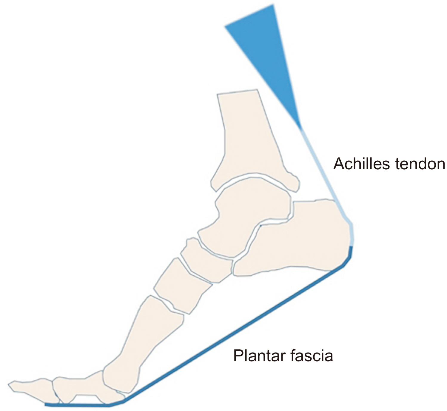
Figure. 1 Plantar fascia is connected to the proximal part through the periosteum of the calcaneus, and is connected to the peritendinous tissue of the Achilles tendon.
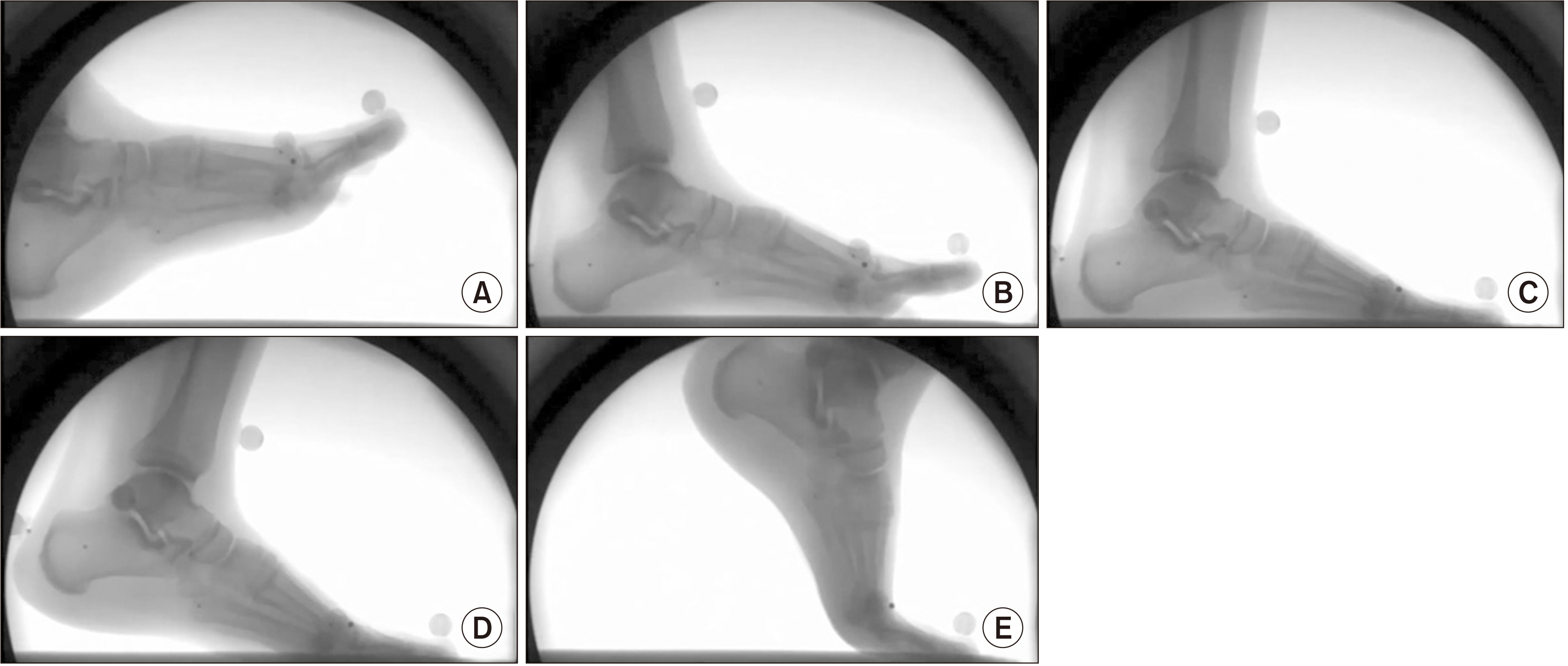
Figure. 2 Dynamic real-time fluoroscopic image of foot is taken during heel strike (A), foot flat (B), mid-stance (C), heel off (D), and toe off (E) phase.

Figure. 3 Achilles tendon tightness leads to midfoot destruction by increasing body moment on mid foot structure, while hindfoot destruction leads to tightness on triceps surae by midfoot flattening and hindfoot eversion. And these processes induce pathologic changes such as Achilles tendinitis, plantar fasciitis, metatarsalgia, hallux valgus, flatfoot, and midfoot arthropathy.
Reference
-
1. Arandes R, Viladot A. 1954; [Biomecánica del calcáneo]. Minerva Chir. 9:439–46. Spanish.2. DiGiovanni CW, Kuo R, Tejwani N, Price R, Hansen ST Jr, Cziernecki J, et al. 2002; Isolated gastrocnemius tightness. J Bone Joint Surg Am. 84:962–70. doi: 10.2106/00004623-200206000-00010. DOI: 10.2106/00004623-200206000-00010. PMID: 12063330.
Article3. Meyer DB, O'Rahilly R. 1976; The onset of ossification in the human calcaneus. Anat Embryol (Berl). 150:19–33. doi: 10.1007/BF00346283. DOI: 10.1007/BF00346283. PMID: 1015627.4. Vieira L. 2020; Phylogenetics of the fascial system. Cureus. 12:e10787. doi: 10.7759/cureus.10787. DOI: 10.7759/cureus.10787. PMID: 33154854. PMCID: PMC7606207.
Article5. Zwirner J, Zhang M, Ondruschka B, Akita K, Hammer N. 2020; An ossifying bridge - on the structural continuity between the Achilles tendon and the plantar fascia. Sci Rep. 10:14523. doi: 10.1038/s41598-020-71316-z. DOI: 10.1038/s41598-020-71316-z. PMID: 32884015. PMCID: PMC7471908.
Article6. Singh A, Zwirner J, Templer F, Kieser D, Klima S, Hammer N. 2021; On the morphological relations of the Achilles tendon and plantar fascia via the calcaneus: a cadaveric study. Sci Rep. 11:5986. doi: 10.1038/s41598-021-85251-0. DOI: 10.1038/s41598-021-85251-0. PMID: 33727610. PMCID: PMC7966405.
Article7. Stecco C, Corradin M, Macchi V, Morra A, Porzionato A, Biz C, et al. 2013; Plantar fascia anatomy and its relationship with Achilles tendon and paratenon. J Anat. 223:665–76. doi: 10.1111/joa.12111. DOI: 10.1111/joa.12111. PMID: 24028383. PMCID: PMC3842207.
Article8. Thordarson DB, Schmotzer H, Chon J, Peters J. 1995; Dynamic support of the human longitudinal arch. A biomechanical evaluation. Clin Orthop Relat Res. (316):165–72. DOI: 10.1097/00003086-199507000-00022. PMID: 7634700.9. Antoniak G, Biswas T, Cortes N, Sikdar S, Chun C, Bhandawat V. 2019; Spring-loaded inverted pendulum goes through two contraction-extension cycles during the single-support phase of walking. Biol Open. 8:bio043695. doi: 10.1242/bio.043695. DOI: 10.1242/bio.043695. PMID: 31097445. PMCID: PMC6602329.10. Ballal MS, Walker CR, Molloy AP. 2014; The anatomical footprint of the Achilles tendon: a cadaveric study. Bone Joint J. 96-B:1344–8. doi: 10.1302/0301-620X.96B10.33771. DOI: 10.1302/0301-620X.96B10.33771. PMID: 25274919.11. DiGiovanni CW, Langer P. 2007; The role of isolated gastrocnemius and combined Achilles contractures in the flatfoot. Foot Ankle Clin. 12:363–79. viiidoi: 10.1016/j.fcl.2007.03.005. DOI: 10.1016/j.fcl.2007.03.005. PMID: 17561207.
Article12. Johnson CH, Christensen JC. 2005; Biomechanics of the first ray part V: the effect of equinus deformity. A 3-dimensional kinematic study on a cadaver model. J Foot Ankle Surg. 44:114–20. doi: 10.1053/j.jfas.2005.01.003. DOI: 10.1053/j.jfas.2005.01.003. PMID: 15768359.13. Johnson KA, Strom DE. 1989; Tibialis posterior tendon dysfunction. Clin Orthop Relat Res. (239):196–206. DOI: 10.1097/00003086-198902000-00022. PMID: 2912622.
Article14. Blackman AJ, Blevins JJ, Sangeorzan BJ, Ledoux WR. 2009; Cadaveric flatfoot model: ligament attenuation and Achilles tendon overpull. J Orthop Res. 27:1547–54. doi: 10.1002/jor.20930. DOI: 10.1002/jor.20930. PMID: 19530145.
Article15. Engkananuwat P, Kanlayanaphotporn R, Purepong N. 2018; Effectiveness of the simultaneous stretching of the Achilles tendon and plantar fascia in individuals with plantar fasciitis. Foot Ankle Int. 39:75–82. doi: 10.1177/1071100717732762. DOI: 10.1177/1071100717732762. PMID: 28985685.
Article16. Patel A, DiGiovanni B. 2011; Association between plantar fasciitis and isolated contracture of the gastrocnemius. Foot Ankle Int. 32:5–8. doi: 10.3113/FAI.2011.0005. DOI: 10.3113/FAI.2011.0005. PMID: 21288428.
Article17. Snow SW, Bohne WH, DiCarlo E, Chang VK. 1995; Anatomy of the Achilles tendon and plantar fascia in relation to the calcaneus in various age groups. Foot Ankle Int. 16:418–21. doi: 10.1177/107110079501600707. DOI: 10.1177/107110079501600707. PMID: 7550955.
Article18. Ramanujam CL, Zgonis T. 2017; Surgical correction of the Achilles tendon for diabetic foot ulcerations and Charcot neuroarthropathy. Clin Podiatr Med Surg. 34:275–80. doi: 10.1016/j.cpm.2016.10.013. DOI: 10.1016/j.cpm.2016.10.013. PMID: 28257680.
Article
- Full Text Links
-
- Actions
-
Cited
- CITED
-
- Close
- Share
-
- Similar articles
-
- Heterotopic Ossification of a Partially Ruptured Achilles Tendon (A Case Report)
- Treatment of Massive Defect in Achilles Tendon with Tendon Allograft: A Case Report
- Long Term Result of Four Cases without a Staged Reconstruction of an Infected Achilles Tendon Following Repair
- Achilles Tendon Rupture Associated With Ipsilateral Medial Malleolar Fracture (A Case Report)
- Reconstruction of Chronic Achilles Tendon Rupture Using Interposed Scar Tissue (A Report of Two Cases)
