J Stroke.
2021 May;23(2):281-284. 10.5853/jos.2020.04784.
7T Magnetic Resonance Imaging Quantification of Brain Glutamate in Acute Ischaemic Stroke
- Nicolo JP
 1,2,3,4
 - Moffat B5
- Wright DK4
- Sinclair B4
- Neal A1,2,3,4
- Lui E6,7
- Desmond P6,7
- Glarin R6,7
- Davis KA8
- Reddy R9
- Yan B1,3
- O’Brien TJ1,2,3,4
- Kwan P1,2,3,4
- Affiliations
-
- 1Department of Neurology, Royal Melbourne Hospital, Parkville, Australia
- 2Department of Neurology, Alfred Hospital, Melbourne, Australia
- 3Department of Medicine, Royal Melbourne Hospital, University of Melbourne, Parkville, Australia
- 4Department of Neurosciences, The Central Clinical School, Monash University, Melbourne, Australia
- 5Melbourne Node of the National Imaging Facility, Department of Radiology, University of Melbourne, Parkville, Australia
- 6Department of Radiology, Royal Melbourne Hospital, Parkville, Australia
- 7Department of Medicine & Radiology, University of Melbourne, Parkville, Australia
- 8Penn Epilepsy Center, Department of Neurology, Hospital of the University of Pennsylvania, Philadelphia, PA, USA
- 9Center for Magnetic Resonance & Optical Imaging, Department of Radiology, University of Pennsylvania, Philadelphia, PA, USA
- KMID: 2516418
- DOI: http://doi.org/10.5853/jos.2020.04784
Figure
-
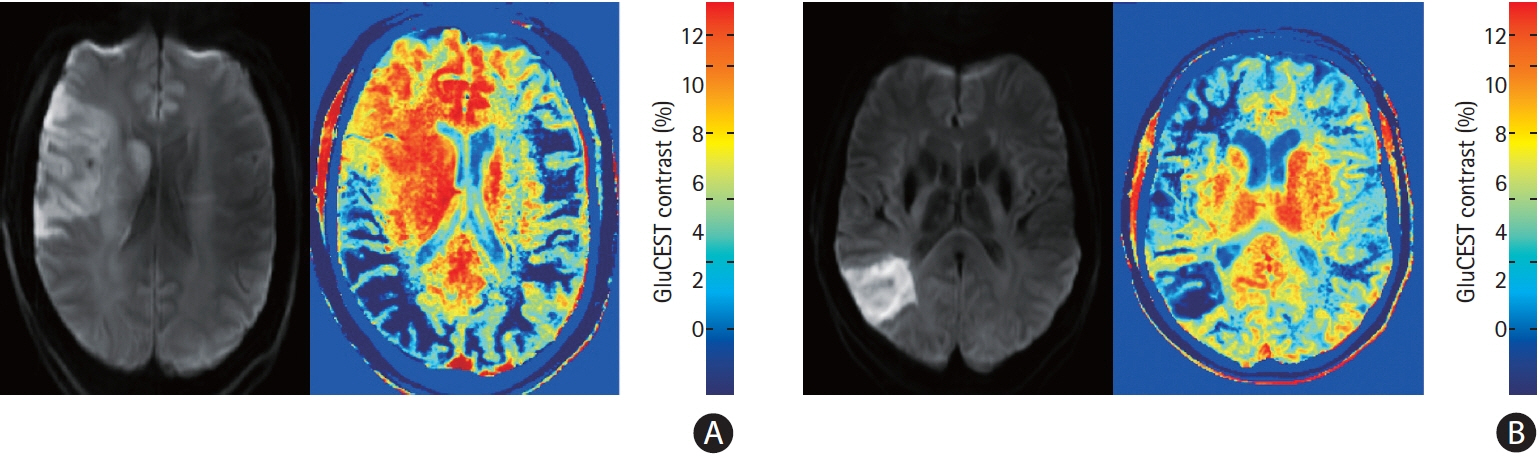
Figure 1. Diffusion weighted imaging and chemical exchange saturation transfer (CEST) images from patient 8 (A) and patient 11 (B) demonstrating regionally increased and decreased glutamate weighted chemical exchange saturation transfer (GluCEST) contrast ipsilateral to the infarction. The asymmetry in GluCEST contrast in regions distant to the infarction may be due to brain positioning asymmetry.
Reference
-
References
1. Nicolo JP, O’Brien TJ, Kwan P. Role of cerebral glutamate in post-stroke epileptogenesis. Neuroimage Clin. 2019; 24:102069.
Article2. Cai K, Haris M, Singh A, Kogan F, Greenberg JH, Hariharan H, et al. Magnetic resonance imaging of glutamate. Nat Med. 2012; 18:302–306.
Article3. Neal A, Moffat BA, Stein JM, Nanga RPR, Desmond P, Shinohara RT, et al. Glutamate weighted imaging contrast in gliomas with 7 Tesla magnetic resonance imaging. Neuroimage Clin. 2019; 22:101694.4. Provencher SW. Estimation of metabolite concentrations from localized in vivo proton NMR spectra. Magn Reson Med. 1993; 30:672–679.5. Bivard A, Yassi N, Krishnamurthy V, Lin L, Levi C, Spratt NJ, et al. A comprehensive analysis of metabolic changes in the salvaged penumbra. Neuroradiology. 2016; 58:409–415.
Article6. Dávalos A, Castillo J, Serena J, Noya M. Duration of glutamate release after acute ischemic stroke. Stroke. 1997; 28:708–710.
Article7. Higuchi T, Fernandez EJ, Maudsley AA, Shimizu H, Weiner MW, Weinstein PR. Mapping of lactate and N-acetyl-L-aspartate predicts infarction during acute focal ischemia: in vivo 1H magnetic resonance spectroscopy in rats. Neurosurgery. 1996; 38:121–129.
Article8. Singh A, Cai K, Haris M, Hariharan H, Reddy R. On B1 inhomogeneity correction of in vivo human brain glutamate chemical exchange saturation transfer contrast at 7T. Magn Reson Med. 2013; 69:818–824.9. Zaiss M, Ehses P, Scheffler K. Snapshot-CEST: optimizing spiral- centric-reordered gradient echo acquisition for fast and robust 3D CEST MRI at 9.4 T. NMR Biomed. 2018; 31:e3879.10. Sun PZ. Fast correction of B0 field inhomogeneity for pHspecific magnetization transfer and relaxation normalized amide proton transfer imaging of acute ischemic stroke without Z-spectrum. Magn Reson Med. 2020; 83:1688–1697.
- Full Text Links
-
- Actions
-
Cited
- CITED
-
- Close
- Share
-
- Similar articles
-
- Comparison of 3 and 7 Tesla Magnetic Resonance Imaging of Obstructive Hydrocephalus Caused by Tectal Glioma
- Silent New Brain Lesions: Innocent Bystander or Guilty Party?
- Functional Magnetic Resonance Imaging of the Brain: Principle and Practical Application
- Fast MRI in Acute Ischemic Stroke: Applications of MRI Acceleration Techniques for MR-Based Comprehensive Stroke Imaging
- Advanced Magnetic Resonance Imaging for Pediatric Brain Tumors: Current Imaging Techniques and Interpretation Algorithms
