Clin Endosc.
2021 May;54(3):447-448. 10.5946/ce.2021.134.
Diagnosis of Gastric Subepithelial Tumor: Role of Endoscopic Ultrasound-Guided Fine-Needle Biopsy
- Affiliations
-
- 1Department of Gastroenterology, Asan Medical Center, University of Ulsan College of Medicine
- KMID: 2516330
- DOI: http://doi.org/10.5946/ce.2021.134
Figure
-
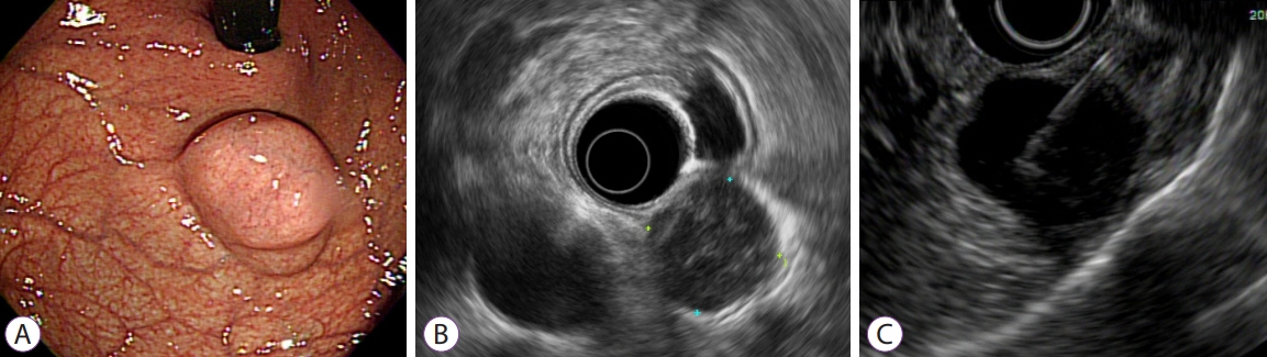
Fig. 1. (A) A 3-cm sized subepithelial tumor was noted at the fundus of the stomach. (B) Endoscopic ultrasonography showed a 2.4×2.3 cm round, hypoechoic mass in the proper muscle layer. (C) Endoscopic ultrasound-guided fine needle biopsy was performed.
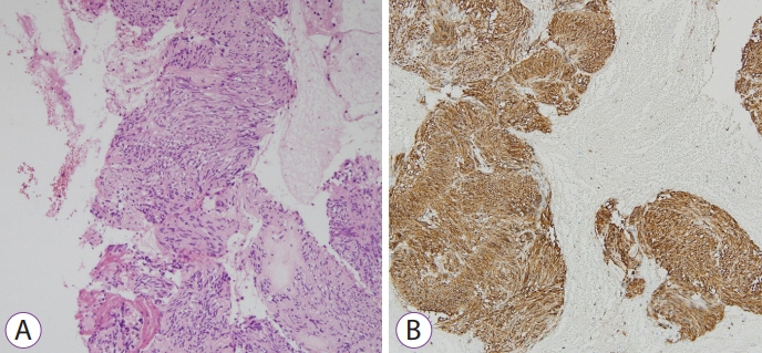
Fig. 2. Pathologic findings of the biopsy specimens. (A) Histopathological examination reveals uniform spindle cells with high cellularity (hematoxylin and eosin ×200). (B) The tumor cells are strongly and diffusely positive for CD117 (×200).
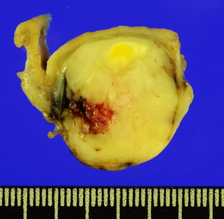
Fig. 3. Macroscopic specimen from surgical resection.
Reference
-
1. Kim GH, Ahn JY, Gong CS, et al. Efficacy of endoscopic ultrasound-guided fine-needle biopsy in gastric subepithelial tumors located in the cardia. Dig Dis Sci. 2020; 65:583–590.
Article2. Lee JH, Cho CJ, Park YS, et al. EUS-guided 22-gauge fine needle biopsy for the diagnosis of gastric subepithelial tumors larger than 2 cm. Scand J Gastroenterol. 2016; 51:486–493.
Article3. Iglesias-Garcia J, Poley J-W, Larghi A, et al. Feasibility and yield of a new EUS histology needle: results from a multicenter, pooled, cohort study. Gastrointest Endosc. 2011; 73:1189–1196.
Article
- Full Text Links
-
- Actions
-
Cited
- CITED
-
- Close
- Share
-
- Similar articles
-
- Endoscopic Ultrasound-Fine Needle Aspiration versus Core Biopsy for the Diagnosis of Subepithelial Tumors
- Tuberculous Lymphadenitis Mimicking Gastric Subepithelial Tumor Diagnosed Using Endoscopic Ultrasound-guided Fine-needle Aspiration
- Fine-Needle Biopsy: Should This Be the First Choice in Endoscopic Ultrasound-Guided Tissue Acquisition?
- Endoscopic Ultrasound-Guided Fine Needle Aspiration in Submucosal Lesion
- Endoscopic Management of Gastric Subepithelial Tumor
