Contrast Harmonic Endoscopic Ultrasound in Pancreatic Diseases
- Affiliations
-
- 1Department of Gastroenterology and Hepatology, Health Research Institute (IDIS), University Hospital of Santiago de Compostela, Santiago de Compostela, Spain
- KMID: 2516309
- DOI: http://doi.org/10.5946/ce.2020.048
Abstract
- Endoscopic ultrasound (EUS) was first described in 1986, with the aim of overcoming the problems affecting transabdominal ultrasound imaging, mainly problems related to the interposition of gas, and artifacts produced by bone or fat. Now, EUS can be considered as the best method for the analysis of pancreatic diseases, overtaking the diagnostic accuracy of computed tomography and magnetic resonance imaging. However, fundamental B-mode imaging is limited for the diagnosis of solid pancreatic lesions, because most of them are depicted as heterogeneous and hypo-echoic, and it is difficult to differentiate between benign and malignant lesions. Similar to how perfusion patterns obtained by computed tomography or magnetic resonance imaging after injection of contrast agents allow for the characterization of focal lesions, EUS has also recently been introduced to the use of contrast agents for performing contrast-enhanced harmonic EUS (CEH-EUS), which has the capability to distinguish the type of perfusion between lesions and surrounding tissue. CEH-EUS has shown its usefulness for the diagnosis and characterization of solid pancreatic lesions. Moreover, CEH-EUS is also highly accurate for distinguishing non-neoplastic from neoplastic cysts in pancreatic lesions. Another potential role of CEH-EUS is its ability to direct EUS-guided tissue acquisition.
Figure
-
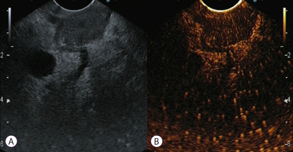
Fig. 1. Typical contrast-enhanced harmonic endoscopic ultrasound (CEH-EUS) images of pancreatic tumors: pancreatic cancer with hypoenhancement. The pancreatic lesion is detected as a low-echoic lesion with fundamental B-mode EUS (A). CEH-EUS detects the pancreatic lesion with hypoenhancement in comparison with the surrounding pancreatic tissue (B).
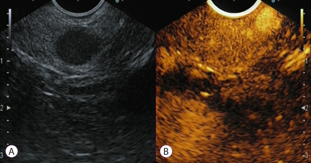
Fig. 2. Contrast-enhanced harmonic endoscopic ultrasound (CEH-EUS) evaluation of pancreatic solid mass, at the end of the arterial phase and beginning of the venous phase (00:40 minutes). The pancreatic lesion is detected as a low-echoic lesion with fundamental B-mode EUS (A). CEH-EUS detects a pancreatic lesion with hyperenhancement in comparison with the surrounding pancreatic tissue (B).

Fig. 3. Contrast-enhanced harmonic endoscopic ultrasound (CEH-EUS) for an intraductal papillary mucinous neoplasm with a suspicion of mural lesion: a representative mucous clot case. Fundamental B-mode EUS (A) shows a hyperechoic lesion (arrow) in a cyst cavity. CEH-EUS (B) detects no vascularity in the lesion (arrow).
Reference
-
1. Iglesias-Garcia J, Lariño-Noia J, Domínguez-Muñoz JE. Contrast harmonic endoscopic ultrasound: instrumentation, echoprocessors, and echoendoscopes. Endosc Ultrasound. 2017; 6:37–42.
Article2. Kitano M, Yamashita Y. New imaging techniques for endoscopic ultrasonography: contrast-enhanced endoscopic ultrasonography. Gastrointest Endosc Clin N Am. 2017; 27:569–583.3. Alvarez-Sánchez MV, Napoléon B. New horizons in the endoscopic ultrasonography-based diagnosis of pancreatic cystic lesions. World J Gastroenterol. 2018; 24:2853–2866.
Article4. Sanchez MV, Varadarajulu S, Napoleon B. EUS contrast agents: what is available, how do they work, and are they effective? Gastrointest Endosc. 2009; 69(2 Suppl):S71–S77.5. Sidhu PS, Cantisani V, Dietrich CF, et al. The EFSUMB guidelines and recommendations for the clinical practice of contrast-enhanced ultrasound (CEUS) in non-hepatic applications: update 2017 (long version). Ultraschall Med. 2018; 39:e2–e44.
Article6. Iglesias-Garcia J, Lindkvist B, Lariño-Noia J, Abdulkader-Nallib I, Dominguez-Muñoz JE. Differential diagnosis of solid pancreatic masses: contrast-enhanced harmonic (CEH-EUS), quantitative-elastography (QE-EUS), or both? United European Gastroenterol J. 2017; 5:236–246.7. Harima H, Kaino S, Shinoda S, Kawano M, Suenaga S, Sakaida I. Differential diagnosis of benign and malignant branch duct intraductal papillary mucinous neoplasm using contrast-enhanced endoscopic ultrasonography. World J Gastroenterol. 2015; 21:6252–6260.
Article8. Kamata K, Kitano M, Omoto S, et al. Contrast-enhanced harmonic endoscopic ultrasonography for differential diagnosis of pancreatic cysts. Endoscopy. 2016; 48:35–41.
Article9. Fujita M, Itoi T, Ikeuchi N, et al. Effectiveness of contrast-enhanced endoscopic ultrasound for detecting mural nodules in intraductal papillary mucinous neoplasm of the pancreas and for making therapeutic decisions. Endosc Ultrasound. 2016; 5:377–383.
Article10. Fusaroli P, Serrani M, De Giorgio R, et al. Contrast harmonic-endoscopic ultrasound is useful to identify neoplastic features of pancreatic cysts (with videos). Pancreas. 2016; 45:265–268.
Article
- Full Text Links
-
- Actions
-
Cited
- CITED
-
- Close
- Share
-
- Similar articles
-
- Role of contrast-enhanced harmonic endoscopic ultrasonography (EUS) and EUS elastography in pancreatic lesions
- Clinical role of contrast-enhanced harmonic endoscopic ultrasound in differentiating pancreatic solid lesions
- Clinical Role of Contrast-Enhanced Harmonic Endoscopic Ultrasound in Differentiating Solid Lesions of the Pancreas: A Single-Center Experience in Korea
- Contrast Enhanced Endoscopic Ultrasound Imaging for Gastrointestinal Subepithelial Tumors
- Current Status of Endoscopic Ultrasound Techniques for Pancreatic Neoplasms
