Desmoid type fibromatosis of the distal pancreas: A case report
- Affiliations
-
- 1Department of Surgery, Presbyterian Medical Center, Jeonju, Korea
- KMID: 2516252
- DOI: http://doi.org/10.14701/ahbps.2021.25.2.276
Abstract
- A 23-year-old Korean female presented epigastric pain of two-months’ duration. She had a laparoscopic ovarian cyst excision 8 months previously. Clinical examination was normal. An abdominal computed tomogram (CT) demonstrated a 10-cm solid mass in the distal pancreas, with signs of splenic artery and vein occlusion, gastric and transverse colon invasion. Operative findings showed a mass involving distal pancreas, invasive to the posterior wall of the antrum of the stomach and transverse colon and 4th portion of the duodenum without lymph node involvement. The surgery consisted of a distal pancreatectomy, splenectomy and combined partial resection of the stomach, transverse colon and 4th portion of the duodenum. The immunohistochemistry and histopathological features were consistent with a confirmed diagnosis of intra-abdominal desmoid type fibromatosis (DTF). The prognosis of pancreatic DTF is not known and she showed no recurrence or distant metastasis during a 3 year follow-up. Herein we report a rare case with an isolated, sporadic, and non-trauma-related DTF, located at the pancreatic body and tail.
Figure
-
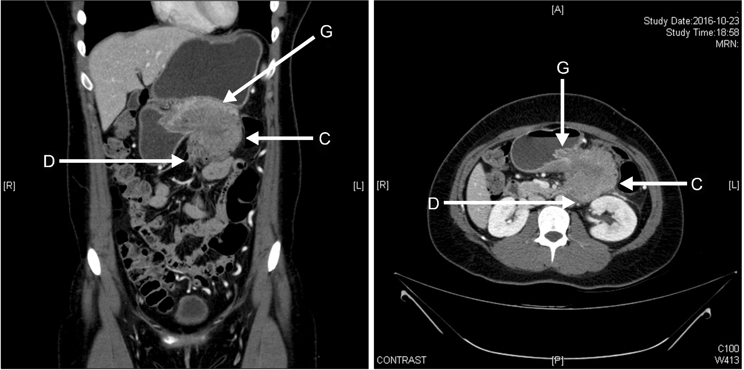
Fig. 1 Abdominal computed to-mogram (CT) showed a 7.7-cm solid mass in the distal pancreas, with signs of splenic artery and vein occlusion, duodenum ‘D’, gastric ‘G’ and transverse colon invasion ‘C’.

Fig. 2 A huge lobulated con-tour mass was found in the body and tail of the pancreas, showing low signal intensity on T1-weighted image (A) and slightly high T2-weighted image (B) with diffusion restriction (C) and delayed enhancement pattern (D).
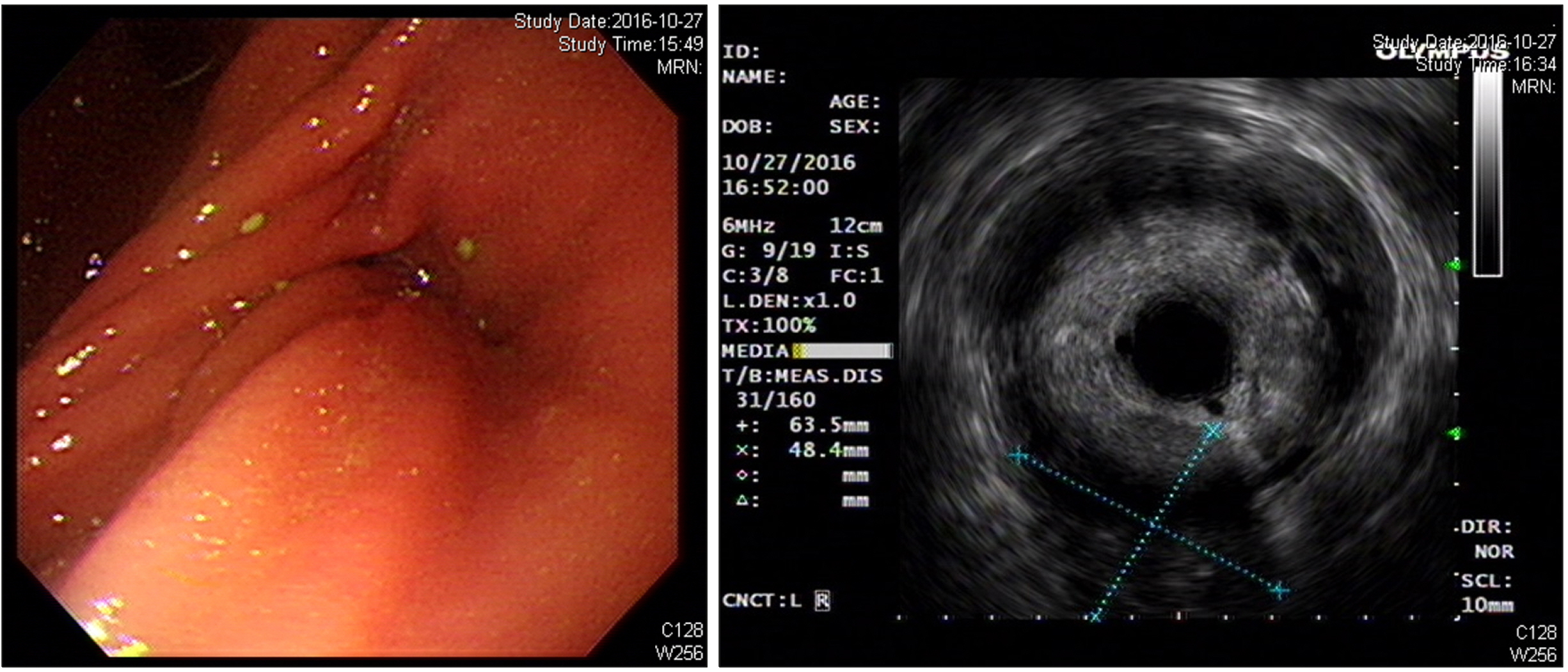
Fig. 3 Endoscopic ultrasono-gram shows a huge soft tissue mass with no cystic portion but multiple echogenic foci in the distal pancreas.
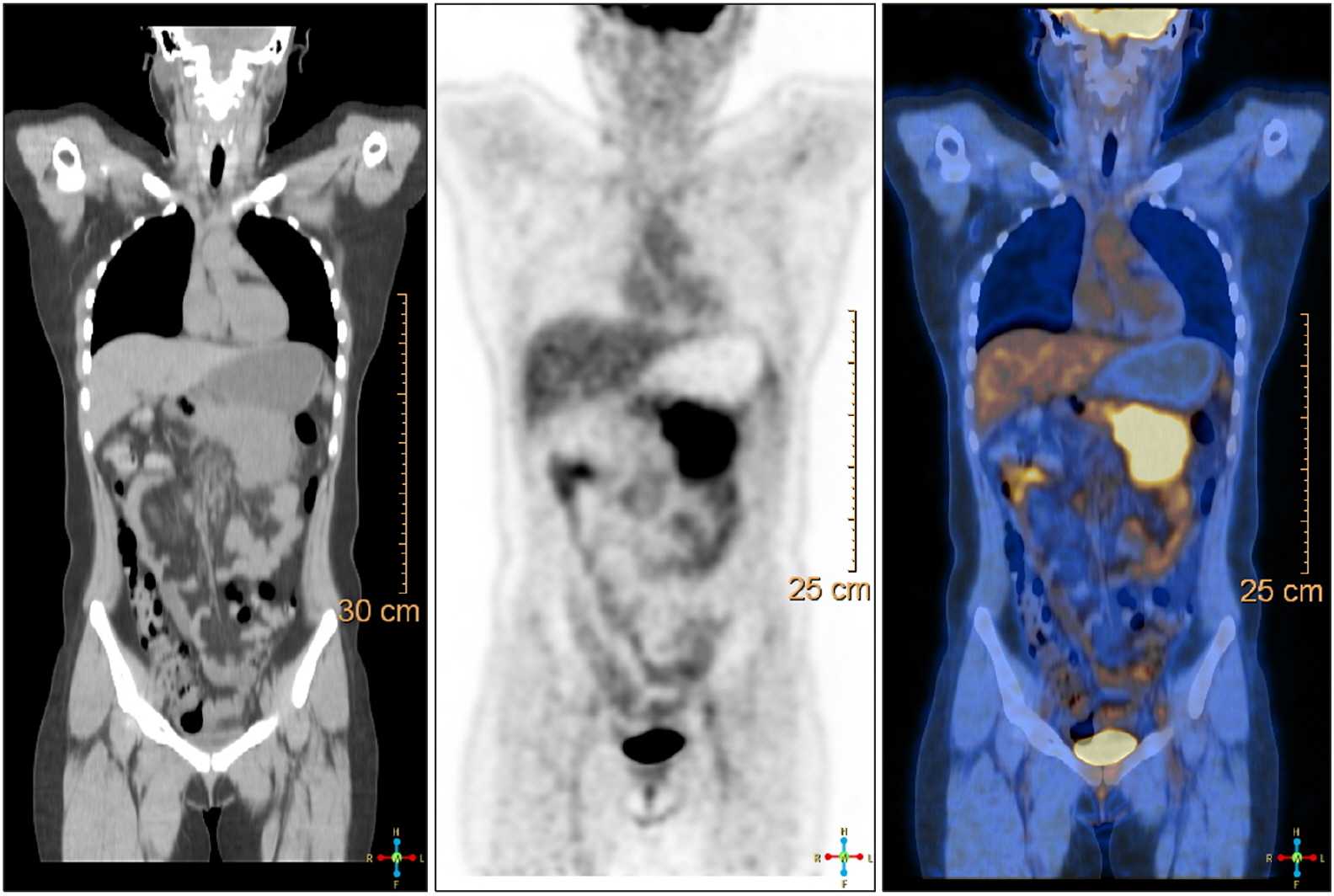
Fig. 4 PET-CT showed heterogeneous hypermetabolic tumor in left upper abdomen.
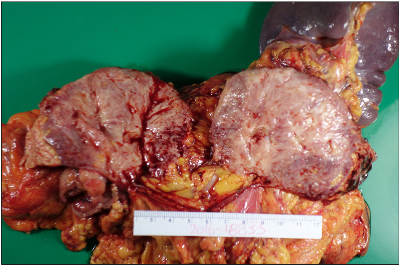
Fig. 5 Gross specimen of the pancreatic body solid mass measuring 10 cm in size.
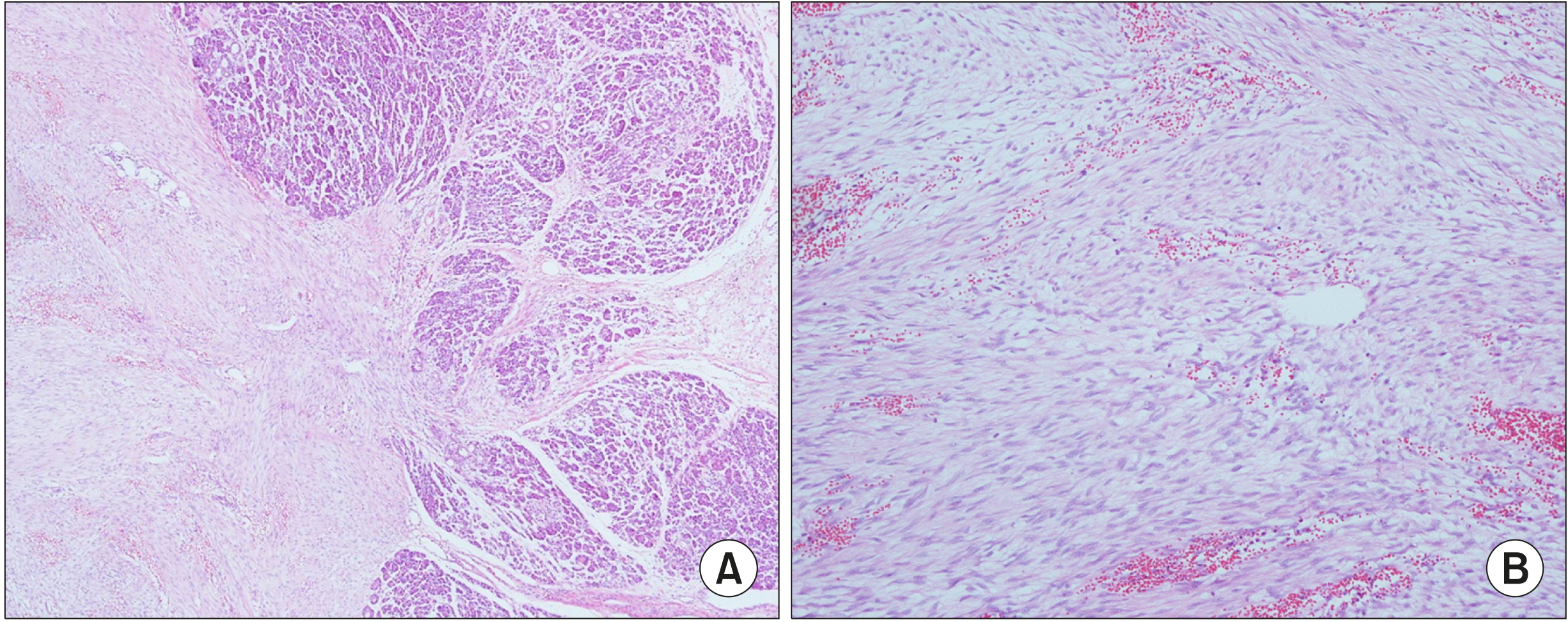
Fig. 6 H&E staining showed spindle cell mass in the pan-creatic gland (A: H&E ×40 mag-nification, B: H&E ×100 magni-fication).
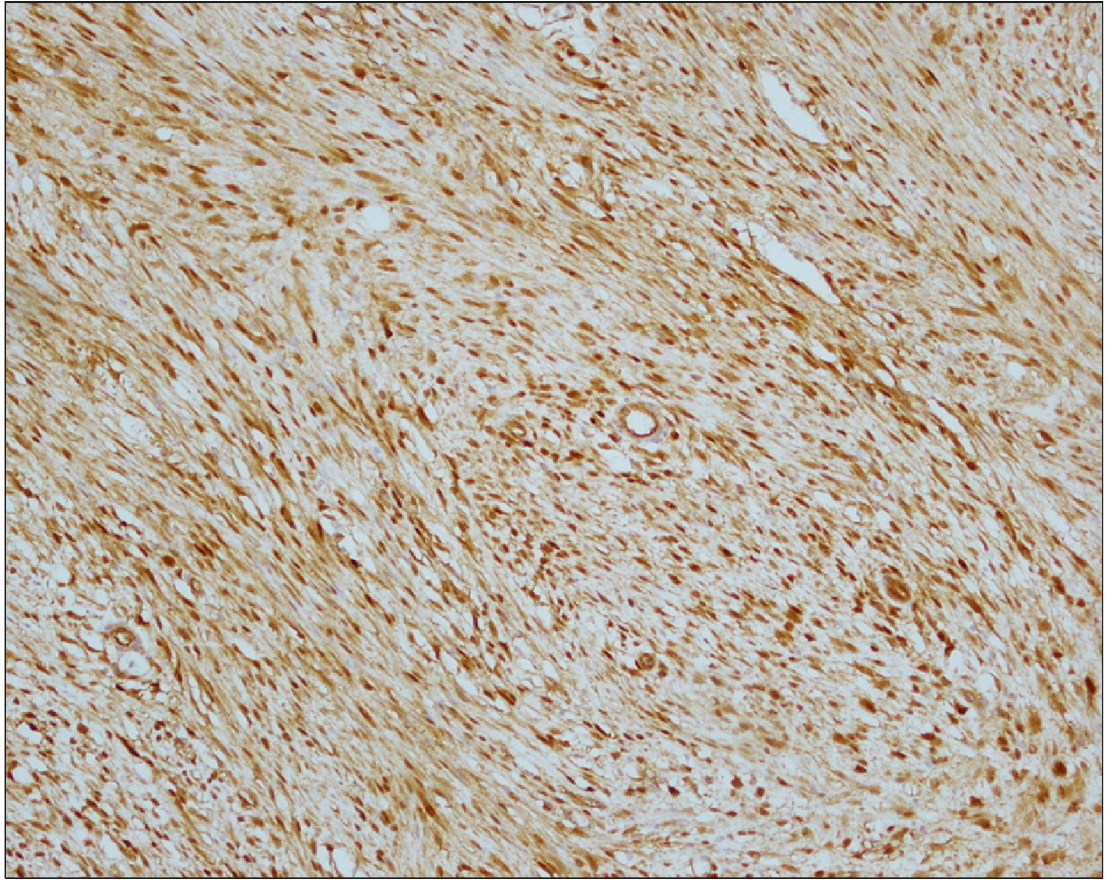
Fig. 7 Immunohistochemical staining showed in strong positive beta-catenin.
Reference
-
1. Wu C, Amini-Nik S, Nadesan P, Stanford WL, Alman BA. 2011; Correction: aggressive fibromatosis (desmoid tumor) is derived from mesenchymal progenitor cells. Cancer Res. 71:6084. DOI: 10.1158/0008-5472.CAN-11-2654.
Article2. Gounder MM, Lefkowitz RA, Keohan ML, D'Adamo DR, Hameed M, Antonescu CR, et al. 2011; Activity of sorafenib against desmoid tumor/deep fibromatosis. Clin Cancer Res. 17:4082–4090. DOI: 10.1158/1078-0432.CCR-10-3322. PMID: 21447727. PMCID: PMC3152981.
Article3. Fisher C, Thway K. 2014; Aggressive fibromatosis. Pathology. 46:135–140. DOI: 10.1097/PAT.0000000000000045. PMID: 24378386.
Article4. Micke O, Seegenschmiedt MH. German Cooperative Group on Radiotherapy for Benign Diseases. 2005; Radiation therapy for aggressive fibromatosis (desmoid tumors): results of a national Patterns of Care Study. Int J Radiat Oncol Biol Phys. 61:882–891. DOI: 10.1016/j.ijrobp.2004.07.705. PMID: 15708271.
Article5. Roggli VL, Kim HS, Hawkins E. 1980; Congenital generalized fibromatosis with visceral involvement. A case report. Cancer. 45:954–960. DOI: 10.1002/1097-0142(19800301)45:5<954::AID-CNCR2820450520>3.0.CO;2-Q.
Article6. Bruce JM, Bradley EL 3rd, Satchidanand SK. 1996; A desmoid tumor of the pancreas. Sporadic intra-abdominal desmoids revisited. Int J Pancreatol. 19:197–203.7. Sedivy R, Ba-Ssalamah A, Gnant M, Hammer J, Klöppel G. 2002; Intraductal papillary-mucinous adenoma associated with unusual focal fibromatosis: a "postoperative" stromal nodule. Virchows Arch. 441:308–311. DOI: 10.1007/s00428-002-0686-x. PMID: 12242530.
Article8. Nursal TZ, Abbasoglu O. 2003; Sporadic hereditary pancreatic desmoid tumor: a new entity? J Clin Gastroenterol. 37:186–188. DOI: 10.1097/00004836-200308000-00019. PMID: 12869894.9. Pho LN, Coffin CM, Burt RW. 2005; Abdominal desmoid in familial adenomatous polyposis presenting as a pancreatic cystic lesion. Fam Cancer. 4:135–138. DOI: 10.1007/s10689-004-1923-z. PMID: 15951964.
Article10. Weiss ES, Burkart AL, Yeo CJ. 2006; Fibromatosis of the remnant pancreas after pylorus-preserving pancreaticoduodenectomy. J Gastrointest Surg. 10:679–688. DOI: 10.1016/j.gassur.2005.09.029. PMID: 16773761.
Article11. Amiot A, Dokmak S, Sauvanet A, Vilgrain V, Bringuier PP, Scoazec JY, et al. 2008; Sporadic desmoid tumor. An exceptional cause of cystic pancreatic lesion. JOP. 9:339–345.12. Polistina F, Costantin G, D'Amore E, Ambrosino G. 2010; Sporadic, nontrauma-related, desmoid tumor of the pancreas: a rare disease-case report and literature review. Case Rep Med. 2010:272760. DOI: 10.1155/2010/272760. PMID: 20300597. PMCID: PMC2838224.
Article13. Jia C, Tian B, Dai C, Wang X, Bu X, Xu F. 2014; Idiopathic desmoid-type fibromatosis of the pancreatic head: case report and literature review. World J Surg Oncol. 12:103. DOI: 10.1186/1477-7819-12-103. PMID: 24755337. PMCID: PMC4032157.
Article14. Xu B, Zhu LH, Wu JG, Wang XF, Matro E, Ni JJ. 2013; Pancreatic solid cystic desmoid tumor: case report and literature review. World J Gastroenterol. 19:8793–8798. DOI: 10.3748/wjg.v19.i46.8793. PMID: 24379602. PMCID: PMC3870530.
Article15. Słowik-Moczydłowska Ż, Rogulski R, Piotrowska A, Małdyk J, Kluge P, Kamiński A. 2015; Desmoid tumor of the pancreas: a case report. J Med Case Rep. 9:104. DOI: 10.1186/s13256-015-0591-y. PMID: 25943401. PMCID: PMC4437747.
Article16. Wang YC, Wong JU. 2016; Complete remission of pancreatic head desmoid tumor treated by COX-2 inhibitor-a case report. World J Surg Oncol. 14:190. DOI: 10.1186/s12957-016-0944-z. PMID: 27450394. PMCID: PMC4957301.
Article17. Lopez R, Kemalyan N, Moseley HS, Dennis D, Vetto RM. 1990; Problems in diagnosis and management of desmoid tumors. Am J Surg. 159:450–453. DOI: 10.1016/S0002-9610(05)81243-7.
Article18. McKinnon JG, Neifeld JP, Kay S, Parker GA, Foster WC, Lawrence W Jr. 1989; Management of desmoid tumors. Surg Gynecol Obstet. 169:104–106.19. Joyce M, Mignanelli E, Church J. 2010; Ureteric obstruction in familial adenomatous polyposis-associated desmoid disease. Dis Colon Rectum. 53:327–332. DOI: 10.1007/DCR.0b013e3181c52894. PMID: 20173481.
Article20. Venkat D, Levine E, Wise WE. 2010; Abdominal pain and colonic obstruction from an intra-abdominal desmoid tumor. Gastroenterol Hepatol (N Y). 6:662–665.21. Einstein DM, Tagliabue JR, Desai RK. 1991; Abdominal desmoids: CT findings in 25 patients. AJR Am J Roentgenol. 157:275–279. DOI: 10.2214/ajr.157.2.1853806. PMID: 1853806.
Article22. de Bree E, Keus R, Melissas J, Tsiftsis D, van Coevorden F. 2009; Desmoid tumors: need for an individualized approach. Expert Rev Anticancer Ther. 9:525–535. DOI: 10.1586/era.09.9. PMID: 19374605.
Article23. Lips DJ, Barker N, Clevers H, Hennipman A. 2009; The role of APC and beta-catenin in the aetiology of aggressive fibromatosis (desmoid tumors). Eur J Surg Oncol. 35:3–10. DOI: 10.1016/j.ejso.2008.07.003. PMID: 18722078.
Article24. Shimada S, Ishizawa T, Ishizawa K, Matsumura T, Hasegawa T, Hirose T. 2006; The value of MDM2 and CDK4 amplification levels using real-time polymerase chain reaction for the differential diagnosis of liposarcomas and their histologic mimickers. Hum Pathol. 37:1123–1129. DOI: 10.1016/j.humpath.2006.04.010. PMID: 16938516.
Article25. Miettinen M, Lasota J. 2006; Gastrointestinal stromal tumors: review on morphology, molecular pathology, prognosis, and differential diagnosis. Arch Pathol Lab Med. 130:1466–1478. DOI: 10.5858/2006-130-1466-GSTROM. PMID: 17090188.
Article26. Bhattacharya B, Dilworth HP, Iacobuzio-Donahue C, Ricci F, Weber K, Furlong MA, et al. 2005; Nuclear beta-catenin expression distinguishes deep fibromatosis from other benign and malignant fibroblastic and myofibroblastic lesions. Am J Surg Pathol. 29:653–659. DOI: 10.1097/01.pas.0000157938.95785.da. PMID: 15832090.
- Full Text Links
-
- Actions
-
Cited
- CITED
-
- Close
- Share
-
- Similar articles
-
- Pancreatic Collision Tumor of Desmoid-Type Fibromatosis and Mucinous Cystic Neoplasm: A Case Report
- Desmoid Tumor of the Facet Joint: A Case Report
- Desmoid Type Fibromatosis in the Facet Joint of Lumbar Spine: Case Report and Review of Literature
- A Case of Nasal Desmoid Tumor
- Recurring Fibromatosis of Breast Following Tumorectomy: A Case Report
