Hepatoid thymic carcinoma: a case report of a rare subtype of thymic carcinoma
- Affiliations
-
- 1Department of Pathology, Asan Medical Center, University of Ulsan College of Medicine, Seoul, Korea
- 2Department of Pathology, Kyung Hee University Hospital at Gangdong, Kyung Hee University College of Medicine, Seoul, Korea
- 3Department of Otolaryngology, Asan Medical Center, University of Ulsan College of medicine, Seoul, Korea
- KMID: 2515928
- DOI: http://doi.org/10.4132/jptm.2021.03.10
Abstract
- Hepatoid thymic carcinoma is an extremely rare subtype of primary thymus tumor resembling “pure” hepatoid adenocarcinomas with hepatocyte paraffin 1 (Hep-Par-1) expression. A 53-year-old man presented with voice change and a neck mass. Multiple masses involving the thyroid, cervical and mediastinal lymph nodes, and lung were detected on computed tomography. Papillary thyroid carcinoma was confirmed by biopsy, and the patient underwent neoadjuvant chemoradiation therapy. However, the anterior mediastinal mass was enlarged after the treatment whereas the multiple masses in the thyroid and neck decreased in size. Microscopically, polygonal tumor cells formed solid sheets or trabeculae resembling hepatocytes and infiltrated remnant thymus. The tumor cells showed immunopositivity for cytokeratin 7, cytokeratin 19, and Hep-Par-1 and negativity for α-fetoprotein. Possibilities of germ cell tumor, squamous cell carcinoma, and metastasis of thyroid papillary carcinoma were excluded by immunohistochemistry. This report on the new subtype of thymic carcinoma is the third in English literature thus far.
Keyword
Figure
-
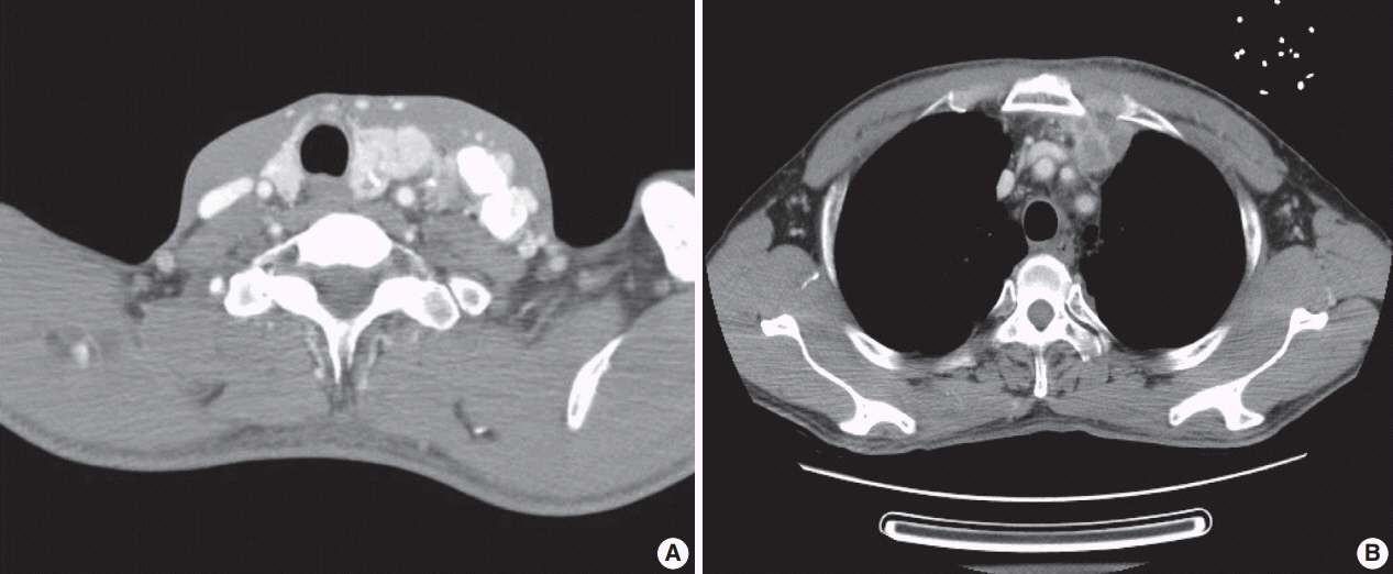
Fig. 1. Radiologic findings. (A) Neck computed tomography (CT) shows a large infiltrating mass with calcification in left thyroid lobe and multiple hypervascular masses in the left level II–VI, left supraclavicular area and right lower neck. (B) The chest CT shows anterior mediastinal mass, measuring 6 cm.

Fig. 2. Pathologic findings. (A) Grossly, an ill-demarcated anterior mediastinal mass, measuring 6 cm shows a heterogeneous, tan colored, firm cut surface. (B, C) Microscopically, the polygonal tumor cells formed solid sheet or trabeculae resembling hepatocytes and infiltrated remnant thymus. Part of the tumor shows clear cytoplasm. (D) The tumor cells show distinct cell borders, abundant eosinophilic cytoplasm, and pleomorphic vesicular nuclei.
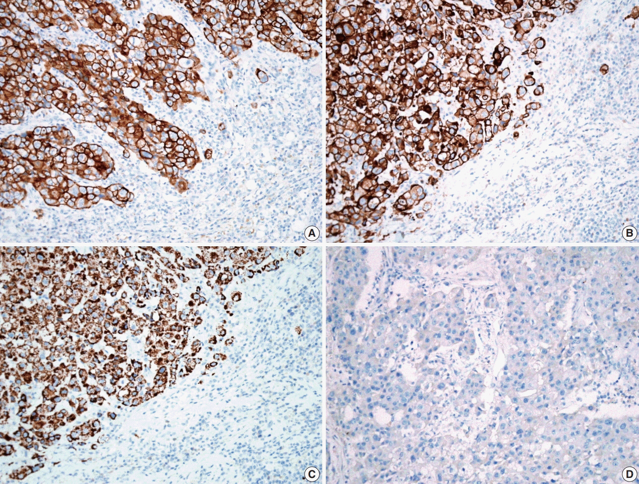
Fig. 3. Representative image of immunohistochemistry. The tumor cells show immune-positivity for cytokeratin 7 (A), cytokeratin 19 (B), and Hep-Par-1 (hepatocyte, C) and negativity for α-fetoprotein (D) by immunohistochemistry
Reference
-
References
1. Travis WD, Brambilla E, Burke AP, Marx A, Nicholson AG. WHO classification of tumours of the lung, pleura, thymus and heart. 4th ed. Lyon: IARC Press;2015.2. Franke A, Strobel P, Fackeldey V, et al. Hepatoid thymic carcinoma: report of a case. Am J Surg Pathol. 2004; 28:250–6.3. Lee JH, Kim H, Chae YS, Won NH, Choi JS, Kim CH. Hepatoid thymic carcinoma: a case report. Korean J Pathol. 2009; 43:562–5.4. Nonaka D, Tang Y, Chiriboga L, Rivera M, Ghossein R. Diagnostic utility of thyroid transcription factors Pax8 and TTF-2 (FoxE1) in thyroid epithelial neoplasms. Mod Pathol. 2008; 21:192–200.
Article5. Su JS, Chen YT, Wang RC, Wu CY, Lee SW, Lee TY. Clinicopathological characteristics in the differential diagnosis of hepatoid adenocarcinoma: a literature review. World J Gastroenterol. 2013; 19:321–7.
Article6. Chu PG, Ishizawa S, Wu E, Weiss LM. Hepatocyte antigen as a marker of hepatocellular carcinoma: an immunohistochemical comparison to carcinoembryonic antigen, CD10, and alpha-fetoprotein. Am J Surg Pathol. 2002; 26:978–88.7. Liu X, Cheng Y, Sheng W, et al. Analysis of clinicopathologic features and prognostic factors in hepatoid adenocarcinoma of the stomach. Am J Surg Pathol. 2010; 34:1465–71.
Article8. Engels EA. Epidemiology of thymoma and associated malignancies. J Thorac Oncol. 2010; 5(10 Suppl 4):S260–5.
Article9. Ishikura H, Kirimoto K, Shamoto M, et al. Hepatoid adenocarcinomas of the stomach: an analysis of seven cases. Cancer. 1986; 58:119–26.
Article10. Ishikura H, Kanda M, Ito M, Nosaka K, Mizuno K. Hepatoid adenocarcinoma: a distinctive histological subtype of alpha-fetoproteinproducing lung carcinoma. Virchows Arch A Pathol Anat Histopathol. 1990; 417:73–80.
Article
