Ann Rehabil Med.
2021 Apr;45(2):116-122. 10.5535/arm.20172.
Is Abnormal Electrodiagnostic Finding Related to the Cross-Sectional Area of the Nerve Root in Cervical Radiculopathy?
- Affiliations
-
- 1Department of Physical Medicine and Rehabilitation, Korea University Guro Hospital, Seoul, Korea
- KMID: 2515392
- DOI: http://doi.org/10.5535/arm.20172
Abstract
Objective
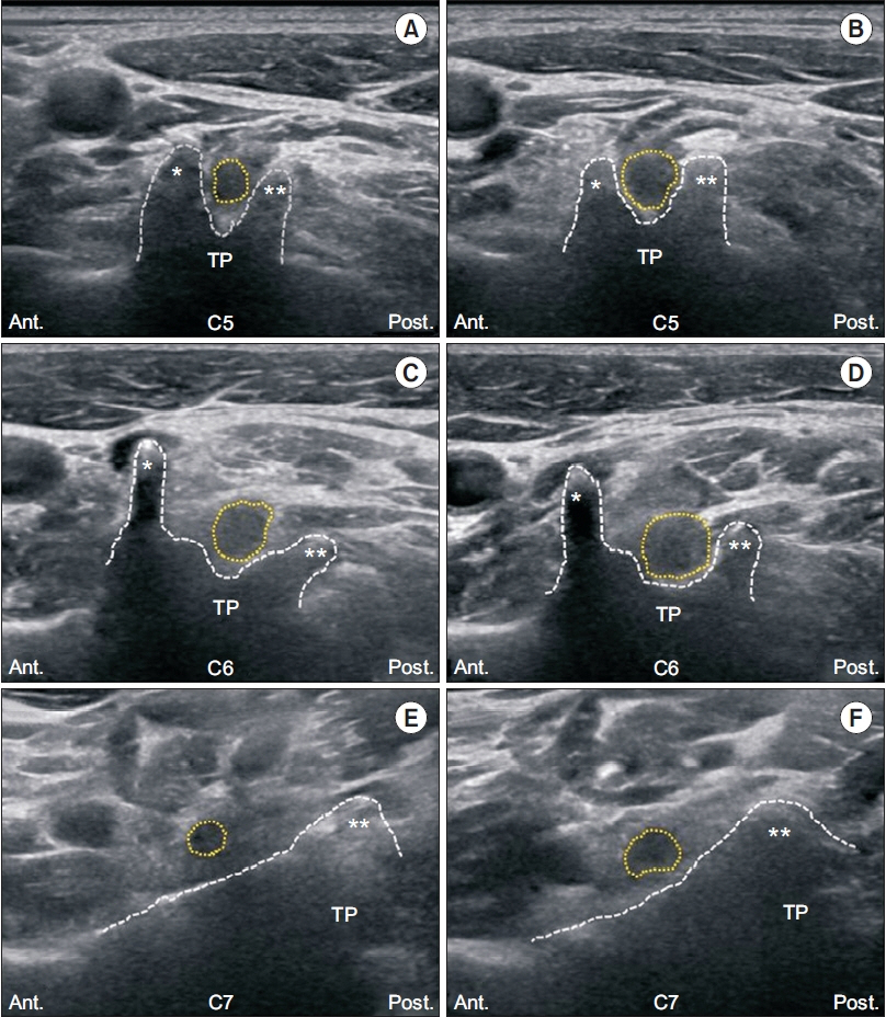
To assess the relevance of electrodiagnosis (EDX) in the cross-sectional area (CSA) of the nerve root of patients with cervical radiculopathy (CR) by using high-resolution ultrasonography (HRUS).
Methods
The CSAs of the cervical nerve roots at C5, C6, and C7 were measured bilaterally using HRUS in 29 patients with unilateral CR whose clinical symptoms, magnetic resonance imaging (MRI) findings, and EDX
results
corresponded with each other (CR-A group), and in 26 patients with unilateral CR whose clinical symptoms and MRI findings matched with each other but did not correspond with the EDX findings (CR-B group). Results The CSA of the affected side in each nerve root was significantly larger than that of the unaffected side in both the CR-A and CR-B groups. The side-to-side difference in the bilateral CSAs of the nerve root and the ratio of the CSAs between the unaffected and affected sides were statistically larger in the CR-A group than in the CR-B group.
Conclusion
The increased CSAs in the CR-A group reflect the physiological changes of the cervical nerve root, which is supported by the EDX findings.
Keyword
Figure
Reference
-
1. Abbed KM, Coumans JV. Cervical radiculopathy: pathophysiology, presentation, and clinical evaluation. Neurosurgery. 2007; 60(1 Supp1 1):S28–34.2. Ellenberg MR, Honet JC, Treanor WJ. Cervical radiculopathy. Arch Phys Med Rehabil. 1994; 75:342–52.
Article3. Van Zundert J, Huntoon M, Patijn J, Lataster A, Mekhail N, van Kleef M, et al. Cervical radicular pain. Pain Pract. 2010; 10:1–17.
Article4. Manelfe C. Imaging of the spine and spinal cord. Curr Opin Radiol. 1991; 3:5–15.
Article5. Malanga GA. The diagnosis and treatment of cervical radiculopathy. Med Sci Sports Exerc. 1997; 29(7 Suppl):S236–45.
Article6. Bianchi S. Ultrasound of the peripheral nerves. Joint Bone Spine. 2008; 75:643–9.
Article7. Haun DW, Cho JC, Kettner NW. Normative crosssectional area of the C5-C8 nerve roots using ultrasonography. Ultrasound Med Biol. 2010; 36:1422–30.
Article8. Tazawa K, Matsuda M, Yoshida T, Shimojima Y, Gono T, Morita H, et al. Spinal nerve root hypertrophy on MRI: clinical significance in the diagnosis of chronic inflammatory demyelinating polyradiculoneuropathy. Intern Med. 2008; 47:2019–24.
Article9. Kim E, Yoon JS, Kang HJ. Ultrasonographic crosssectional area of spinal nerve roots in cervical radiculopathy: a pilot study. Am J Phys Med Rehabil. 2015; 94:159–64.10. Takeuchi M, Wakao N, Hirasawa A, Murotani K, Kamiya M, Osuka K, et al. Ultrasonography has a diagnostic value in the assessment of cervical radiculopathy: a prospective pilot study. Eur Radiol. 2017; 27:3467–73.
Article11. Lance JW, Burke D. Mechanisms of spasticity. Arch Phys Med Rehabil. 1974; 55:332–7.12. Reza Soltani Z, Sajadi S, Tavana B. A comparison of magnetic resonance imaging with electrodiagnostic findings in the evaluation of clinical radiculopathy: a cross-sectional study. Eur Spine J. 2014; 23:916–21.13. Partanen J, Partanen K, Oikarinen H, Niemitukia L, Hernesniemi J. Preoperative electroneuromyography and myelography in cervical root compression. Electromyogr Clin Neurophysiol. 1991; 31:21–6.14. Ashkan K, Johnston P, Moore AJ. A comparison of magnetic resonance imaging and neurophysiological studies in the assessment of cervical radiculopathy. Br J Neurosurg. 2002; 16:146–8.
Article15. Won SJ, Kim BJ, Park KS, Kim SH, Yoon JS. Measurement of cross-sectional area of cervical roots and brachial plexus trunks. Muscle Nerve. 2012; 46:711–6.
Article16. Jensen MC, Brant-Zawadzki MN, Obuchowski N, Modic MT, Malkasian D, Ross JS. Magnetic resonance imaging of the lumbar spine in people without back pain. N Engl J Med. 1994; 331:69–73.
Article17. Nicotra A, Khalil NM, O’Neill K. Cervical radiculopathy: discrepancy or concordance between electromyography and magnetic resonance imaging? Br J Neurosurg. 2011; 25:789–90.
Article18. Bianchi S, Montet X, Martinoli C, Bonvin F, Fasel J. High-resolution sonography of compressive neuropathies of the wrist. J Clin Ultrasound. 2004; 32:451–61.
Article19. G Bodner. Nerve compression syndromes. In : Peer S, Bodner G, editors. High-resolution sonography of the peripheral nervous system. 2nd ed. Berlin, Germany: Springer;2008. p. 72–122.
- Full Text Links
- Actions
-
Cited
- CITED
-
- Close
- Share
- Similar articles
-
- Acute Spinal Cord Injury after Cervical Nerve Root Block
- Anatomical and Electrophysiological Myotomes Corresponding to the Flexor Carpi Ulnaris Muscle
- Normal Sonographic Peripheral Nerve Cross-Sectional Area Based on the Results of Meta-Analysis Studies
- Cervical Radiculopathy: Selective Nerve Root Injection of Steroids
- Correlation between Severity of Intervertebral Disc Herniation and Electrodiagnostic Findings in the S1 Radiculopathy


