Giant Intradiploic Epidermoid Cyst in the Occipital Bone: A Case Report
- Affiliations
-
- 1Departments of Neurosurgery, Yeungnam University Hospital, Yeungnam University College of Medicine, Daegu, Korea
- 2Departments of Pathology, Yeungnam University Hospital, Yeungnam University College of Medicine, Daegu, Korea
- KMID: 2515078
- DOI: http://doi.org/10.14791/btrt.2021.9.e3
Abstract
- Epidermoid cysts are uncommon intracranial tumors. As one of the extradural types of epidermoid cysts, intradiploic epidermoid cysts are even rarer tumors and occur in any part of the skull. We herein report a rare case of a giant intradiploic epidermoid cyst of the occipital bone. A 57-year-old woman presented with a 1-year history of localized headache in the occipital area. CT and MRI showed an extradural mass measuring 50×70 mm in the occipital bone with bony destruction. The patient underwent surgical resection. The tumor was completely removed with its capsule. There was no extension to the intradural space. The pathological report confirmed that the tumor was an epidermoid cyst. Follow-up MRI 24 months after the operation showed no recurrence. The headache was well controlled without any medications. We report a rare case of intradiploic epidermoid cyst with clinical and radiologic features and surgical treatment. It is important to consider this diagnosis for a patient with persistent regional headache with or without a growing scalp mass.
Keyword
Figure
-
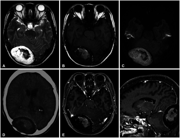
Fig. 1 CT and MRI shows an extradural mass measuring 50×70 mm in the occipital bone. MRI also reveals compression in the right occipital lobe and cerebellar hemisphere. A: T2-weighted MRI shows hyperintensity with a low intratumoral signal. B: T1-weighted MRI shows hypointensity with a high intratumoral signal. C: Diffusion-weighted imaging shows restricted diffusion with characteristic hyperintensity. D: CT scanning shows a large hypodense mass with sharply demarcated bony defects in the right occipital bone. E and F: T1-weighted contrast MRI shows no enhancement of the mass. There is no abnormal signal change in the brain.
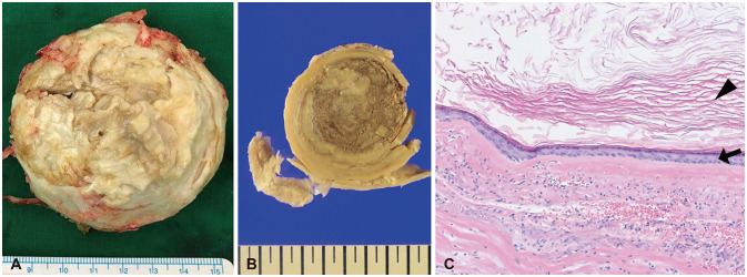
Fig. 2 The cyst is unilocular and consisted of a grayish brown, sticky material. A and B: The cyst wall is gray-white and smooth. C: Histologic examination shows that the cyst is lined by mature squamous epithelium (arrow) and filled with anucleate laminated keratin material (arrowhead) (hematoxylin-eosin stain, ×100).
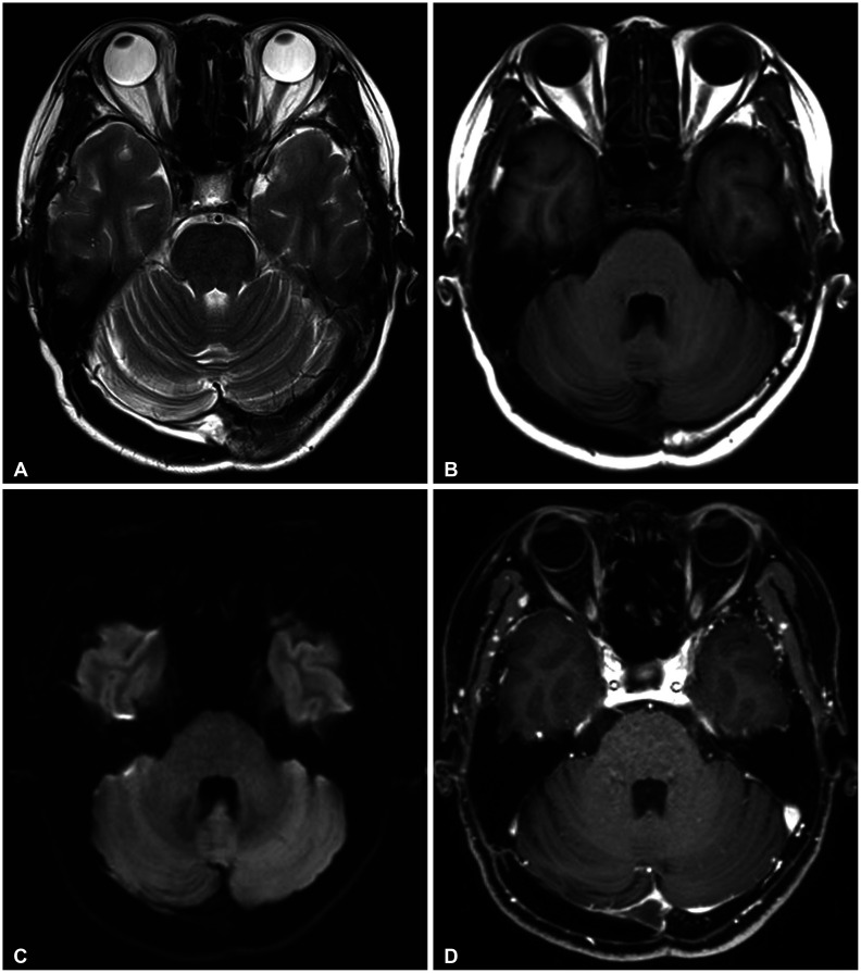
Fig. 3 A follow-up MRI at 24 months after surgery shows no residual or recurrent mass. A: T2-weighted MRI. B: T1-weighted MRI. C: Diffusion-weighted imaging. D: T1-weighed gadolinium-enhanced MRI.
Reference
-
1. Rubin G, Scienza R, Pasqualin A, Rosta L, Da Pian R. Craniocerebral epidermoids and dermoids. A review of 44 cases. Acta Neurochir (Wien). 1989; 97:1–16. PMID: 2718791.2. Tan TI. Epidermoids and dermoids of the central nervous system. (With two exceptional cases not represented in the literature). Acta Neurochir (Wien). 1972; 26:13–24. PMID: 5043165.3. Ciappetta P, Artico M, Salvati M, Raco A, Gagliardi FM. Intradiploic epidermoid cysts of the skull: report of 10 cases and review of the literature. Acta Neurochir (Wien). 1990; 102:33–37. PMID: 2407051.4. Prall JA, Lloyd GL, Breeze RE. Traumatic brain injury associated with an intradiploic epidermoid cyst: case report. Neurosurgery. 1995; 37:523–525. PMID: 7501121.5. Ulrich J. Intracranial epidermoids. A study on their distribution and spread. J Neurosurg. 1964; 21:1051–1058. PMID: 14279825.6. Singh N, Symons SP, Montanera W, et al. Hemorrhagic epidermoid cyst in a patient with generalized tonic clonic seizure. J Clin Neurosci. 2013; 20:750–752. PMID: 23352350.7. Kveton JF, Glasscock ME 3rd, Christiansen SG. Malignant degeneration of an epidermoid of the temporal bone. Otolaryngol Head Neck Surg. 1986; 94:633–636. PMID: 3088529.8. Dupre DA, Pu C, Yu A, Tomycz N. Traumatic intradiploic epidermoid cyst manifest as scalp papule. BMJ Case Rep. 2015; 2015:bcr2014207968.9. Constans JP, Meder JF, De Divitiis E, Donzelli R, Maiuri F. Giant intradiploic epidermoid cysts of the skull. Report of two cases. J Neurosurg. 1985; 62:445–448. PMID: 3973714.10. Krupp W, Heckert A, Holland H, Meixensberger J, Fritzsch D. Giant intradiploic epidermoid cyst with large osteolytic lesions of the skull: a case report. J Med Case Rep. 2012; 6:85. PMID: 22439665.11. Hasturk AE, Basmaci M, Yilmaz ER, Kertmen H, Gurer B, Atilgan AO. Giant intradiploic epidermoid cyst presenting as solitary skull mass with intracranial extension. J Craniofac Surg. 2013; 24:2169–2171. PMID: 24220431.12. Jaiswal AK, Mahapatra AK. Giant intradiploic epidermoid cysts of the skull. A report of eight cases. Br J Neurosurg. 2000; 14:225–228. PMID: 10912199.13. Bhaskar MS, Char G, Bhatt RP. Giant cranial intradiploic epidermoid cyst. West Indian Med J. 1988; 37:119–122. PMID: 3218226.14. Maiuri F, Del Basso De Caro M, D'Acunzi G, Tortora F, Esposito F. Giant intradiploic epidermoid cyst of the occipital bone. Zentralbl Neurochir. 2004; 65:32–35. PMID: 14981574.15. Duan ZX, Chu SH, Ma YB, Zhang H, Zhu JL. Giant intradiploic epidermoid cyst of the occipital bone. J Clin Neurosci. 2009; 16:1478–1480. PMID: 19586771.16. Oommen A, Govindan J, Peroor DS, Azeez CR, Rashmi R, Abdul Jalal MJ. Giant occipital intradiploic epidermoid cyst. Asian J Neurosurg. 2018; 13:514–517. PMID: 29682075.17. Arko L 4th, Berry CT, Desai AS, Weaver M. Intradiploic epidermoid tumors of the cranium: case report with review of the literature. J Neurol Surg A Cent Eur Neurosurg. 2017; 78:167–179. PMID: 27556641.18. Smirniotopoulos JG, Chiechi MV. Teratomas, dermoids, and epidermoids of the head and neck. Radiographics. 1995; 15:1437–1455. PMID: 8577967.19. Bikmaz K, Cosar M, Bek S, Gokduman CA, Arslan M, Iplikcioglu AC. Intradiploic epidermoid cysts of the skull: a report of four cases. Clin Neurol Neurosurg. 2005; 107:262–267. PMID: 15884157.20. Dunn RC Jr, Archer CA, Rapport RL 2nd, Looi LM. Unusual CT-dense posterior fossa epidermoid cyst: case report. J Neurosurg. 1981; 55:654–656. PMID: 7277016.21. Hasegawa H, Bitoh S, Nakata M, Fujiwara M, Yasuda H. Intracranial epidermoid mimicking meningioma. Surg Neurol. 1981; 15:372–374. PMID: 9760977.22. Timmer FA, Sluzewski M, Treskes M, van Rooij WJ, Teepen JL, Wijnalda D. Chemical analysis of an epidermoid cyst with unusual CT and MR characteristics. AJNR Am J Neuroradiol. 1998; 19:1111–1112. PMID: 9672020.23. Inoue Y, Ohata K, Nakayama K, Haba T, Shakudo M. An unusual middle fossa interdural epidermoid tumor. Case report. J Neurosurg. 2001; 95:902–904. PMID: 11702885.24. Arana E, Martí-Bonmatí L. CT and MR imaging of focal calvarial lesions. AJR Am J Roentgenol. 1999; 172:1683–1688. PMID: 10350315.25. Hakyemez B, Aksoy U, Yildiz H, Ergin N. Intracranial epidermoid cysts: diffusion-weighted, FLAIR and conventional MR findings. Eur J Radiol. 2005; 54:214–220. PMID: 15837401.26. Juglard R, Elfikri A, Fesselet J, et al. Intradiploic epidermoid cyst: diagnosis by ultrasonography and MRI. J Radiol. 2003; 84(2 Pt 1):155–157. PMID: 12717289.27. Yamaguchi S, Hirohata T, Sumida M, Arita K, Kurisu K. Intradiploic arachnoid cyst identified by diffusion-weighted magnetic resonance imaging--case report. Neurol Med Chir (Tokyo). 2002; 42:137–139. PMID: 11936057.28. Guridi J, Ollier J, Aguilera F. Giant intradiploic epidermoid tumor of the occipital bone: case report. Neurosurgery. 1990; 27:978–980. discussion 980–1. PMID: 2274141.
- Full Text Links
-
- Actions
-
Cited
- CITED
-
- Close
- Share
-
- Similar articles
-
- Posttraumatic Giant Extradural Intradiploic Epidermoid Cysts of Posterior Cranial Fossa : Case Report and Review of the Literature
- A Case of Giant Intradiploic Epidermoid Cyst of the Temporal Bone
- Intradiploic Epidermoid Cyst of the Skull
- Occipital Intradiploic Epidermoid Cyst with Intracranial Hypertension
- A Case of Large Intradiploic Epidermoid Cyst of the Temporal Bone
