Basic Physical Principles and Clinical Applications of Computed Tomography
- Affiliations
-
- 1Development Headquarter, FutureChem Co., Ltd, Korean Institute of Radiological & Medical Sciences (KIRAMS), Seoul, Korea
- 2Division of Applied RI, Korean Institute of Radiological & Medical Sciences (KIRAMS), Seoul, Korea
- KMID: 2514821
- DOI: http://doi.org/10.14316/pmp.2021.32.1.1
Abstract
- The evolution of X-ray computed tomography (CT) has been based on the discovery of X-rays, the inception of the Radon transform, and the development of X-ray digital data acquisition systems and computer technology. Unlike conventional X-ray imaging (general radiography), CT reconstructs cross-sectional anatomical images of the internal structures according to X-ray attenuation coefficients (approximate tissue density) for almost every region in the body. This article reviews the essential physical principles and technical aspects of the CT scanner, including several notable evolutions in CT technology that resulted in the emergence of helical, multidetector, cone beam, portable, dual-energy, and phase-contrast CT, in integrated imaging modalities, such as positronemission-tomography一CT and single-photon-emission-computed-tomography一CT, and in clinical applications, including image acquisition parameters, CT angiography, image adjustment, versatile image visualizations, volumetric/surface rendering on a computer workstation, radiation treatment planning, and target localization in radiotherapy. The understanding of CT characteristics will provide more effective and accurate patient care in the fields of diagnostics and radiotherapy, and can lead to the improvement of image quality and the optimization of exposure doses.
Keyword
Figure
-
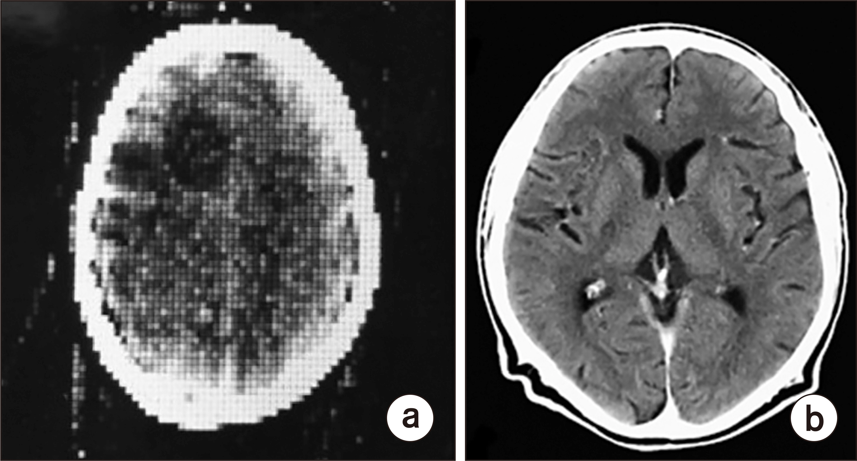
Fig. 1 First clinical computed tomography (CT) image (80×80 image matrix obtained from the Atkinson Morley Hospital, London, UK) of the brain acquired in 1971 (a) [9,10], brain CT image acquired recently (512×512 image matrix, Siemens SOMATOM Plus 4 [Siemens medical systems, Erlangen, Germany]) with a more advanced CT scanner (b).
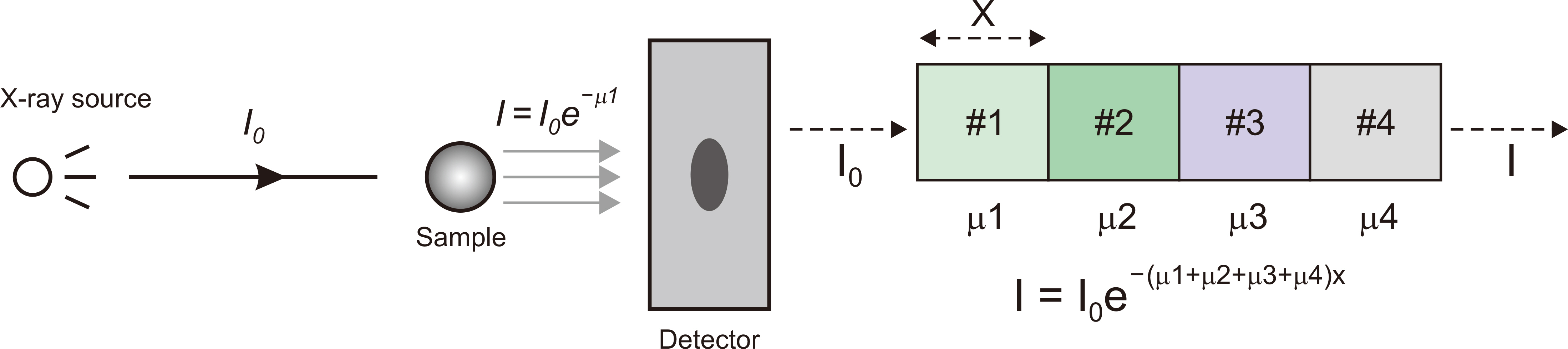
Fig. 2 X-ray beam attenuations passing through of an object (left) and intensity of an X-ray beam passing through an object with multiple different linear attenuation coefficients (μ1, μ2, μ3, μ4) (right).
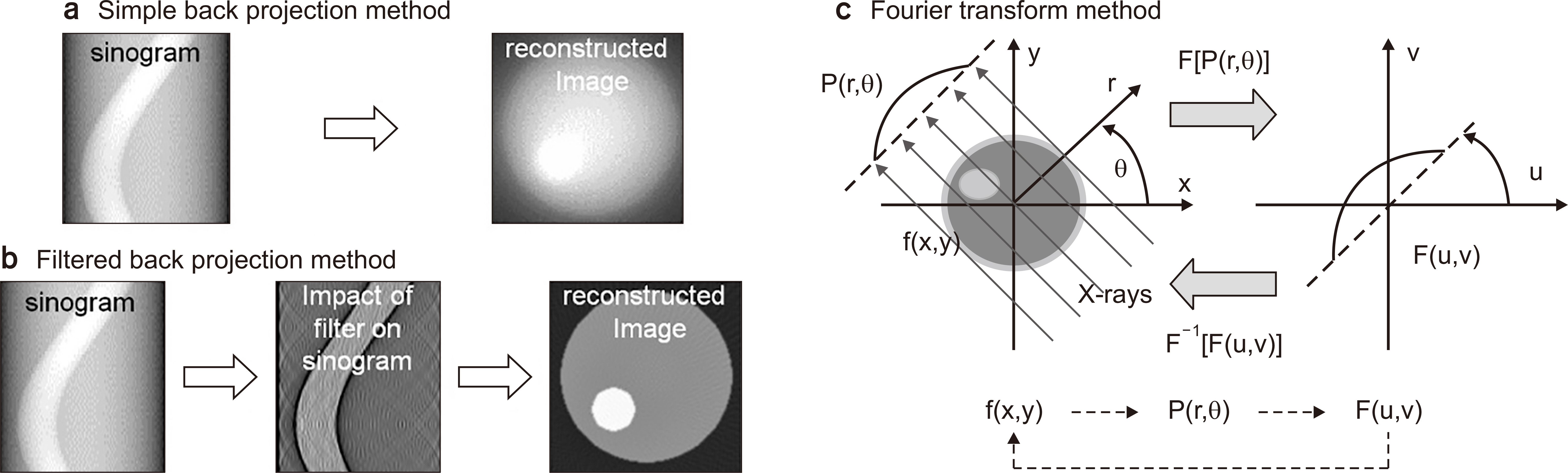
Fig. 3 Computed tomography image reconstruction methods. (a) Simple back projection algorithm method, (b) filtered back projection algorithm method, and (c) Fourier transform algorithm method.
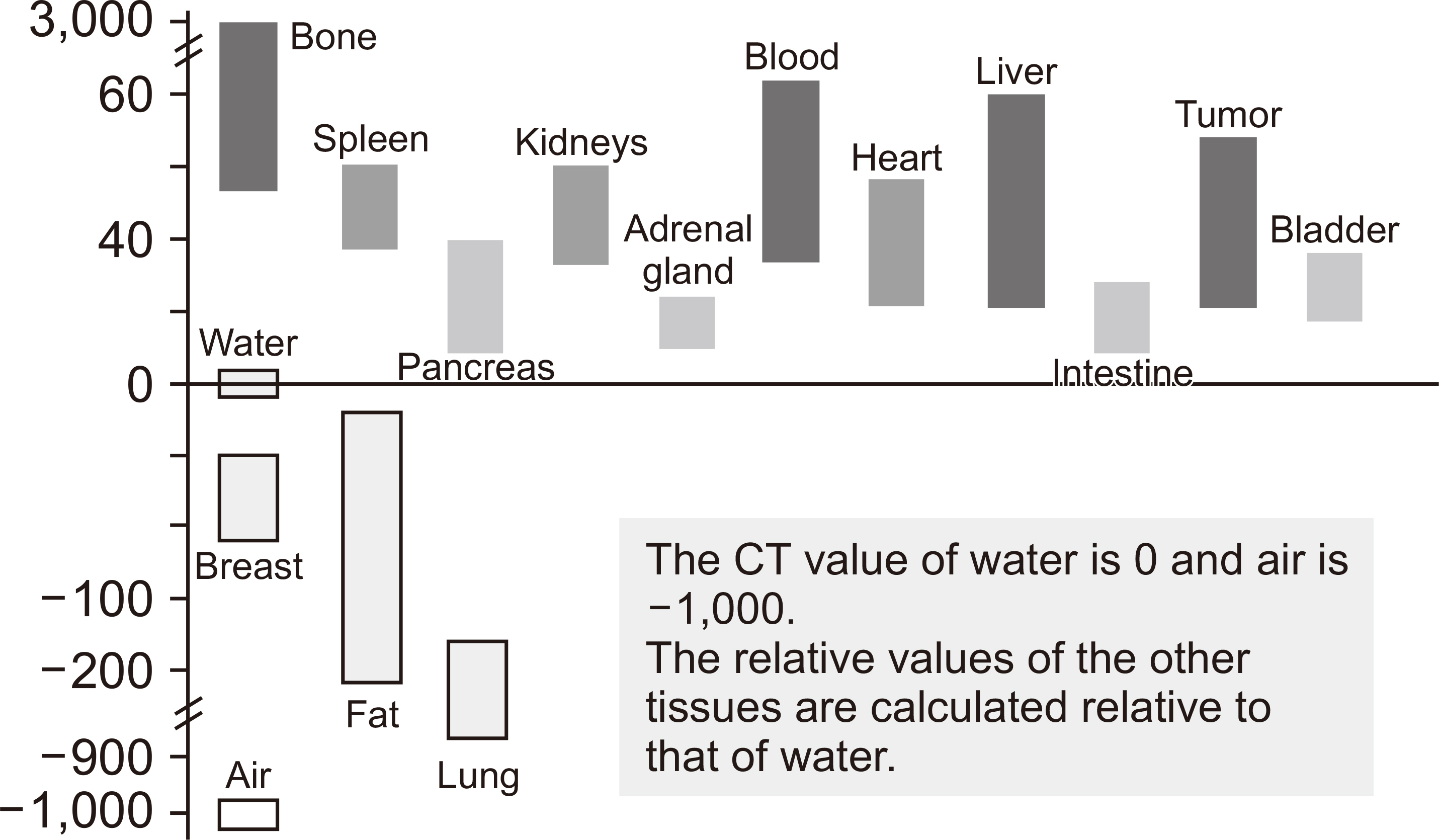
Fig. 4 Hounsfield scale of computed tomography (CT) numbers for various tissues.
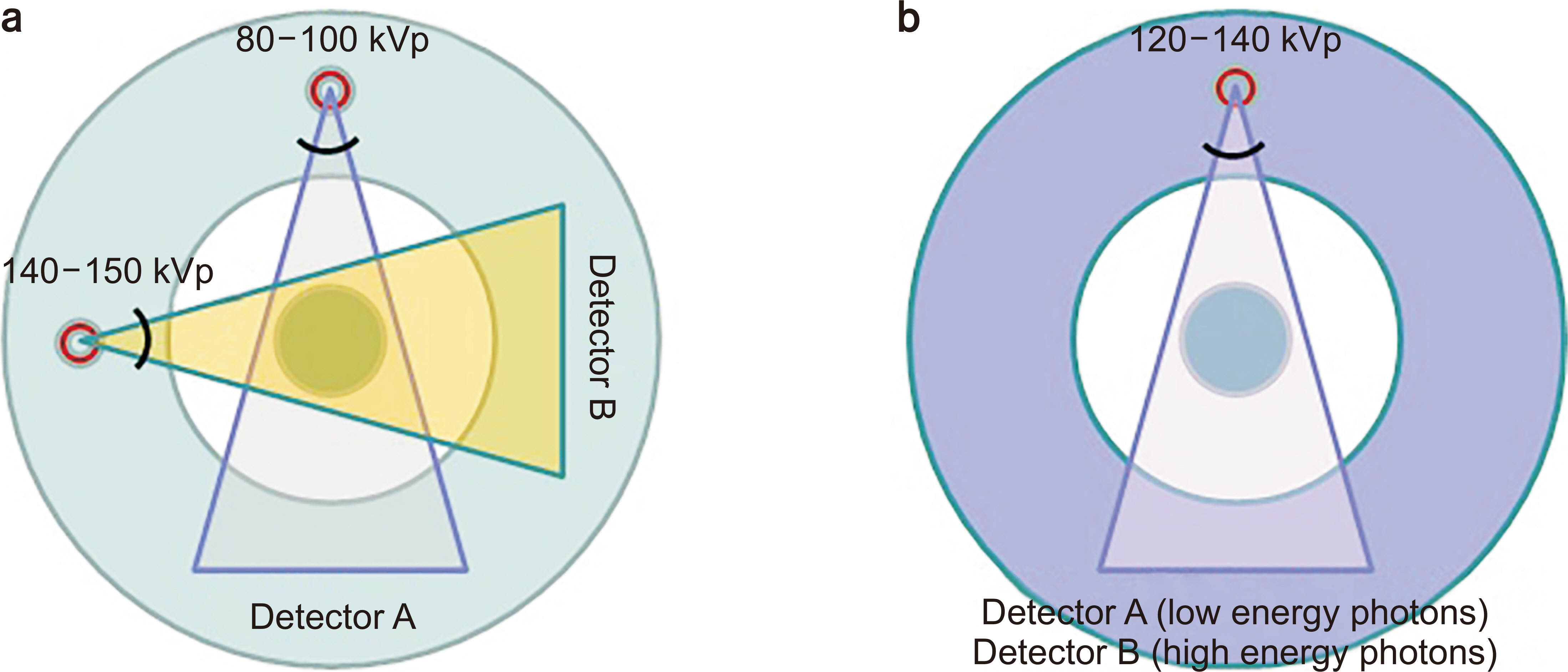
Fig. 5 Structure and mechanism of (a) dual-energy computed tomography (CT) system (adopt two X-ray tubes and corresponding detector arrays) and (b) detector-based spectral CT system (single X-ray tube and two layers within detector arrays).
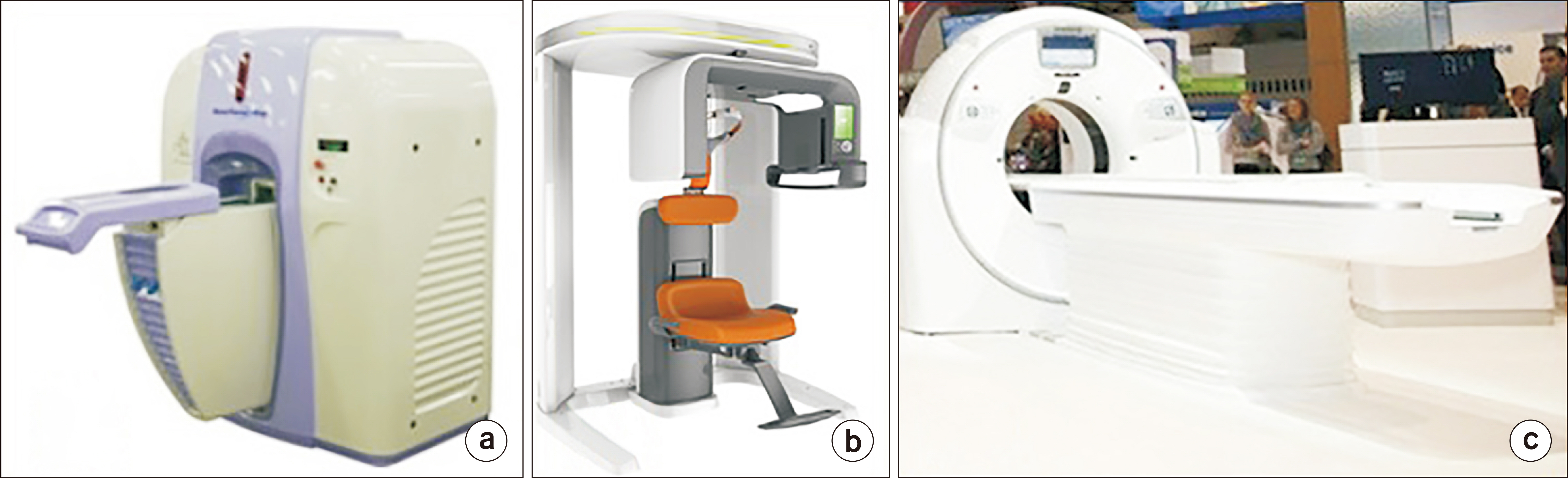
Fig. 6 Domestic commercial computed tomography (CT) scanners. (a) NanoFocusRay’s gantry of the rotational microct system (NanoFocusRay Co., Ltd., Seoul, Korea; 2008). (b) Vatech’s conical beam dental CT (PaX-13D Green Premium) (Vatech Co., Ltd., Seoul, Korea; 2012). (c) Diagnostic CT Scanner (NExCT 7) (Samsung Electronics Co. Ltd., Suwon, Korea; 2015).
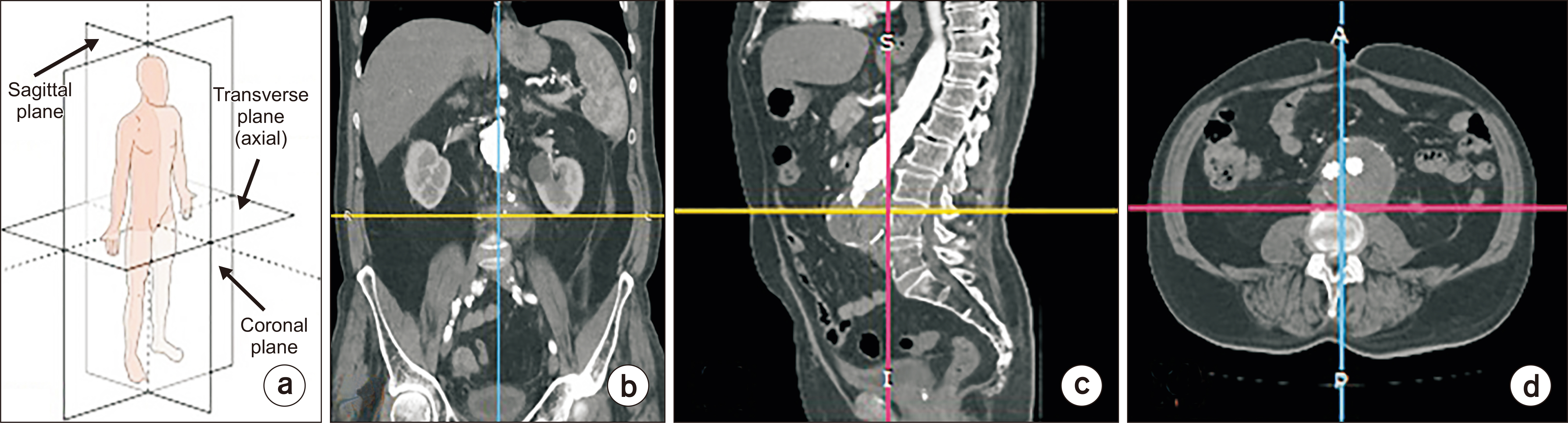
Fig. 7 (a) Anatomical planes in human, multiplanar reconstruction computed tomography images, such as (b) coronal and (c) sagittal converted from (d) axial.
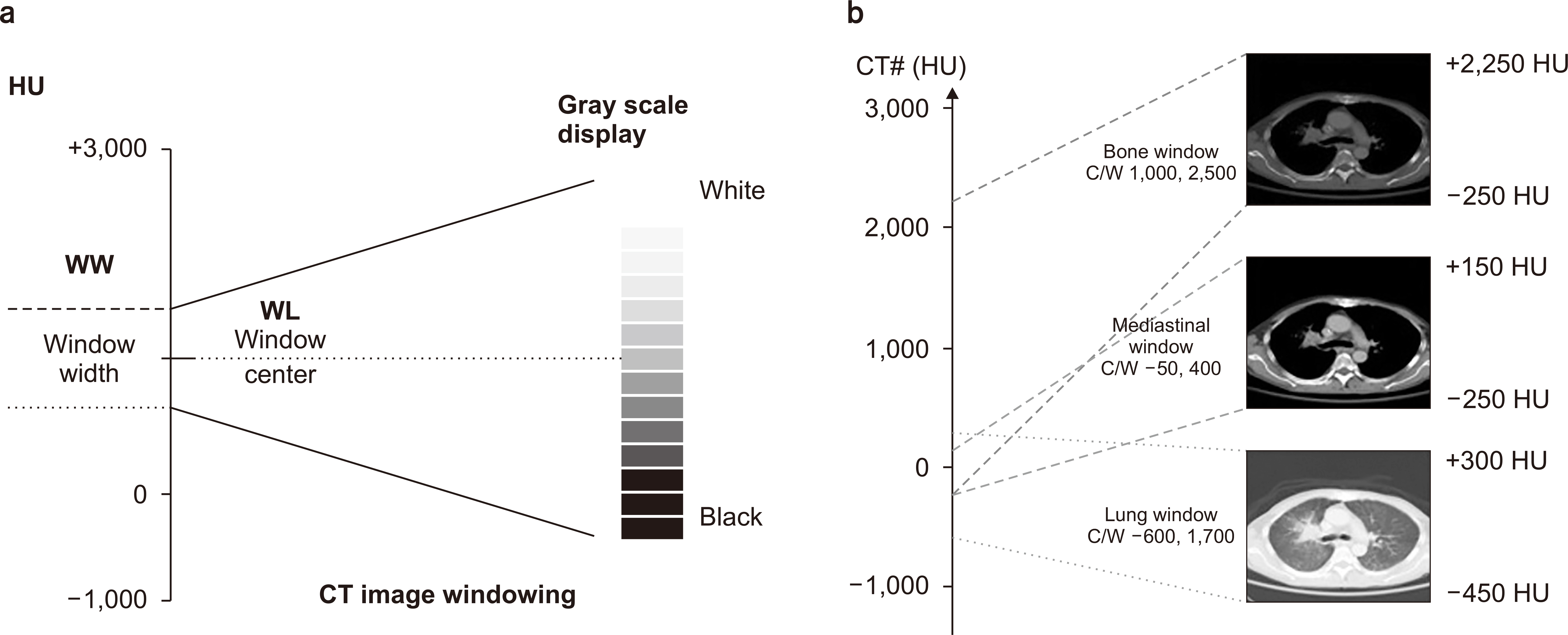
Fig. 8 Computed tomography (CT) image windowing (a), chest CT scan displayed according to the adjustment of bone, mediastinal, and lung windows (b). HU, Hounsfield unit; WW, window width; WL, window level.
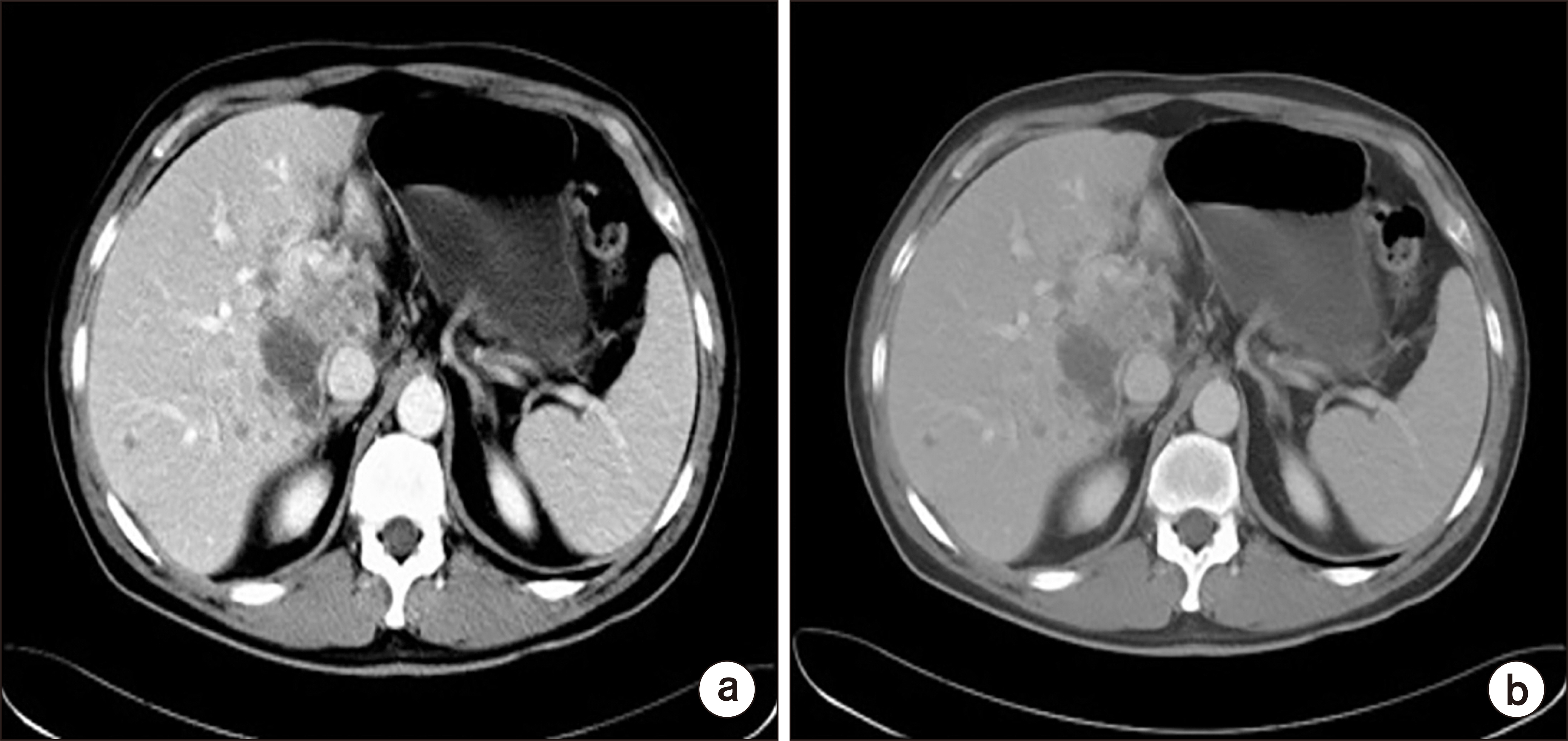
Fig. 9 Computed tomography images shown in Fig. 8 above indicate how the adjustment of the window width changes the contrast of the image, narrow window width (a) and wide window width (b).
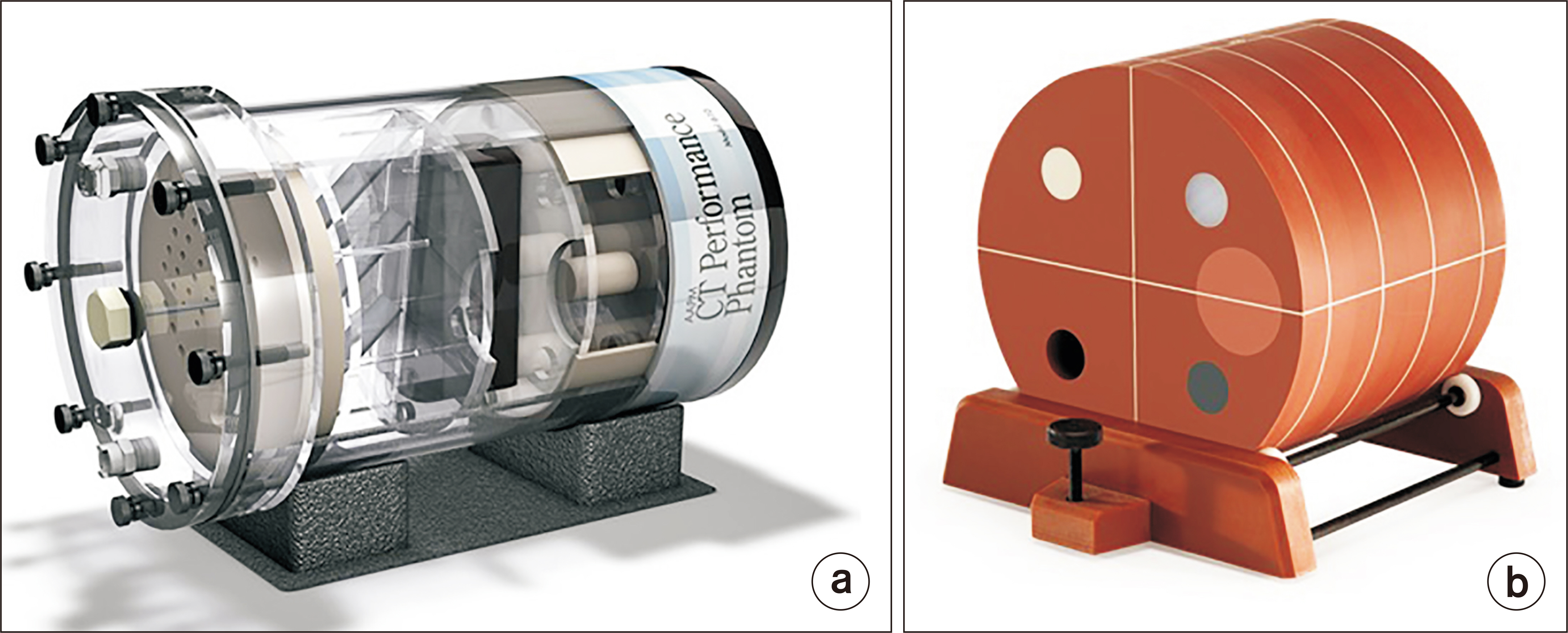
Fig. 10 . (a) American Association of Physicists in Medicine CT performance phantom model 76-410. (b) American College of Radiology CT 464 phantom designed to examine a broad range of image quality parameters.
Reference
-
References
1. Goodman PC. 1995; The new light: discovery and introduction of the X-ray. AJR Am J Roentgenol. 165:1041–1045. DOI: 10.2214/ajr.165.5.7572473. PMID: 7572473.
Article2. Mould RF. 1995; Invited review: Röntgen and the discovery of X-rays. Br J Radiol. 68:1145–1176. DOI: 10.1259/0007-1285-68-815-1145. PMID: 8542220.3. Beatty J. 2012. The radon transform and the mathematics of medical imaging. Colby College;Waterville: Available from: https://digitalcommons.colby.edu/cgi/viewcontent.cgi?article=1649&context=honorstheses . cited 2021 Feb 15.4. Barrett HH. 1984; III The radon transform and its applications. Prog Opt. 21:217–286.
Article5. Hornegger J, Maier A, Kowarschik M. CT Image reconstruction basics. Hornegger J, Maier A, Kowarschik M, CT Image reconstruction basics. Radiology Key, 2018;Available from: https://radiologykey.com/ct-image-reconstruction-basics/ . cited 2021 Feb 15.6. Maier A, Steidl S, Christlein V, Hornegger J. 2018. Medical imaging systems: an introductory guide. Springer Nature Switzerland AG;Cham: p. 148–167.7. Goldman LW. 2007; Principles of CT and CT technology. J Nucl Med Technol. 35:115–128. quiz 129-130. DOI: 10.2967/jnmt.107.042978. PMID: 17823453.
Article8. Bae KT, Whiting BR. Basic principles of computed tomography physics and technical considerations. Radiology Key, 2016;Available from: https://radiologykey.com/basic-principles-of-computed-tomography-physics-and-technical-considerations/ . cited 2021 Feb 15.9. Hounsfield GN. 1973; Computerized transverse axial scanning (tomography). 1. Description of system. Br J Radiol. 46:1016–1022. DOI: 10.1259/0007-1285-46-552-1016. PMID: 4757352.10. Rincon-Guio C, Gómez AM, Charry JD. 2017; The role of computed tomography as a prognostic tool in traumatic brain trauma. Imaging Med. 9:171–178.11. Kachelriess M, Schaller S, Kalender WA. 2000; Advanced single-slice rebinning in cone-beam spiral CT. Med Phys. 27:754–772. DOI: 10.1118/1.598938. PMID: 10798698.12. Kak AC, Slaney M. 1987. Principles of computerized tomographic imaging. Society of Industrial and Applied Mathematics;Philadelphia: p. 49–112.13. Stierstorfer K, Rauscher A, Boese J, Bruder H, Schaller S, Flohr T. 2004; Weighted FBP--a simple approximate 3D FBP algorithm for multislice spiral CT with good dose usage for arbitrary pitch. Phys Med Biol. 49:2209–2218. DOI: 10.1088/0031-9155/49/11/007. PMID: 15248573.
Article14. Flohr TG, Schaller S, Stierstorfer K, Bruder H, Ohnesorge BM, Schoepf UJ. 2005; Multi-detector row CT systems and image-reconstruction techniques. Radiology. 235:756–773. DOI: 10.1148/radiol.2353040037. PMID: 15833981.
Article15. Hata A, Yanagawa M, Honda O, Kikuchi N, Miyata T, Tsukagoshi S, et al. 2018; Effect of matrix size on the image quality of ultra-high-resolution CT of the lung: comparison of 512 × 512, 1024 × 1024, and 2048 × 2048. Acad Radiol. 25:869–876. DOI: 10.1016/j.acra.2017.11.017. PMID: 29373211.16. Ginat DT, Gupta R. 2014; Advances in computed tomography imaging technology. Annu Rev Biomed Eng. 16:431–453. DOI: 10.1146/annurev-bioeng-121813-113601. PMID: 25014788.
Article17. Garvey CJ, Hanlon R. 2002; Computed tomography in clinical practice. BMJ. 324:1077–1080. DOI: 10.1136/bmj.324.7345.1077. PMID: 11991915. PMCID: PMC1123029.
Article18. Flohr T. 2013; CT systems. Curr Radiol Rep. 1:52–63.
Article19. van Ooijen PM, van Geuns RJ, Rensing BJ, Bongaerts AH, de Feyter PJ, Oudkerk M. 2003; Noninvasive coronary imaging using electron beam CT: surface rendering versus volume rendering. AJR Am J Roentgenol. 180:223–226. DOI: 10.2214/ajr.180.1.1800223. PMID: 12490509.20. Kopp AF, Klingenbeck-Regn K, Heuschmid M, Küttner A, Ohnesorge B, Flohr T, et al. 2000; Multislice computed tomography: basic principles and clinical applications. Electromedica. 68:94–105.21. Grajo JR, Patino M, Prochowski A, Sahani DV. 2016; Dual energy CT in practice: basic principles and applications. Appl Radiol. 45:6–12.22. Sajja S, Lee Y, Eriksson M, Nordström H, Sahgal A, Hashemi M, et al. 2019; Technical principles of dual-energy cone beam computed tomography and clinical applications for radiation therapy. Adv Radiat Oncol. 5:1–16. DOI: 10.1016/j.adro.2019.07.013. PMID: 32051885. PMCID: PMC7004939.
Article23. Ohana M, Jeung MY, Labani A, El Ghannudi S, Roy C. 2014; Thoracic dual energy CT: acquisition protocols, current applications and future developments. Diagn Interv Imaging. 95:1017–1026. DOI: 10.1016/j.diii.2014.01.001. PMID: 24780370.
Article24. Coursey CA, Nelson RC, Boll DT, Paulson EK, Ho LM, Neville AM, et al. 2010; Dual-energy multidetector CT: how does it work, what can it tell us, and when can we use it in abdominopelvic imaging? Radiographics. 30:1037–1055. DOI: 10.1148/rg.304095175. PMID: 20631367.
Article25. Rassouli N, Etesami M, Dhanantwari A, Rajiah P. 2017; Detector-based spectral CT with a novel dual-layer technology: principles and applications. Insights Imaging. 8:589–598. DOI: 10.1007/s13244-017-0571-4. PMID: 28986761. PMCID: PMC5707218.
Article26. Silva AC, Morse BG, Hara AK, Paden RG, Hongo N, Pavlicek W. 2011; Dual-energy (spectral) CT: applications in abdominal imaging. Radiographics. 31:1031–1046. discussion 1047-1050. DOI: 10.1148/rg.314105159. PMID: 21768237.
Article27. Lee SC, Kim HK, Chun IK, Cho MH, Cho MH, Lee SY. 2004; Development of a micro-CT system for small animal imaging. J Biomed Eng Res. 25:97–102.28. Nam KY, Kim KW, Kim JH, Son HH, Ryu JH, Kang SH, et al. 2008; Micro-CT system for small animal imaging. Korean J Med Phys. 19:102–112. DOI: 10.1117/12.772303,. PMID: 22049304. PMCID: PMC3204786.29. Goldman LW. 2007; Principles of CT: radiation dose and image quality. J Nucl Med Technol. 35:213–225. quiz 226-228.DOI: 10.2967/jnmt.106.037846. PMID: 18006597.
Article30. Calhoun PS, Kuszyk BS, Heath DG, Carley JC, Fishman EK. 1999; Three-dimensional volume rendering of spiral CT data: theory and method. Radiographics. 19:745–764. DOI: 10.1148/radiographics.19.3.g99ma14745. PMID: 10336201.
Article31. Duran AH, Duran MN, Masood I, Maciolek LM, Hussain H. 2019; The additional diagnostic value of the three-dimensional volume rendering imaging in routine radiology practice. Cureus. 11:e5579. DOI: 10.7759/cureus.5579. PMID: 31695998. PMCID: PMC6820665.
Article32. Grunert P, Müller-Forell W, Darabi K, Reisch R, Busert C, Hopf N, et al. 1998; Basic principles and clinical applications of neuronavigation and intraoperative computed tomography. Comput Aided Surg. 3:166–173. DOI: 10.1002/(SICI)1097-0150(1998)3:4<166::AID-IGS6>3.0.CO;2-E. PMID: 10027490.
Article33. Liguori C, Frauenfelder G, Massaroni C, Saccomandi P, Giurazza F, Pitocco F, et al. 2015; Emerging clinical applications of computed tomography. Med Devices (Auckl). 8:265–278. DOI: 10.2147/MDER.S70630. PMID: 26089707. PMCID: PMC4467659.34. Davis AT, Palmer AL, Nisbet A. 2017; Can CT scan protocols used for radiotherapy treatment planning be adjusted to optimize image quality and patient dose? A systematic review. Br J Radiol. 90:20160406. DOI: 10.1259/bjr.20160406. PMID: 28452568. PMCID: PMC5603945.
Article35. Posiewnik M, Piotrowski T. 2019; A review of cone-beam CT applications for adaptive radiotherapy of prostate cancer. Phys Med. 59:13–21. DOI: 10.1016/j.ejmp.2019.02.014. PMID: 30928061.
Article36. Srinivasan K, Mohammadi M, Shepherd J. 2014; Applications of linac-mounted kilovoltage Cone-beam Computed Tomography in modern radiation therapy: a review. Pol J Radiol. 79:181–193. DOI: 10.12659/PJR.890745. PMID: 25006356. PMCID: PMC4085117.
Article37. Abramovitch K, Rice DD. 2014; Basic principles of cone beam computed tomography. Dent Clin North Am. 58:463–484. DOI: 10.1016/j.cden.2014.03.002. PMID: 24993919.
Article38. Scarfe WC, Farman AG. 2008; What is cone-beam CT and how does it work? Dent Clin North Am. 52:707–730. DOI: 10.1016/j.cden.2008.05.005. PMID: 18805225.
Article39. Kumar M, Shanavas M, Sidappa A, Kiran M. 2015; Cone beam computed tomography - know its secrets. J Int Oral Health. 7:64–68. PMID: 25859112. PMCID: PMC4377156.40. Srinivasan K, Mohammadi M, Shepherd J. 2014; Cone beam computed tomography for adaptive radiotherapy treatment planning. J Med Biol Eng. 34:377–385.41. Miracle AC, Mukherji SK. 2009; Conebeam CT of the head and neck, part 1: physical principles. AJNR Am J Neuroradiol. 30:1088–1095. DOI: 10.3174/ajnr.A1653. PMID: 19439484. PMCID: PMC7051341.
Article42. Sepulcri M, Paronetto C, El Khouzai B, Novo A, Aldegheri V, Scaggion A, et al. 2020; Effectiveness of cone beam computed tomography imaging during radiation therapy for the detection of initial coronavirus lung disease 2019. Adv Radiat Oncol. 5:697–699. DOI: 10.1016/j.adro.2020.04.019. PMID: 32395669. PMCID: PMC7207168.
Article43. Guckenberger M. 2011; Image-guided radiotherapy based on kilovoltage cone-beam computed tomography - a review of technology and clinical outcome. Eur Oncol Haematol. 7:121–124.
Article44. Maher MM, Hahn PF, Gervais DA, Seoighe B, Ravenscroft JB, Mueller PR. 2004; Portable abdominal CT: analysis of quality and clinical impact in more than 100 consecutive cases. AJR Am J Roentgenol. 183:663–670. DOI: 10.2214/ajr.183.3.1830663. PMID: 15333353.
Article45. Rumboldt Z, Huda W, All JW. 2009; Review of portable CT with assessment of a dedicated head CT scanner. AJNR Am J Neuroradiol. 30:1630–1636. DOI: 10.3174/ajnr.A1603. PMID: 19661166. PMCID: PMC7051518.
Article46. Niu T, Zhu L. 2012. Low-dose quantitative cone-beam CT imaging in radiation therapy. Paper presented at: 2012 IEEE Nuclear Science Symposium and Medical Imaging Conference Record (NSS/MIC). Anaheim;USA: p. 2907–2909. 2012 Oct 27-Nov 3.47. Larke FJ, Kruger RL, Cagnon CH, Flynn MJ, McNitt-Gray MM, Wu X, et al. 2011; Estimated radiation dose associated with low-dose chest CT of average-size participants in the National Lung Screening Trial. AJR Am J Roentgenol. 197:1165–1169. DOI: 10.2214/AJR.11.6533. PMID: 22021510.
Article48. Ono K, Hiraoka T, Ono A, Komatsu E, Shigenaga T, Takaki H, et al. 2013; Low-dose CT scan screening for lung cancer: comparison of images and radiation doses between low-dose CT and follow-up standard diagnostic CT. Springerplus. 2:393. DOI: 10.1186/2193-1801-2-393. PMID: 24010047. PMCID: PMC3755805.
Article49. Su AW, Hillen TJ, Eutsler EP, Bedi A, Ross JR, Larson CM, et al. 2019; Low-dose computed tomography reduces radiation exposure by 90% compared with traditional computed tomography among patients undergoing hip-preservation surgery. Arthroscopy. 35:1385–1392. DOI: 10.1016/j.arthro.2018.11.013. PMID: 30987906. PMCID: PMC6500754.
Article50. Rampinelli C, Origgi D, Bellomi M. 2013; Low-dose CT: technique, reading methods and image interpretation. Cancer Imaging. 12:548–556. DOI: 10.1102/1470-7330.2012.0049. PMID: 23400217. PMCID: PMC3569671.
Article51. Kubo T, Ohno Y, Takenaka D, Nishino M, Gautam S, Sugimura K, et al. 2016; Standard-dose vs. low-dose CT protocols in the evaluation of localized lung lesions: capability for lesion characterization-iLEAD study. Eur J Radiol Open. 3:67–73. DOI: 10.1016/j.ejro.2016.03.002. PMID: 27957516. PMCID: PMC5144111.
Article52. Costello JE, Cecava ND, Tucker JE, Bau JL. 2013; CT radiation dose: current controversies and dose reduction strategies. AJR Am J Roentgenol. 201:1283–1290. DOI: 10.2214/AJR.12.9720. PMID: 24261368.
Article53. Willekens I, Buls N, Lahoutte T, Baeyens L, Vanhove C, Caveliers V, et al. 2010; Evaluation of the radiation dose in micro-CT with optimization of the scan protocol. Contrast Media Mol Imaging. 5:201–207. DOI: 10.1002/cmmi.394. PMID: 20665903.
Article54. Lell MM, Wildberger JE, Alkadhi H, Damilakis J, Kachelriess M. 2015; Evolution in computed tomography: the battle for speed and dose. Invest Radiol. 50:629–644. DOI: 10.1097/RLI.0000000000000172. PMID: 26135019.55. Smith-Bindman R, Lipson J, Marcus R, Kim KP, Mahesh M, Gould R, et al. 2009; Radiation dose associated with common computed tomography examinations and the associated lifetime attributable risk of cancer. Arch Intern Med. 169:2078–2086. DOI: 10.1001/archinternmed.2009.427. PMID: 20008690. PMCID: PMC4635397.
Article56. Obenaus A, Smith A. 2004; Radiation dose in rodent tissues during micro-CT imaging. J X-Ray Sci Technol. 12:241–249.57. Goo HW. 2012; CT radiation dose optimization and estimation: an update for radiologists. Korean J Radiol. 13:1–11. DOI: 10.3348/kjr.2012.13.1.1. PMID: 22247630. PMCID: PMC3253393.
Article58. American Association of Physicists in Medicine. 1993. Specification and acceptance testing of computed tomography scanners. American Association of Physicists in Medicine;Alexandria: p. 93.59. An HJ, Son JM, Jin HM, Sung JW, Chun MS. 2019; Acceptance test and clinical commissioning of CT simulator. Prog Med Phys. 30:160–166.
Article60. American College of Radiology. 2017. The 2017 Computed Tomography Quality Control manual. American College of Radiology.61. Mutic S, Palta JR, Butker EK, Das IJ, Huq MS, Loo LN, et al. 2003; Quality assurance for computed-tomography simulators and the computed-tomography-simulation process: report of the AAPM Radiation Therapy Committee Task Group No. 66. Med Phys. 30:2762–2792. DOI: 10.1118/1.1609271. PMID: 14596315.
Article62. Appel E, Kröpil P, Bethge OT, Aissa J, Thomas C, Antoch G, et al. 2018; Quality assurance in CT: implementation of the updated national diagnostic reference levels using an automated CT dose monitoring system. Clin Radiol. 73:677.e13–e677.e20. DOI: 10.1016/j.crad.2018.02.012. PMID: 29567269.
Article63. Mansour Z, Mokhtar A, Sarhan A, Ahmed MT, El-Diasty T. 2016; Quality control of CT image using American College of Radiology (ACR) phantom. Egypt J Radiol Nucl Med. 47:1665–1671.
Article64. McCollough CH, Bruesewitz MR, McNitt-Gray MF, Bush K, Ruckdeschel T, Payne JT, et al. 2004; The phantom portion of the American College of Radiology (ACR) computed tomography (CT) accreditation program: practical tips, artifact examples, and pitfalls to avoid. Med Phys. 31:2423–2442. DOI: 10.1118/1.1769632. PMID: 15487722.
Article
- Full Text Links
-
- Actions
-
Cited
- CITED
-
- Close
- Share
-
- Similar articles
-
- Basic principles and applications of 18F-FDG-PET/CT in oral and maxillofacial imaging: A pictorial essay
- Study on application to the field of dentistry using optical coherence tomography (OCT)
- Basic principle of cone beam computed tomography
- Dermatological Applications of Iontophoresis
- The principles of artificial intelligence and its applications in dentistry
