Anat Cell Biol.
2021 Mar;54(1):128-131. 10.5115/acb.20.264.
Chiari 1.5 malformation, accessory odontoid synchondrosis, and ventral compression: case report
- Affiliations
-
- 1Department of Neurosurgery, Tulane Center for Clinical Neurosciences, Tulane University School of Medicine, New Orleans, LA, USA
- 2Department of Neurology, Tulane Center for Clinical Neurosciences, Tulane University School of Medicine, New Orleans, LA, USA
- 3Department of Structural & Cellular Biology, Tulane University School of Medicine, New Orleans, LA, USA
- 4Department of Neurosurgery and Ochsner Neuroscience Institute, Ochsner Health System, New Orleans, LA, USA
- 5Department of Anatomical Sciences, St. George’s University, St. George’s, Grenada, West Indies
- KMID: 2514593
- DOI: http://doi.org/10.5115/acb.20.264
Abstract
- The pathogenesis of Chiari 1 malformations has been explained in several different ways, but extensive evidence suggests a relationship between loss of volume within the posterior cranial fossa and Chiari 1 presentations. It is important to be able to differentiate Chiari 1.5 from Chiari 1 malformations as they have similar clinical presentations, but the latter have progressed further and are characterized by caudal herniations of the brain stem through the foramen magnum. Despite the similarities of presentation, Chiari 1.5 malformations have greater rates of complications following posterior decompression surgeries, which are typically performed to relieve ventral compression. An improved understanding of the odontoid synchondroses could lead to better understanding of Chiari malformations and lead to improved treatment of patients with these presentations. Here we present a rare case of an accessory odontoid synchondrosis in a patient with a Chiari 1.5 malformation and ventral compression.
Figure
-
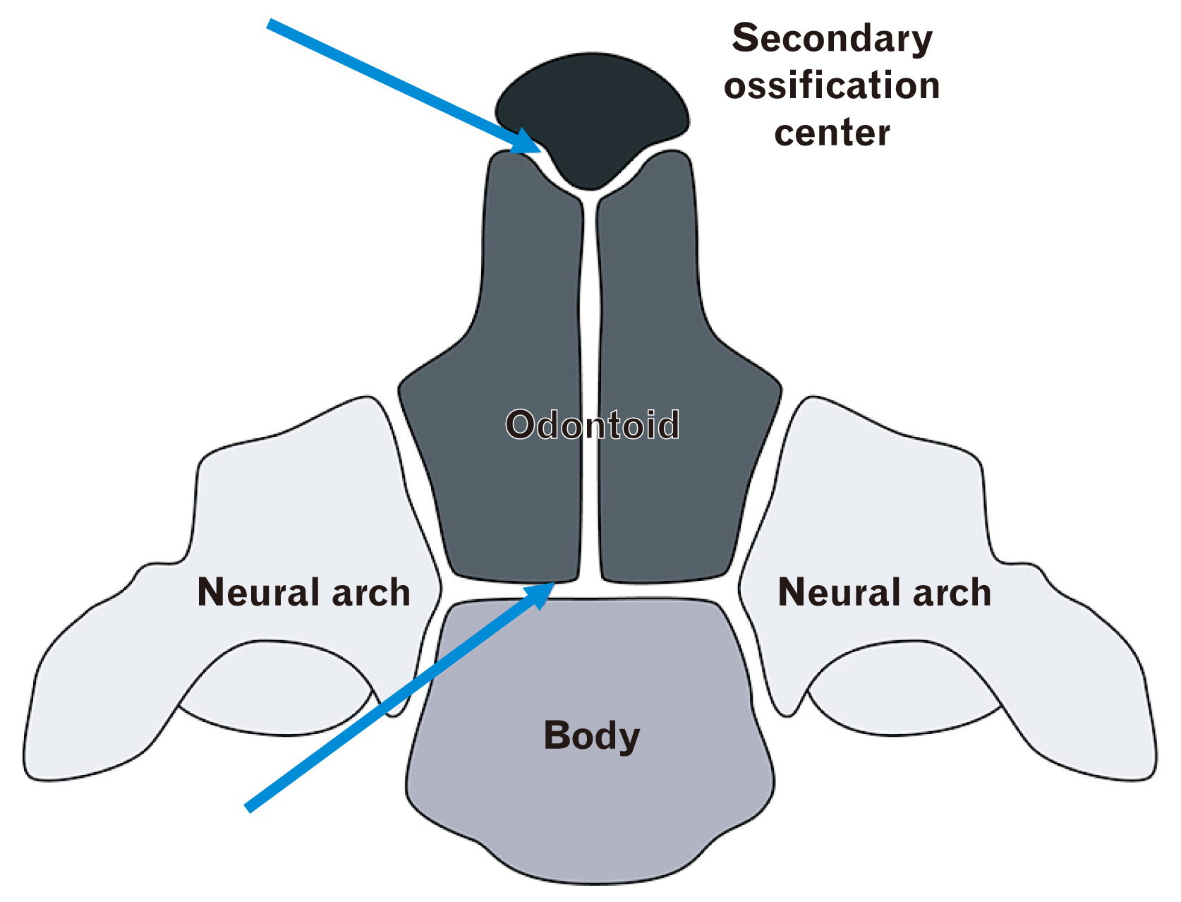
Fig. 1 Schematic drawing of the ossification centers and synchondroses (arrows) of the C2 vertebra.
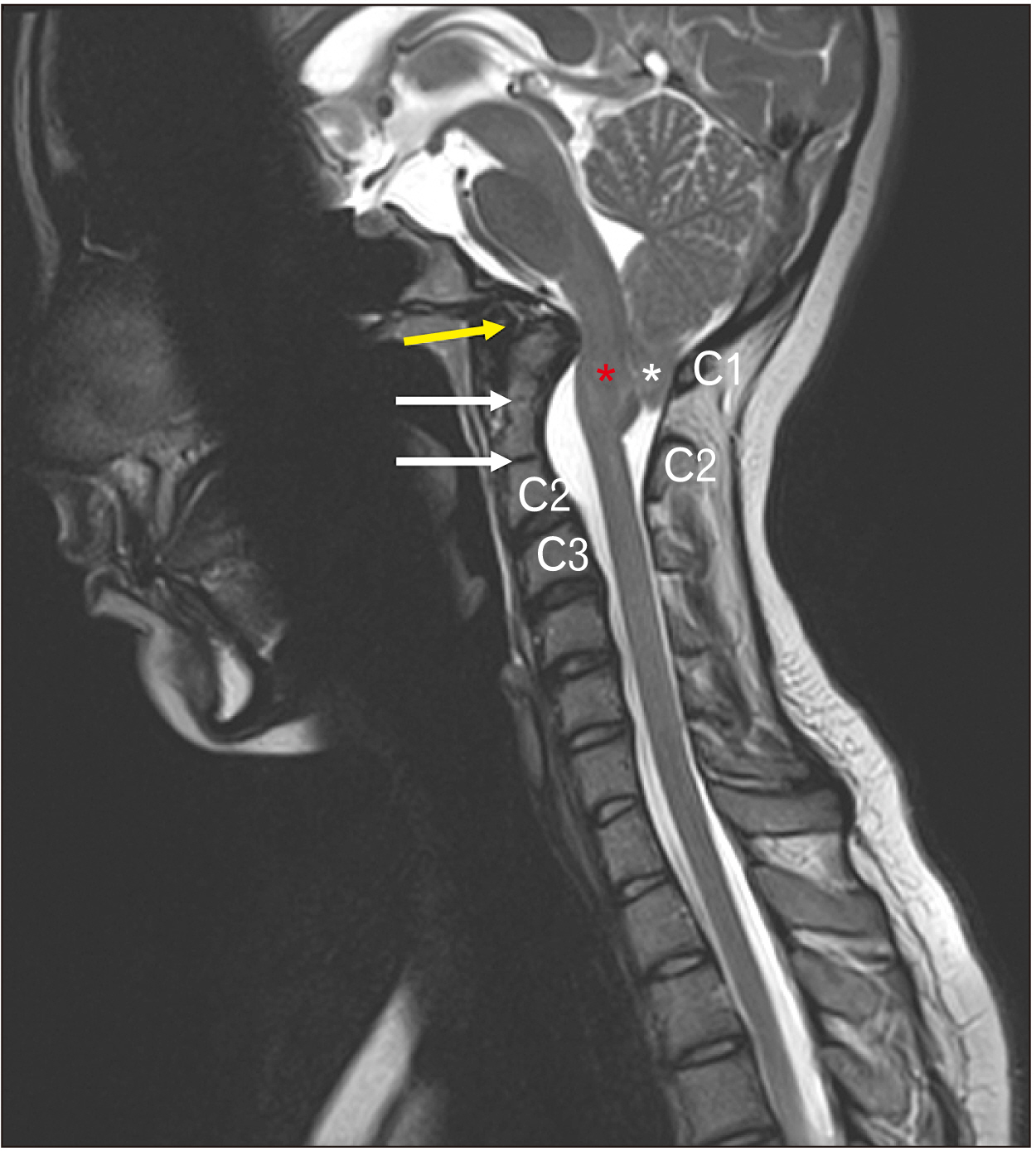
Fig. 2 Two odontoid synchondroses in the same C2 vertebra (white arrows) shown in a midsagittal magnetic resonance imaging image. The upper white arrow notes the accessory synchondrosis. Note the cerebellar tonsils (asterisk), and the lower part of the brainstem (red star) located below the foramen magnum i.e., Chiari 1.5 malformation. The anterior arch of the atlas (yellow arrow) is not ossified. The posterior arch of C1 and spinous process of C2 are also shown.
Reference
-
References
1. Akobo S, Rizk E, Loukas M, Chapman JR, Oskouian RJ, Tubbs RS. 2015; The odontoid process: a comprehensive review of its anatomy, embryology, and variations. Childs Nerv Syst. 31:2025–34. DOI: 10.1007/s00381-015-2866-4. PMID: 26254085.
Article2. Gebauer M, Lohse C, Barvencik F, Pogoda P, Rueger JM, Püschel K, Amling M. 2006; Subdental synchondrosis and anatomy of the axis in aging: a histomorphometric study on 30 autopsy cases. Eur Spine J. 15:292–8. DOI: 10.1007/s00586-005-0990-7. PMID: 16167152. PMCID: PMC3489288.
Article3. Muthukumar N. 2014; Odontoid synchondrosis fracture treated by c1-2 polyaxial screw-rod fixation. J Korean Neurosurg Soc. 55:212–4. DOI: 10.3340/jkns.2014.55.4.212. PMID: 25024826. PMCID: PMC4094747.
Article4. Cramer GD, Darby SA. 2013. Clinical anatomy of the spine, spinal cord, and ANS. 3rd ed. Mosby;St. Louis: p. 688.5. Kim IK, Wang KC, Kim IO, Cho BK. 2010; Chiari 1.5 malformation: an advanced form of Chiari I malformation. J Korean Neurosurg Soc. 48:375–9. DOI: 10.3340/jkns.2010.48.4.375. PMID: 21113370. PMCID: PMC2982921.6. Azahraa Haddad F, Qaisi I, Joudeh N, Dajani H, Jumah F, Elmashala A, Adeeb N, Chern JJ, Tubbs RS. 2018; The newer classifications of the chiari malformations with clarifications: an anatomical review. Clin Anat. 31:314–22. DOI: 10.1002/ca.23051. PMID: 29344999.
Article7. Tubbs RS, Iskandar BJ, Bartolucci AA, Oakes WJ. 2004; A critical analysis of the Chiari 1.5 malformation. J Neurosurg. 101(2 Suppl):179–83. DOI: 10.3171/ped.2004.101.2.0179. PMID: 15835105.
Article8. Capra V, De Marco P, Merello E, Baffico AM, Baldi M, Divizia MT, Gimelli S, Mallet D, Raso A, Mascelli S, Tomà P, Rossi A, Pavanello M, Cama A, Magnani C. 2009; Craniosynostosis, hydrocephalus, Chiari I malformation and radioulnar synostosis: probably a new syndrome. Eur J Med Genet. 52:17–22. DOI: 10.1016/j.ejmg.2008.10.005. PMID: 19022412.
Article9. Tubbs RS, McGirt MJ, Oakes WJ. 2003; Surgical experience in 130 pediatric patients with Chiari I malformations. J Neurosurg. 99:291–6. DOI: 10.3171/jns.2003.99.2.0291. PMID: 12924703.
Article10. Shoja MM, Johal J, Oakes WJ, Tubbs RS. 2018; Embryology and pathophysiology of the Chiari I and II malformations: a comprehensive review. Clin Anat. 31:202–15. DOI: 10.1002/ca.22939. PMID: 28612426.
Article11. Shoja MM, Ramdhan R, Jensen CJ, Chern JJ, Oakes WJ, Tubbs RS. 2018; Embryology of the craniocervical junction and posterior cranial fossa, part II: embryogenesis of the hindbrain. Clin Anat. 31:488–500. DOI: 10.1002/ca.23048. PMID: 29344994.
Article12. Shoja MM, Ramdhan R, Jensen CJ, Chern JJ, Oakes WJ, Tubbs RS. 2018; Embryology of the craniocervical junction and posterior cranial fossa, part I: development of the upper vertebrae and skull. Clin Anat. 31:466–87. DOI: 10.1002/ca.23049. PMID: 29345006.
Article13. Goel A. 2009; Basilar invagination, Chiari malformation, syringomyelia: a review. Neurol India. 57:235–46. DOI: 10.4103/0028-3886.53260. PMID: 19587461.
Article14. Cesmebasi A, Loukas M, Hogan E, Kralovic S, Tubbs RS, Cohen-Gadol AA. 2015; The Chiari malformations: a review with emphasis on anatomical traits. Clin Anat. 28:184–94. DOI: 10.1002/ca.22442. PMID: 25065525.
Article15. Besachio DA, Khaleel Z, Shah LM. 2015; Odontoid process inclination in normal adults and in an adult population with Chiari malformation Type I. J Neurosurg Spine. 23:701–6. DOI: 10.3171/2015.3.SPINE14926. PMID: 26315958.
Article
