Anat Cell Biol.
2021 Mar;54(1):1-9. 10.5115/acb.20.258.
Morphometric analysis of vastus medialis oblique muscle and its influence on anterior knee pain
- Affiliations
-
- 1Department of Anatomy, Faculty of Medicine, Ain Shams University, Cairo, Egypt
- 2Department of Physical Medicine, Rheumatology and Rehabilitation, Faculty of Medicine, Ain Shams University, Cairo, Egypt
- KMID: 2514576
- DOI: http://doi.org/10.5115/acb.20.258
Abstract
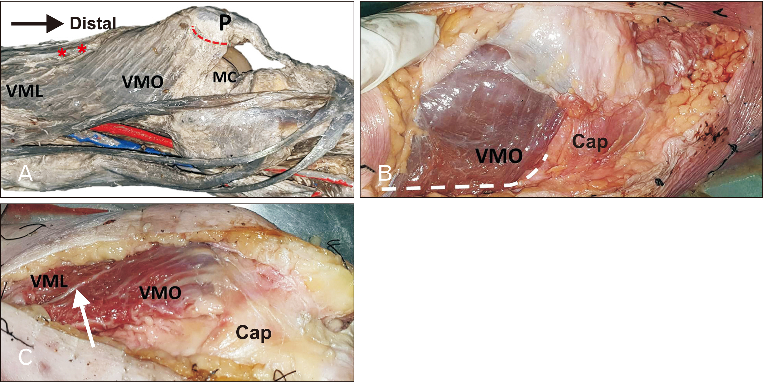
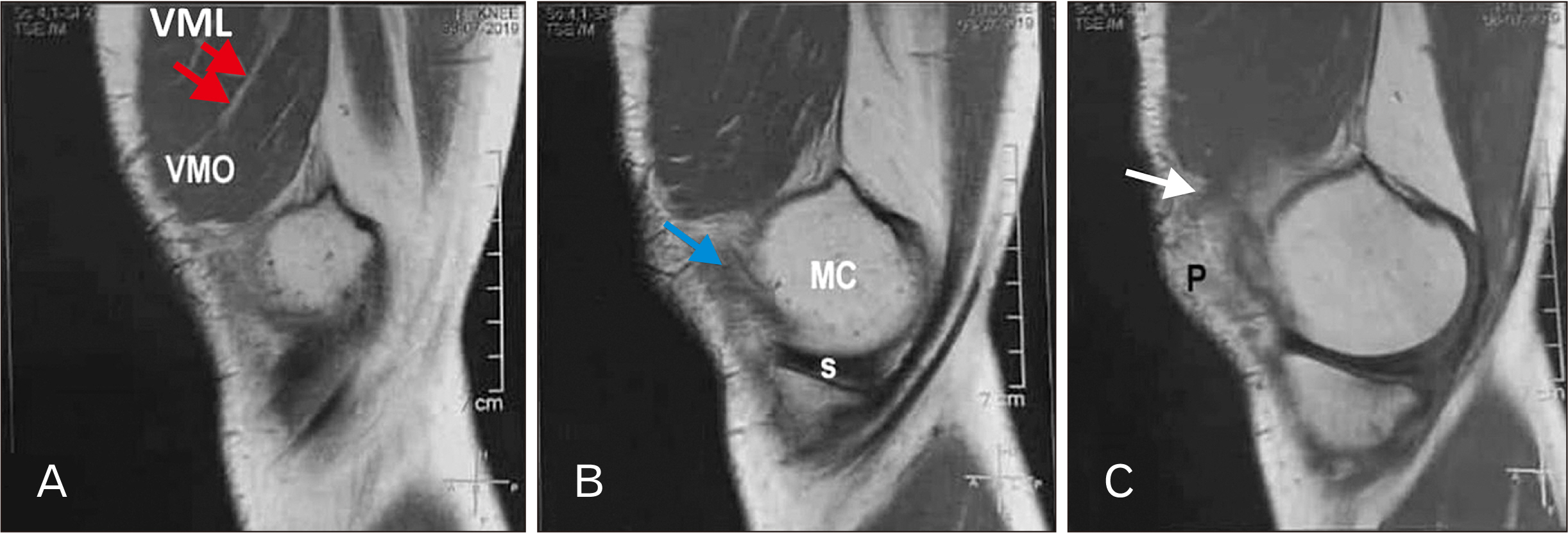
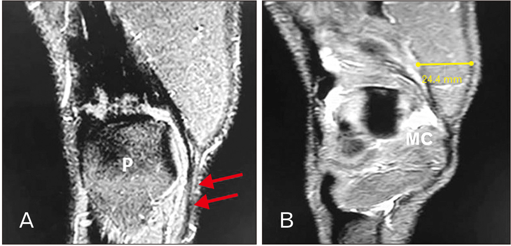
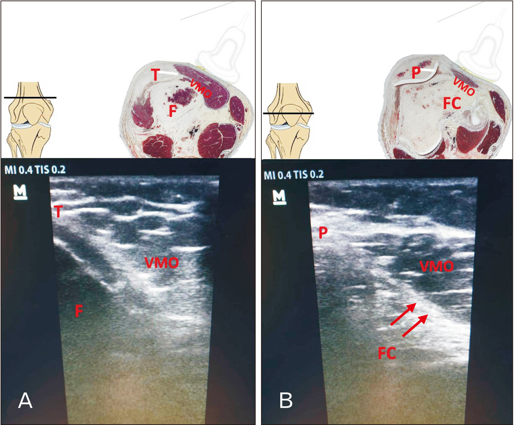
- Healthy knees require full range squatting movements. Vastus medialis (VM) muscle regulates and adjusts the extensor apparatus that inf luences the patellofemoral function. This work was designed to investigate the anatomy and morphometry of vastus medialis oblique (VMO) muscle by widely used imaging techniques and investigate how VMO muscle participates in anterior knee pain. Ten dissected cadaveric specimens were examined, focusing on fiber orientations, origin, insertions and nerve supply of VMO muscle. Magnetic resonance imaging and ultrasound of VMO muscle were recorded. Anatomical cross-sectional areas of VMO muscle were determined in painless and painful knees and statistically analyzed. In cadaveric specimens, there was distinct separation between VM longus and VMO (change in fiber angle or fibrofascial plane). VMO inserted directly into the medial proximal margin of the patella, capsule of the knee joint and continuous with the patellar tendon. Separate branch of femoral nerve run along the anteromedial border of the muscle. Anatomical cross-sectional area was significantly decreased in painful knee by –17.2%±11.0% at lower end of shaft of femur, –21.1%±6.0% at upper border of patella, –36.7%±11.0% at mid-patellar level. VMO is distinct muscle within quadriceps femoris group. VMO muscle would track the patella medially and participate in last phase of knee extension. Assessment of the VMO muscle anatomical cross-sectional area by ultrasonography may constitute promising and reliable tool to evaluate patellofemoral pain syndrome staging.
Keyword
Figure
Reference
-
References
1. Myer GD, Ford KR, Barber Foss KD, Goodman A, Ceasar A, Rauh MJ, Divine JG, Hewett TE. 2010; The incidence and potential pathomechanics of patellofemoral pain in female athletes. Clin Biomech (Bristol, Avon). 25:700–7. DOI: 10.1016/j.clinbiomech.2010.04.001. PMID: 20466469. PMCID: PMC2900391.
Article2. Engelina S, Antonios T, Robertson CJ, Killingback A, Adds PJ. 2014; Ultrasound investigation of vastus medialis oblique muscle architecture: an in vivo study. Clin Anat. 27:1076–84. DOI: 10.1002/ca.22413. PMID: 24797580.3. Arazpour M, Bahramian F, Abutorabi A, Nourbakhsh ST, Alidousti A, Aslani H. 2016; The effect of patellofemoral pain syndrome on gait parameters: a literature review. Arch Bone Jt Surg. 4:298–306. PMID: 27847840. PMCID: PMC5100443.4. Arazpour M, Notarki TT, Salimi A, Bani MA, Nabavi H, Hutchins SW. 2013; The effect of patellofemoral bracing on walking in individuals with patellofemoral pain syndrome. Prosthet Orthot Int. 37:465–70. DOI: 10.1177/0309364613476535. PMID: 23436695.
Article5. Johnston LB, Gross MT. 2004; Effects of foot orthoses on quality of life for individuals with patellofemoral pain syndrome. J Orthop Sports Phys Ther. 34:440–8. DOI: 10.2519/jospt.2004.34.8.440. PMID: 15373007.
Article6. Dixit S, DiFiori JP, Burton M, Mines B. 2007; Management of patellofemoral pain syndrome. Am Fam Physician. 75:194–202. PMID: 17263214.7. Murphy AC, Muldoon SF, Baker D, Lastowka A, Bennett B, Yang M, Bassett DS. 2018; Structure, function, and control of the human musculoskeletal network. PLoS Biol. 16:e2002811. DOI: 10.1371/journal.pbio.2002811. PMID: 29346370. PMCID: PMC5773011.
Article8. Hortobágyi T, Garry J, Holbert D, Devita P. 2004; Aberrations in the control of quadriceps muscle force in patients with knee osteoarthritis. Arthritis Rheum. 51:562–9. DOI: 10.1002/art.20545. PMID: 15334428.
Article9. Lefebvre R, Leroux A, Poumarat G, Galtier B, Guillot M, Vanneuville G, Boucher JP. 2006; Vastus medialis: anatomical and functional considerations and implications based upon human and cadaveric studies. J Manipulative Physiol Ther. 29:139–44. DOI: 10.1016/j.jmpt.2005.12.006. PMID: 16461173.
Article10. Sattler M, Dannhauer T, Ring-Dimitriou S, Sänger AM, Wirth W, Hudelmaier M, Eckstein F. 2014; Relative distribution of quadriceps head anatomical cross-sectional areas and volumes--sensitivity to pain and to training intervention. Ann Anat. 196:464–70. DOI: 10.1016/j.aanat.2014.07.005. PMID: 25153247. PMCID: PMC4250421.
Article11. Castanov V, Hassan SA, Shakeri S, Vienneau M, Zabjek K, Richardson D, McKee NH, Agur AMR. 2019; Muscle architecture of vastus medialis obliquus and longus and its functional implications: a three-dimensional investigation. Clin Anat. 32:515–23. DOI: 10.1002/ca.23344. PMID: 30701597.
Article12. Sattler M, Dannhauer T, Hudelmaier M, Wirth W, Sänger AM, Kwoh CK, Hunter DJ, Eckstein F. 2012; Side differences of thigh muscle cross-sectional areas and maximal isometric muscle force in bilateral knees with the same radiographic disease stage, but unilateral frequent pain- data from the osteoarthritis initiative. Osteoarthritis Cartilage. 20:532–40. DOI: 10.1016/j.joca.2012.02.635. PMID: 22395037. PMCID: PMC3350840.13. Glass NA, Torner JC, Frey Law LA, Wang K, Yang T, Nevitt MC, Felson DT, Lewis CE, Segal NA. 2013; The relationship between quadriceps muscle weakness and worsening of knee pain in the MOST cohort: a 5-year longitudinal study. Osteoarthritis Cartilage. 21:1154–9. DOI: 10.1016/j.joca.2013.05.016. PMID: 23973125. PMCID: PMC3774035.
Article14. Villafañe JH, Bissolotti L, La Touche R, Pedersini P, Negrini S. 2019; Effect of muscle strengthening on perceived pain and static knee angles in young subjects with patellofemoral pain syndrome. J Exerc Rehabil. 15:454–9. DOI: 10.12965/jer.1938224.112. PMID: 31316941. PMCID: PMC6614779.
Article15. Abe T, Loenneke JP, Thiebaud RS. 2015; Morphological and functional relationships with ultrasound measured muscle thickness of the lower extremity: a brief review. Ultrasound. 23:166–73. DOI: 10.1177/1742271X15587599. PMID: 27433253. PMCID: PMC4760590.
Article16. Parry SM, El-Ansary D, Cartwright MS, Sarwal A, Berney S, Koopman R, Annoni R, Puthucheary Z, Gordon IR, Morris PE, Denehy L. 2015; Ultrasonography in the intensive care setting can be used to detect changes in the quality and quantity of muscle and is related to muscle strength and function. J Crit Care. 30:1151.e9–14. DOI: 10.1016/j.jcrc.2015.05.024. PMID: 26211979.
Article17. Grob K, Manestar M, Filgueira L, Ackland T, Gilbey H, Kuster MS. 2016; New insight in the architecture of the quadriceps tendon. J Exp Orthop. 3:32. DOI: 10.1186/s40634-016-0068-y. PMID: 27813020. PMCID: PMC5095096.
Article18. Ayala-Mejias JD, Garcia-Gonzalez B, Alcocer-Perez-España L, Villafañe JH, Berjano P. 2017; Relationship between widening and position of the tunnels and clinical results of anterior cruciate ligament reconstruction to knee osteoarthritis: 30 patients at a minimum follow-up of 10 years. J Knee Surg. 30:501–8. DOI: 10.1055/s-0036-1593367. PMID: 27685765.
Article19. Ema R, Wakahara T, Hirayama K, Kawakami Y. 2017; Effect of knee alignment on the quadriceps femoris muscularity: cross-sectional comparison of trained versus untrained individuals in both sexes. PLoS One. 12:e0183148. DOI: 10.1371/journal.pone.0183148. PMID: 28806771. PMCID: PMC5555710.
Article20. Rueden CT, Schindelin J, Hiner MC, DeZonia BE, Walter AE, Arena ET, Eliceiri KW. 2017; ImageJ2: ImageJ for the next generation of scientific image data. BMC Bioinformatics. 18:529. DOI: 10.1186/s12859-017-1934-z. PMID: 29187165. PMCID: PMC5708080.
Article21. Franchi MV, Longo S, Mallinson J, Quinlan JI, Taylor T, Greenhaff PL, Narici MV. 2018; Muscle thickness correlates to muscle cross-sectional area in the assessment of strength training-induced hypertrophy. Scand J Med Sci Sports. 28:846–53. DOI: 10.1111/sms.12961. PMID: 28805932. PMCID: PMC5873262.
Article22. Winby CR, Lloyd DG, Besier TF, Kirk TB. 2009; Muscle and external load contribution to knee joint contact loads during normal gait. J Biomech. 42:2294–300. DOI: 10.1016/j.jbiomech.2009.06.019. PMID: 19647257.
Article23. Andriacchi TP, Koo S, Scanlan SF. 2009; Gait mechanics influence healthy cartilage morphology and osteoarthritis of the knee. J Bone Joint Surg Am. 91(Suppl 1):95–101. DOI: 10.2106/JBJS.H.01408. PMID: 19182033. PMCID: PMC2663350.
Article24. Englund M. 2010; The role of biomechanics in the initiation and progression of OA of the knee. Best Pract Res Clin Rheumatol. 24:39–46. DOI: 10.1016/j.berh.2009.08.008. PMID: 20129198.
Article25. Kim D, Park G, Kuo LT, Park W. 2018; The effects of pain on quadriceps strength, joint proprioception and dynamic balance among women aged 65 to 75 years with knee osteoarthritis. BMC Geriatr. 18:245. DOI: 10.1186/s12877-018-0932-y. PMID: 30332992. PMCID: PMC6192068.
Article26. Chopp-Hurley JN, Langenderfer JE, Dickerson CR. 2014; Probabilistic evaluation of predicted force sensitivity to muscle attachment and glenohumeral stability uncertainty. Ann Biomed Eng. 42:1867–79. DOI: 10.1007/s10439-014-1035-3. PMID: 24866570.
Article27. Grob K, Manestar M, Filgueira L, Kuster MS, Gilbey H, Ackland T. 2018; The interaction between the vastus medialis and vastus intermedius and its influence on the extensor apparatus of the knee joint. Knee Surg Sports Traumatol Arthrosc. 26:727–38. DOI: 10.1007/s00167-016-4396-3. PMID: 28124107.
Article28. Belli G, Vitali L, Botteghi M, Vittori LN, Petracci E, Latessa PM. 2015; Electromyographic analysis of leg extension exercise during different ankle and knee positions. J Mech Med Biol. 15:1540037. DOI: 10.1142/S0219519415400370.
Article29. Kang JI, Park JS, Choi H, Jeong DK, Kwon HM, Moon YJ. 2017; A study on muscle activity and ratio of the knee extensor depending on the types of squat exercise. J Phys Ther Sci. 29:43–7. DOI: 10.1589/jpts.29.43. PMID: 28210036. PMCID: PMC5300802.
Article30. Muhamed R, Saralaya VV, Murlimanju BV, Chettiar GK. 2017; In vivo magnetic resonance imaging morphometry of the patella bone in South Indian population. Anat Cell Biol. 50:99–103. DOI: 10.5115/acb.2017.50.2.99. PMID: 28713612. PMCID: PMC5509906.
Article31. Page BJ, Mrowczynski OD, Payne RA, Tilden SE, Lopez H, Rizk E, Harbaugh K. 2019; The relative location of the major femoral nerve motor branches in the thigh. Cureus. 11:e3882. DOI: 10.7759/cureus.3882. PMID: 30899633. PMCID: PMC6420329.
Article32. Chaitow L, DeLany J. 2011. Clinical application of neuromuscular techniques volume 2, the lower body. 2nd ed. Churchill Livingstone;Edinburgh:33. Toumi H, Poumarat G, Benjamin M, Best TM, F'Guyer S, Fairclough J. 2007; New insights into the function of the vastus medialis with clinical implications. Med Sci Sports Exerc. 39:1153–9. DOI: 10.1249/01.mss.0b013e31804ec08d. PMID: 17596784.
Article34. Chang WD, Huang WS, Lai PT. 2015; Muscle activation of vastus medialis oblique and vastus lateralis in sling-based exercises in patients with patellofemoral pain syndrome: a cross-over study. Evid Based Complement Alternat Med. 2015:740315. DOI: 10.1155/2015/740315. PMID: 26504480. PMCID: PMC4609425.
Article35. Thomaes T, Thomis M, Onkelinx S, Coudyzer W, Cornelissen V, Vanhees L. 2012; Reliability and validity of the ultrasound technique to measure the rectus femoris muscle diameter in older CAD-patients. BMC Med Imaging. 12:7. DOI: 10.1186/1471-2342-12-7. PMID: 22471726. PMCID: PMC3342139.
Article36. Stevens DE, Smith CB, Harwood B, Rice CL. 2014; In vivo measurement of fascicle length and pennation of the human anconeus muscle at several elbow joint angles. J Anat. 225:502–9. DOI: 10.1111/joa.12233. PMID: 25223934. PMCID: PMC4292751.
Article37. Blazevich AJ, Coleman DR, Horne S, Cannavan D. 2009; Anatomical predictors of maximum isometric and concentric knee extensor moment. Eur J Appl Physiol. 105:869–78. DOI: 10.1007/s00421-008-0972-7. PMID: 19153760.
Article38. Trezise J, Blazevich AJ. 2019; Anatomical and neuromuscular determinants of strength change in previously untrained men following heavy strength training. Front Physiol. 10:1001. DOI: 10.3389/fphys.2019.01001. PMID: 31447693. PMCID: PMC6691166.
Article39. Wang Y, Wluka AE, Berry PA, Siew T, Teichtahl AJ, Urquhart DM, Lloyd DG, Jones G, Cicuttini FM. 2012; Increase in vastus medialis cross-sectional area is associated with reduced pain, cartilage loss, and joint replacement risk in knee osteoarthritis. Arthritis Rheum. 64:3917–25. DOI: 10.1002/art.34681. PMID: 23192791.
Article40. Miyazaki T, Wada M, Kawahara H, Sato M, Baba H, Shimada S. 2002; Dynamic load at baseline can predict radiographic disease progression in medial compartment knee osteoarthritis. Ann Rheum Dis. 61:617–22. DOI: 10.1136/ard.61.7.617. PMID: 12079903. PMCID: PMC1754164.
Article41. Roos EM, Herzog W, Block JA, Bennell KL. 2011; Muscle weakness, afferent sensory dysfunction and exercise in knee osteoarthritis. Nat Rev Rheumatol. 7:57–63. DOI: 10.1038/nrrheum.2010.195. PMID: 21119605.
Article42. Ruhdorfer A, Dannhauer T, Wirth W, Hitzl W, Kwoh CK, Guermazi A, Hunter DJ, Benichou O, Eckstein F. 2013; Thigh muscle cross-sectional areas and strength in advanced versus early painful osteoarthritis: an exploratory between-knee, within-person comparison in osteoarthritis initiative participants. Arthritis Care Res (Hoboken). 65:1034–42. DOI: 10.1002/acr.21965. PMID: 23401316.
Article
- Full Text Links
- Actions
-
Cited
- CITED
-
- Close
- Share
- Similar articles
-
- Preferential Vastus Medialis Oblique Activation Achieved by Isokinetic Cycling at High Angular Velocity
- Comparison of Muscle Activity of Vastus Lateralis and Medialis Oblique among Knee Extension Angles at 90°, 135°, 180° in Sitting Position
- Arthroscopic Reduction of Irreducible Posterolateral Knee Dislocation with Interposition of the Vastus Medialis: A Case Report
- Irreducible Knee Dislocation with Vastus Medialis Muscle Interposition
- Clinical Study on Safety, Clinical Indicators of Polydioxanone Sutures Inserted into Vastus Medialis Muscle in Degenerative Knee Osteoarthritis


