Ann Pediatr Endocrinol Metab.
2021 Mar;26(1):53-59. 10.6065/apem.2040120.060.
Thyroid imaging study in children with suspected thyroid dysgenesis
- Affiliations
-
- 1Department of Pediatrics, Dankook University Hospital, Dankook University College of Medicine, Cheonan, Korea
- 2Department of Radiology, Dankook University Hospital, Dankook University College of Medicine, Cheonan, Korea
- KMID: 2514153
- DOI: http://doi.org/10.6065/apem.2040120.060
Abstract
- Purpose
Thyroid dysgenesis is one of the most common causes of permanent congenital hypothyroidism. Thyroid ultrasonography or scan is used to detect thyroid dysgenesis. We analyzed the sensitivity and specificity of thyroid ultrasonography and scan in diagnosing thyroid dysgenesis to determine the clinical utility of each thyroid imaging method.
Methods
Sixty-one patients younger than 7 years of age were investigated via thyroid scan. Nineteen patients who were initially interpreted as having thyroid dysgenesis, such as ectopia, hemiagenesis, or aplasia, by thyroid scan were included in the study. Clinical characteristics and findings of a thyroid imaging study were reviewed.
Results
Initially, thyroid scan results were interpreted as ectopia (n=9), hemiagenesis (n=1), and nonvisualization (n=9). In contrast, the results of thyroid ultrasonography were normal thyroid gland (n=5), ectopia (n=6), and hypoplasia (n=8). After reviewing the results of both studies, final imaging diagnoses were as follows: normal thyroid gland (n=5), hemiagenesis (n=1), ectopia (n=9) including 2 dual ectopy, hypoplasia (n=3), and aplasia (n=1). Thyroid ultrasonography showed higher sensitivity and specificity in detecting presence of normal thyroid gland. Thyroid scan was better to detect ectopia. Among 8 patients who were initially interpreted as having hypoplasia by ultrasonography, 4 were confirmed as ectopia and one as aplasia.
Conclusion
This study showed that thyroid ultrasonography is useful as the first-line imaging study to detect normal-sized eutopic thyroid gland. Thyroid scan should be performed to investigate the presence of ectopia if hypoplasia or aplasia is suspected by ultrasonography.
Keyword
Figure
-
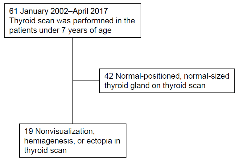
Fig. 1. Flow diagram of the study. A total of 19 participants was included in the study after excluding 42 individuals who revealed normal-positioned, normal-sized thyroid gland on thyroid scan.
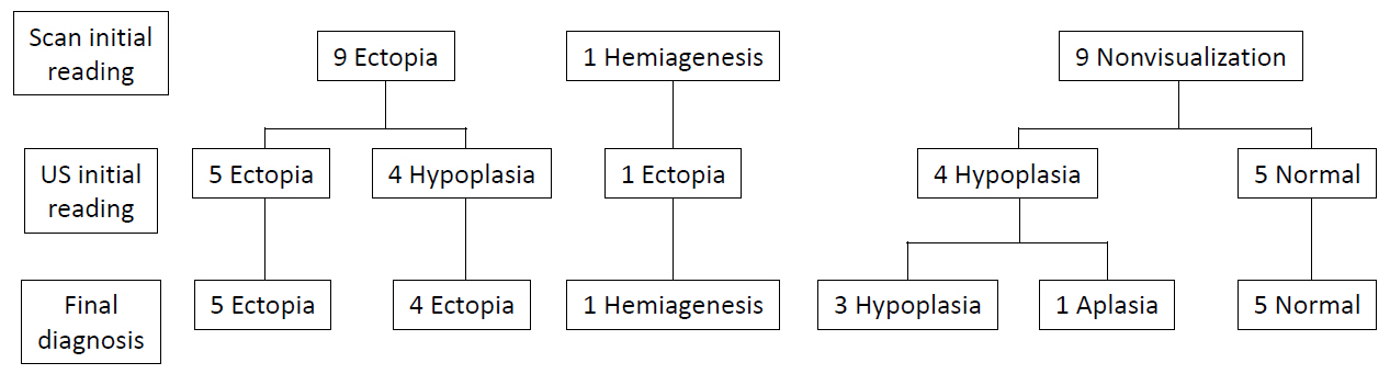
Fig. 2. Grouping of the patients according to the final diagnosis based on the results of thyroid scan and ultrasonography (US). Initial interpretations of thyroid scan and thyroid ultrasonography were changed as follows: ectopia (n=9), hemiagenesis (n=1), hypoplasia (n=3), aplasia (n=1), and normal (n=5).

Fig. 3. There were 2 patients with dual ectopy (A, B) and 1 patient with hemiagenesis (C). Dual ectopia was found at the level of lingual/sublingual and submandibular areas (A) and sublingual areas (B), respectively.
Reference
-
References
1. LaFranchi SH. Approach to the diagnosis and treatment of neonatal hypothyroidism. J Clin Endocrinol Metab. 2011; 96:2959–67.
Article2. Léger J, Olivieri A, Donaldson M, Torresani T, Krude H, van Vliet G, et al. European society for paediatric endocrinolog y consensus guidelines on screening, diagnosis, and management of congenital hypothyroidism. J Clin Endocrinol Metab. 2014; 99:363–84.3. Cherella CE, Wassner AJ. Congenital hypothyroidism: insights into pathogenesis and treatment. Int J Pediatr Endocrinol. 2017; 2017:11.
Article4. Hinton CF, Harris KB, Borgfeld L, Drummond-Borg M, Eaton R, Lorey F, et al. Trends in incidence rates of congenital hypothyroidism related to select demographic fac tors: d at a From t he Unite d St ates, C a liforni a, Massachusetts, New York, and Texas. Pediatrics. 2010; 125 Suppl 2:S37–47.5. De Felice M, Di Lauro R. Thyroid development and its disorders: genetics and molecular mechanisms. Endocr Rev. 2004; 25:722–46.
Article6. Szczepanek-Parulska E, Hernik A, Ruchala M. Thyroid ectopy - diagnostic and therapeutic challenges before and in the era of TSH neonatal screening. Endokrynol Pol. 2017; 68:708–21.7. Sood A, Sood V, Sharma DR, Seam RK, Kumar R. Thyroid scintigraphy in detecting dual ectopic thyroid: a review. Eur J Nucl Med Mol Imaging. 2008; 35:843–6.
Article8. Marković V, Glavina G, Eterović D, Punda A, Brdar D. Dual ectopic thyroid gland: sonography and scintigraphy of lingual and sublingual thyroid. Clin Nucl Med. 2014; 39:556–8.9. Jain TK, Meena RS, Bhatia A, Sood A, Bhattacharya A, Mittal BR. Dual thyroid ectopia-role of thyroid scintigraphy and neck ultrasonography. Indian J Nucl Med. 2015; 30:338–40.
Article10. Nakamura S, Masuda T, Ishimori M. Dual ectopic thyroid associated with thyroid hemiagenesis. Endocrinol Diabetes Metab Case Rep. 2018; 2018:18–0026.
Article11. Castanet M, Polak M, Bonaïti-Pellié C, Lyonnet S, Czernichow P, Léger J, et al. Nineteen years of national screening for congenital hypothyroidism: familial cases with thyroid dysgenesis suggest the involvement of genetic factors. J Clin Endocrinol Metab. 2001; 86:2009–14.
Article12. Karakoc E, Turan S, Akpinar I, Isguven P, Adal E, Haklar G, et al. Screening of parents and siblings of patients with thyroid dysgenesis by thyroid function tests and ultrasound. Horm Res. 2008; 70:329–39.
Article13. Borges MF, Sedassari NA, Sedassari AA, Souza LRMF, Ferreira BP, Lara BHJ, et al. Timing of thyroid ultrasonography in the etiological investigation of congenital hypothyroidism. Arch Endocrinol Metab. 2017; 61:432–7.
Article14. Yeung VT, Loong EP, Cockram CS. Cretinism and lingual thyroid presenting in an adult. Postgrad Med J. 1987; 63:881–3.
Article15. Toso A, Colombani F, Averono G, Aluffi P, Pia F. Lingual thyroid causing dysphagia and dyspnoea. Case reports and review of the literature. Acta Otorhinolaryngol Ital. 2009; 29:213–7.16. Adelchi C, Mara P, Melissa L, De Stefano A, Cesare M. Ectopic thyroid tissue in the head and neck: a case series. BMC Res Notes. 2014; 7:790.
Article17. Chanoine JP, Toppet V, Lagasse R, Spehl M, Delange F. Determination of thyroid volume by ultrasound from the neonatal period to late adolescence. Eur J Pediatr. 1991; 150:395–9.
Article18. Perry RJ, Hollman AS, Wood AM, Donaldson MDC. Ultrasound of the thyroid gland in the newborn: normative data. Arch Dis Child Fetal Neonatal Ed. 2002; 87:F209–11.
Article19. Aydıner Ö, Aydıner EK, Akpınar I, Turan S, Bereket A. Normative data of thyroid volume-ultrasonographic evaluation of 422 subjects aged 0-55 years. J Clin Res Pediatr Endocrinol. 2015; 7:98–101.
Article20. De Bruyn R, Ng WK, Taylor J, Campbell F, Mitton SG, Dicks-Mireaux C, et al. Neonatal hypothyroidism: comparison of radioisotope and ultrasound imaging in 54 cases. Acta Paediatr Scand. 1990; 79:1194–8.
Article21. Takashima S, Nomura N, Tanaka H, Itoh Y, Miki K, Harada T. Congenital hypothyroidism: assessment with ultrasound. Am J Neuroradiol. 1995; 16:1117–23.22. Perry RJ, Maroo S, Maclennan AC, Jones JH, Donaldson MD. Combined ultrasound and isotope scanning is more informative in the diagnosis of congenital hypothyroidism than single scanning. Arch Dis Child. 2006; 91:972–6.
Article23. Ruchala M, Szczepanek E, Sowiński J. Diagnostic value of radionuclide scanning and ultrasonography in thyroid developmental anomaly imaging. Nucl Med Rev. 2011; 14:21–8.
Article24. Kreisner E, Camargo-Neto E, Maia CR, Gross JL. Accuracy of ultrasonography to establish the diagnosis and aetiology of permanent primary congenital hypothyroidism. Clin Endocrinol (Oxf). 2003; 59:361–5.
Article25. Sood A, Kumar R. The ectopic thyroid gland and the role of nuclear medicine techniques in its diagnosis and management. Hell J Nucl Med. 2008; 11:168–71.26. Bubuteishvili L, Garel C, Czernichow P, Léger J. Thyroid abnormalities by ultrasonography in neonates with congenital hypothyroidism. J Pediatr. 2003; 143:759–64.
Article27. Karakoc-Aydiner E, Turan S, Akpinar I, Dede F, Isguven P, Adal E, et al. Pitfalls in the diagnosis of thyroid dysgenesis by thyroid ultrasonography and scintigraphy. Eur J Endocrinol. 2012; 166:43–8.
Article
- Full Text Links
-
- Actions
-
Cited
- CITED
-
- Close
- Share
-
- Similar articles
-
- Ultrasonography of Various Thyroid Diseases in Children and Adolescents: A Pictorial Essay
- A Case of Dual Ectopic Thyroid
- Primary Follicular Carcinoma Arising in Ectopic Thyroid Tissue of the Lateral Neck: A Case Report
- A Case of Lingual Thyroid with Unilateral Thyroid Agenesis and Contralateral Goiter
- Serum Thyroglobulin Concentrations in Congenital Hypothyroidism
