J Rhinol.
2021 Mar;28(1):50-56. 10.18787/jr.2020.00334.
Nasal Septal Deviation and Incidental Paranasal Sinus Opacification: A Role of Computed Tomography
- Affiliations
-
- 1Department of Otorhinolaryngology-Head and Neck Surgery, School of Medicine, Kyung Hee University, Seoul, Korea
- KMID: 2513843
- DOI: http://doi.org/10.18787/jr.2020.00334
Abstract
- Background and Objectives
The purpose of this study was to investigate the prevalence of incidental paranasal sinus (PNS) opacification in nasal septal deviation (NSD) using computed tomography (CT) and to identify contributing factors.
Subjects and Method
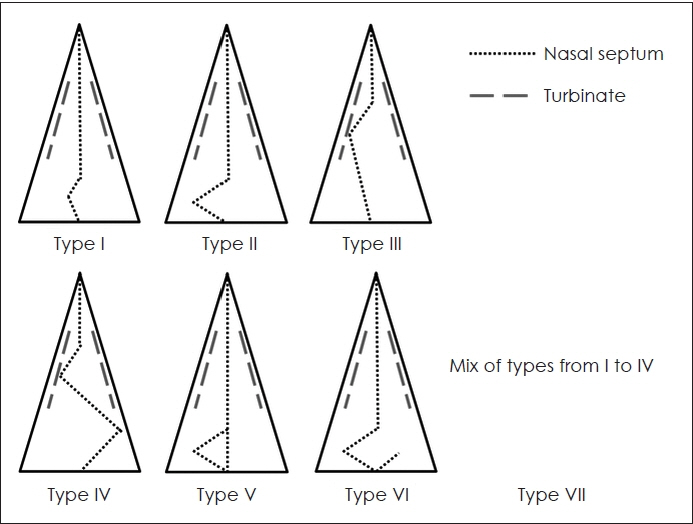
We analyzed 216 patients who underwent septoplasty for the correction of NSD and who underwent preoperative PNS CT. We assessed the prevalence of incidental PNS opacification in these patients and determined the type of NSD according to Mladina classification. We also evaluated whether the direction of NSD affected the presence of PNS opacification on CT, and whether the presence of PNS opacification was associated with other rhinologic symptoms.
Results
Of 216 patients with NSD, 86 showed opacified PNS on CT. According to Mladina classification, NSD patients were classified as type I (24.1%), type II (36.1%), type III (20.8%), type IV (5.6%), type V (9.7%), type VI (2.3%), or type VII (1.4%). Patients with type II NSD showed a significantly higher incidence of PNS opacification compared with other types of NSD (p=0.001). However, the direction of NSD did not significantly influence the presence of incidental PNS opacification. Furthermore, regardless of the presence of PNS opacification, there was no significant difference in rhinologic symptoms such as olfactory dysfunction, among others.
Conclusion
We found that incidental PNS opacification on CT was common in NSD patients, especially in patients with type II NSD. Thus, we suggest that CT evaluation of patients with NSD may be helpful for assessing comorbid PNS pathologies as well as objectively identifying nasal septal deformities.
Keyword
Figure
Reference
-
References
1. Mladina R, Cujic E, Subaric M, Vukovic K. Nasal septal deformities in ear, nose, and throat patients: an international study. Am J Otolaryngol. 2008; 29(2):75–82.2. Min YG, Jung HW, Kim CS. Prevalence study of nasal septal deformities in Korea: results of a nation-wide survey. Rhinology. 1995; 33(2):61–5.3. Blackwell DL, Lucas JW, Clarke TC. Summary health statistics for U.S. adults: national health interview survey, 2012. Vital Health Stat. 2014; 260:1–161.4. Benninger MS, Ferguson BJ, Hadley JA, Hamilos DL, Jacobs M, Kennedy DW, et al. Adult chronic rhinosinusitis: definitions, diagnosis, epidemiology, and pathophysiology. Otolaryngol Head Neck Surg. 2003; 129(3 Suppl):S1–32.5. Karatas D, Yuksel F, Senturk M, Dogan M. The contribution of computed tomography to nasal septoplasty. J Craniofac Surg. 2013; 24(5):1549–51.6. Hansen AG, Helvik AS, Nordgård S, Bugten V, Stovner LJ, Håberg AK, et al. Incidental findings in MRI of the paranasal sinuses in adults: a population-based study (HUNT MRI). BMC Ear Nose Throat Disord. 2014; 14(1):13.7. Mladina R. The role of maxillary morphology in the development of pathological septal deformities. Rhinology. 1987; 25(3):199–205.8. Jonas TJ, Clark AR. Bailey’s Head & Neck Surgery-otolaryngology. 5th ed. Philadelphia: Lippincott Williams & Wilkins;2014. Chapter 42, Septoplasty, Turbinate Reduction, and Correction of Nasal Obstruction; p.612-21.9. Wojas O, Szczesnowicz-Dabrowska P, Grzanka A, Krzych-Falta E, Samolinski B. Nasal septum deviation by age and sex in a study population of Poles. J Laryngorhino-Otologie. 1997; 7:1–6.10. Orlandi RR. A systematic analysis of septal deviation associated with rhinosinusitis. Laryngoscope. 2010; 120(8):1687–95.11. Yasan H, Dogru H, Baykal B, Doner F, Tuz M. What is the relationship between chronic sinus disease and isolated nasal septal deviation? Otolaryngol Head Neck Surg. 2005; 133(2):190–3.12. Mundra RK, Gupta Y, Sinha R, Gupta A. CT Scan Study of Influence of Septal Angle Deviation on Lateral Nasal Wall in Patients of Chronic Rhinosinusitis. Indian J Otolaryngol Head Neck Surg. 2014; 66(2):187–90.13. Ahn JC, Kim JW, Lee CH, Rhee CS. Prevalence and Risk Factors of Chronic Rhinosinusitus, Allergic Rhinitis, and Nasal Septal Deviation: Results of the Korean National Health and Nutrition Survey 2008-2012. JAMA Otolaryngol Head Neck Surg. 2016; 142(2):162–7.14. Harar RPS, Chadha NK, Rogers G. The role of septal deviation in adult chronic rhinosinusitis: a study of 500 patients. Rhinology. 2004; 42(3):126–30.15. Stallman JS, Lobo JN, Som PM. The Incidence of Concha Bullosa and Its Relationship to Nasal Septal Deviation and Paranasal Sinus Disease. Am J Neuroradiol. 2004; 25(9):1613–8.16. Kim SH, Oh JS, Jang YJ. Incidence and Radiological Findings of Incidental Sinus Opacifications in Patients Undergoing Septoplasty or Septorhinoplasty. Ann Otol Rhinol Laryngol. 2020; 129(2):122–7.17. Prasad S, Varshney S, Bist SS, Mishra S, Kabdwal N. Correlation study between nasal septal deviation and rhinosinusitis. Indian J Otolaryngol Head Neck Surg. 2013; 65(4):363–6.18. Huh HB, Suh KS, Kim JY, Hur CH. Comparison of the PNS Series and the OMU-CT in Chronic Paranasal Sinusitis. Korean J Otolaryngol. 1996; 39(11):1767–72.19. Rudmik L, Mace J, Smith T. Low-stage computed tomography chronic rhinosinusitis: what is the role of endoscopic sinus surgery? Laryngoscope. 2011; 121(2):417–21.20. Rosenthal PA, Lundy KC, Massoglia DP, Payne EH, Gilbert G, Gebregziabher M. Incidental paranasal sinusitis on routine brain magnetic resonance scans: association with atherosclerosis. Int Forum Allergy Rhinol. 2016; 6(12):1253–63.21. Yoo JY, Chung MJ, Choi B, Jung HN, Koo JH, Bae YA, et al. Digital tomosynthesis for PNS evaluation: comparisons of patient exposure and image quality with plain radiography.? Korean J Radiol. 2012; 13(2):136–43.
- Full Text Links
- Actions
-
Cited
- CITED
-
- Close
- Share
- Similar articles
-
- The Effect of Radiation Therapy on Paranasal Sinus Opacification in Nasopharyngeal Cancer Patients
- Prevalence of incidental paranasal sinus opacification in dental paediatric patients
- Prevalence of incidental paranasal sinus opacification in an adult dental population
- A case report of incidental finding of fungus ball on CBCT of maxillary sinus in treatment planning of dental implant
- The Relationship between Anatomic Variation of Nasal Cavity and Paranasal Sinusitis


