Selective inhibition of V600E-mutant BRAF gene induces apoptosis in thyroid carcinoma cell lines
- Park KS
 1,2,3
1,2,3 - Saindane M1,2
- Yang EY1
- Jin T3
- Rallabandi HR1,2
- Heil A4
- Nam SE1,2
- Yoo YB1,2
- Yang JH1,2
- Kim JB5
- Park SY6
- Park WS7
- Youn YK8
- Affiliations
-
- 1Department of Surgery, Konkuk University School of Medicine, Seoul, Korea
- 2Department of Surgery, Konkuk University Medical Center, Seoul, Korea
- 3Research Institute of Medical Science, Konkuk University School of Medicine, Seoul, Korea
- 4Institute of Botany and Molecular Genetics, RWTH, Aachen University, Aachen, Germany
- 5Research Centers for Cellular Homeostasis, Ewha Womans University, Seoul, Korea
- 6Cancer Research Institute, Seoul National University College of Medicine, Seoul, Korea
- 7Department of Surgery, School of Medicine, Kyung Hee University, Seoul, Korea
- 8Thyroid Clinic, St. Peter’s Hospital, Seoul, Korea
- KMID: 2513193
- DOI: http://doi.org/10.4174/astr.2021.100.3.127
Abstract
- Purpose
Papillary thyroid cancer (PTC) has a high incidence of BRAF V600E mutation. The purpose of this study was to evaluate the potential relationship between thyroiditis and BRAF V600E mutation status in patients with PTC. We investigated how a selective inhibitor of BRAF V600E PLX4032 affects the proliferation and inflammatory cytokine levels of thyroid cancer.
Methods
Two thyroid cancer cell lines TPC1 and 8505C were treated with PLX4032, an analysis was done on cell growth, cell cycle, the degree of apoptosis, and levels of inflammatory cytokines. To identify the functional links of BRAF, we used the STRING database.
Results
Docking results illustrated PLX4032 blocked the kinase activity by exclusively binding on the serine/threonine kinase domain. STRING results indicated BRAF is functionally linked to mitogen-activated protein kinase. Both cell lines showed a dose-dependent reduction in growth rate but had a different half maximal inhibitory concentration value for PLX4032. The reaction to PLX4032 was more sensitive in the 8505C cells than in the TPC1 cells. PLX4032 induced a G2/ M phase arrest in the TPC1 cells and G0/G1 in the 8505C cells. PLX4032 induced apoptosis only in the 8505C cells. With PLX4032, the TPC1 cells showed decreased levels of vascular endothelial growth factor, granulocyte-macrophage colonystimulating factor, chemokine (C-C motif) ligand 2/monocyte chemoattractant protein 1, whereas the 8505C cells showed significantly decreased levels of IL-8, serpin E1/plasminogen activator inhibitor-1, and matrix metalloproteinase (MMP)-3.
Conclusion
PLX4032 was cytotoxic in both TPC1 and 8505C cells and induced apoptosis. In the 8505C cells, inflammatory cytokines such as IL-8 and MMP-3 were down-regulated. These findings suggest the possibility that the BRAF V600E mutation needs to target inflammatory signaling pathways in the treatment of thyroid cancer.
Keyword
Figure
-
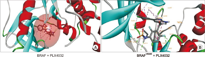
Fig. 1 Docking for PLX4032 in wild type BRAF and V600E mutant binding patterns of PLX4032 on BRAF. Molecular docking of PLX4032 with BRAF wild type and V600E. (A) The first panel illustrates PLX4032 docked into the hydrophobic core of wild type BRAF showing hydrogen, hydrophobic, and cation interactions. (B) The second panel indicates the best-docked confirmation of PLX4032 into BRAFV600E along with binding interactions representation.
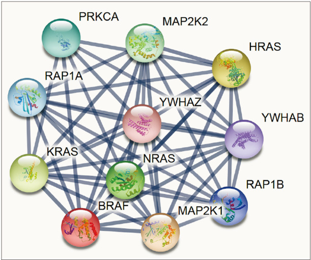
Fig. 2 Predicting the interaction network of BRAF using STRING (https://string-db.org). Interacting nodes are displayed in circles, obtained using STRING. Predicted functional partners of BRAF are shown, taking into consideration functional links, coexpression, colocalization, genetic interactions, pathway, physical interactions, predicted, and shared protein domains.
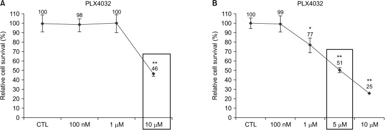
Fig. 3 Growth rate of cells after PLX4032 treatment. TPC1 and 8505C cells cultured in Dulbecco's modified Eagle's medium containing 10% fetal bovine serum. (A) TPC1 cells were treated for 72 hours with 100 nM, 1 µM, and 10 µM of DMSO (CTL) or PLX4032. (B) 8505C cells were treated for 72 hours with 100 nM, 1 µM, 5 µM, and 10 µM of DMSO (CTL) or PLX4032. Relative cell survival rate is shown as percentage of survival vs. control cells after treatment with PLX4032. Values presented here are mean values and standard errors from at least 3 independent experiments. Square box indicates the IC50 value of the TPC1 and 8505C cells. DMSO, dimethyl sulfoxide; CTL, cytotoxic T lymphocyte; IC50, half maximal inhibitory concentration. *P < 0.05, **P < 0.01 vs. control.
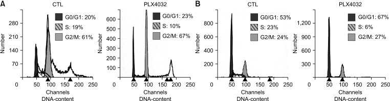
Fig. 4 Cell cycle analysis after PLX4032 treatment. (A) TPC1 cells were treated for 72 hours with 10 µM of DMSO (CTL) or PLX4032. (B) 8505C cells were treated for 72 hours with 5 µM of DMSO (CTL) or PLX4032. Cell cycle was analyzed by flow cytometry after DNA staining with propidium iodide. Data represent percentage of cells at each stage of cell cycle. Data are from representative experiment (out of a total of 3 experiments). DMSO, dimethyl sulfoxide; CTL, cytotoxic T lymphocyte.
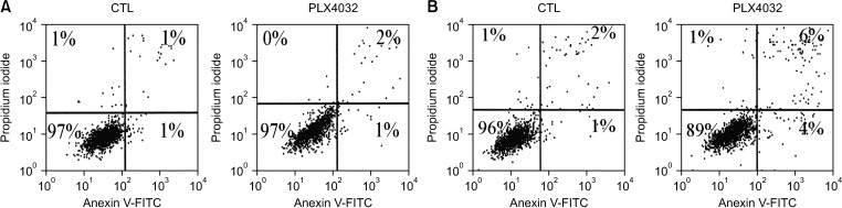
Fig. 5 Apoptosisanalysisafter PLX4032 treatment. (A) TPC1 cells were treated for 72 hours with 10 µM of DMSO (CTL) or PLX4032. (B) 8505C cells were treated for 72 hours with at 5 µM of DMSO (CTL) or PLX4032. The cell cycle was analyzed by flow cytometry after DNA staining with propidium iodide. All cells were stained with FITC-conjugated annexin V in buffer containing propidium iodide and analyzed by flow cytometry. For each treatment, percentage of viable cells is shown in lower left quadrant, which indicates low levels of annexin V and propidium iodide. Data are from representative experiment (out of a total of 3 experiments). DMSO, dimethyl sulfoxide; CTL, cytotoxic T lymphocyte; FITC, fluorescein isothiocyanate.
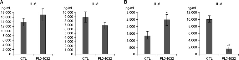
Fig. 6 Expression of IL-6 and IL-8 after PLX4032 treatment. (A) TPC1 cells were treated for 72 hours with 10 µM of DMSO (CTL) or PLX4032. (B) 8505C cells were treated for 72 hours with 5 µM of DMSO (CTL) or PLX4032. Density levels of IL-6 and IL-8 were measured with Bio-Plex 200 system (Bio-Rad, Hercules, CA, USA). DMSO, dimethyl sulfoxide; CTL, cytotoxic T lymphocyte. *P < 0.05, **P < 0.01 vs. control.

Fig. 7 Expression of VEGF and GM-CSF after PLX4032 treatment. (A) TPC1 cells were treated for 72 hours with 10 µM of DMSO (CTL) or PLX4032. (B) 8505C cells were treated for 72 hours with 5 µM of DMSO (CTL) or PLX4032. Density levels of VEGF and GM-CSF were measured with Bio-Plex 200 system (Bio-Rad, Hercules, CA, USA). VEGF, vascular endothelial growth factor; GM-CSF, granulocyte-macrophage colony-stimulating factor; DMSO, dimethyl sulfoxide; CTL, cytotoxic T lymphocyte. *P < 0.05, **P < 0.01 vs. control.
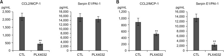
Fig. 8 Expression of CCL2/MCP-1 and serpin E1/PAI-1 after PLX4032 treatment. (A) TPC1 cells were treated for 72 hours with 10 µM of DMSO (CTL) or PLX4032. (B) 8505C cells were treated for 72 hours with 5 µM of DMSO (CTL) or PLX4032. Density levels of CCL2/MCP-1 and serpin E1/PAI-1 were measured with Bio-Plex 200 system (Bio-Rad, Hercules, CA, USA). CCL2, chemokine (C-C motif) ligand 2; MCP-1, monocyte chemoattractant protein 1; PAI-1, plasminogen activator inhibitor 1; DMSO, dimethyl sulfoxide; CTL, cytotoxic T lymphocyte. *P < 0.05, **P < 0.01 vs. control.
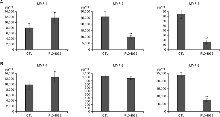
Fig. 9 Expression of MMP-1, 2, and 3 after PLX4032 treatment. (A) TPC1 cells were treated for 72 hours with 10 µM of DMSO (CTL) or PLX4032 and PLX4720. (B) 8505C cells were treated for 72 hours with 5 µM of DMSO (CTL) or PLX4032. Density levels of MMP-1, -2, and -3 were measured with Bio-Plex 200 system (Bio-Rad, Hercules, CA, USA). MMP, matrix metalloproteinase; DMSO, dimethyl sulfoxide; CTL, cytotoxic T lymphocyte. *P < 0.05, **P < 0.01 vs. control.
Reference
-
1. Won YJ, Sung J, Jung KW, Kong HJ, Park S, Shin HR, et al. Nationwide cancer incidence in Korea, 2003–2005. Cancer Res Treat. 2009; 41:122–131. PMID: 19809561.
Article2. Jemal A, Siegel R, Ward E, Murray T, Xu J, Thun MJ. Cancer statistics, 2007. CA Cancer J Clin. 2007; 57:43–66. PMID: 17237035.
Article3. Larson SD, Jackson LN, Riall TS, Uchida T, Thomas RP, Qiu S, et al. Increased incidence of well-differentiated thyroid cancer associated with Hashimoto thyroiditis and the role of the PI3k/Akt pathway. J Am Coll Surg. 2007; 204:764–773. PMID: 17481480.
Article4. Zhang L, Li H, Ji QH, Zhu YX, Wang ZY, Wang Y, et al. The clinical features of papillary thyroid cancer in Hashimoto's thyroiditis patients from an area with a high prevalence of Hashimoto's disease. BMC Cancer. 2012; 12:610. PMID: 23256514.
Article5. Teng W, Shan Z, Teng X, Guan H, Li Y, Teng D, et al. Effect of iodine intake on thyroid diseases in China. N Engl J Med. 2006; 354:2783–2793. PMID: 16807415.
Article6. Kobawala TP, Trivedi TI, Gajjar KK, Patel DH, Patel GH, Ghosh NR. Significance of interleukin-6 in papillary thyroid carcinoma. J Thyroid Res. 2016; 2016:6178921. PMID: 27034885.
Article7. Kobawala TP, Patel GH, Gajjar DR, Patel KN, Thakor PB, Parekh UB, et al. Clinical utility of serum interleukin-8 and interferon-alpha in thyroid diseases. J Thyroid Res. 2011; 2011:270149. PMID: 21461397.
Article8. Stetler-Stevenson WG. The role of matrix metalloproteinases in tumor invasion, metastasis, and angiogenesis. Surg Oncol Clin N Am. 2001; 10:383–392. PMID: 11382593.
Article9. Kebebew E, Weng J, Bauer J, Ranvier G, Clark OH, Duh QY, et al. The prevalence and prognostic value of BRAF mutation in thyroid cancer. Ann Surg. 2007; 246:466–470. PMID: 17717450.
Article10. Nucera C, Lawler J, Parangi S. BRAF(V600E) and microenvironment in thyroid cancer: a functional link to drive cancer progression. Cancer Res. 2011; 71:2417–2422. PMID: 21447745.11. Sala E, Mologni L, Truffa S, Gaetano C, Bollag GE, Gambacorti-Passerini C. BRAF silencing by short hairpin RNA or chemical blockade by PLX4032 leads to different responses in melanoma and thyroid carcinoma cells. Mol Cancer Res. 2008; 6:751–759. PMID: 18458053.
Article12. Meireles AM, Preto A, Rocha AS, Rebocho AP, Máximo V, Pereira-Castro I, et al. Molecular and genotypic characterization of human thyroid follicular cell carcinoma-derived cell lines. Thyroid. 2007; 17:707–715. PMID: 17725429.
Article13. Koh CS, Ku JL, Park SY, Kim KH, Choi JS, Kim IJ, et al. Establishment and characterization of cell lines from three human thyroid carcinomas: responses to all-trans-retinoic acid and mutations in the BRAF gene. Mol Cell Endocrinol. 2007; 264:118–127. PMID: 17134824.
Article14. Nadeau V, Guillemette S, Bélanger LF, Jacob O, Roy S, Charron J. Map2k1 and Map2k2 genes contribute to the normal development of syncytiotrophoblasts during placentation. Development. 2009; 136:1363–1374. PMID: 19304888.15. Dubois T, Rommel C, Howell S, Steinhussen U, Soneji Y, Morrice N, et al. 14-3-3 is phosphorylated by casein kinase I on residue 233. Phosphorylation at this site in vivo regulates Raf/14-3-3 interaction. J Biol Chem. 1997; 272:28882–28888. PMID: 9360956.16. Halaban R, Zhang W, Bacchiocchi A, Cheng E, Parisi F, Ariyan S, et al. PLX4032, a selective BRAF(V600E) kinase inhibitor, activates the ERK pathway and enhances cell migration and proliferation of BRAF melanoma cells. Pigment Cell Melanoma Res. 2010; 23:190–200. PMID: 20149136.17. Emery CM, Vijayendran KG, Zipser MC, Sawyer AM, Niu L, Kim JJ, et al. MEK1 mutations confer resistance to MEK and B-RAF inhibition. Proc Natl Acad Sci U S A. 2009; 106:20411–20416. PMID: 19915144.
Article18. Salerno P, De Falco V, Tamburrino A, Nappi TC, Vecchio G, Schweppe RE, et al. Cytostatic activity of adenosine triphosphate-competitive kinase inhibitors in BRAF mutant thyroid carcinoma cells. J Clin Endocrinol Metab. 2010; 95:450–455. PMID: 19880792.
Article19. Wang L, Yi T, Zhang W, Pardoll DM, Yu H. IL-17 enhances tumor development in carcinogen-induced skin cancer. Cancer Res. 2010; 70:10112–10120. PMID: 21159633.
Article20. Mantovani A, Allavena P, Sica A, Balkwill F. Cancer-related inflammation. Nature. 2008; 454:436–444. PMID: 18650914.
Article21. Pikarsky E, Porat RM, Stein I, Abramovitch R, Amit S, Kasem S, et al. NF-kappaB functions as a tumour promoter in inflammation-associated cancer. Nature. 2004; 431:461–466. PMID: 15329734.22. Muzza M, Degl'Innocenti D, Colombo C, Perrino M, Ravasi E, Rossi S, et al. The tight relationship between papillary thyroid cancer, autoimmunity and inflammation: clinical and molecular studies. Clin Endocrinol (Oxf). 2010; 72:702–708. PMID: 20447069.
Article23. Lumachi F, Basso SM, Orlando R. Cytokines, thyroid diseases and thyroid cancer. Cytokine. 2010; 50:229–233. PMID: 20381375.
Article24. Reynolds JV, Donohoe CL, Doyle SL. Diet, obesity and cancer. Ir J Med Sci. 2011; 180:521–527. PMID: 21174166.
Article25. Crawford S, Belajic D, Wei J, Riley JP, Dunford PJ, Bembenek S, et al. A novel B-RAF inhibitor blocks interleukin-8 (IL-8) synthesis in human melanoma xenografts, revealing IL-8 as a potential pharmacodynamic biomarker. Mol Cancer Ther. 2008; 7:492–499. PMID: 18347137.
Article26. Linkov F, Ferris RL, Yurkovetsky Z, Marrangoni A, Velikokhatnaya L, Gooding W, et al. Multiplex analysis of cytokines as biomarkers that differentiate benign and malignant thyroid diseases. Proteomics Clin Appl. 2008; 2:1575–1585. PMID: 19234619.
Article27. Kim S, Park YW, Schiff BA, Doan DD, Yazici Y, Jasser SA, et al. An orthotopic model of anaplastic thyroid carcinoma in athymic nude mice. Clin Cancer Res. 2005; 11:1713–1721. PMID: 15755992.
Article28. Charafe-Jauffret E, Ginestier C, Iovino F, Wicinski J, Cervera N, Finetti P, et al. Breast cancer cell lines contain functional cancer stem cells with metastatic capacity and a distinct molecular signature. Cancer Res. 2009; 69:1302–1313. PMID: 19190339.
Article
- Full Text Links
-
- Actions
-
Cited
- CITED
-
- Close
- Share
-
- Similar articles
-
- Induction of the BRAFV600E Mutation in Thyroid Cells Leads to Frequent Hypermethylation
- BRAFV600E Mutation Enhances Estrogen-Induced Metastatic Potential of Thyroid Cancer by Regulating the Expression of Estrogen Receptors
- Induction of Resistance to BRAF Inhibitor Is Associated with the Inability of Spry2 to Inhibit BRAF-V600E Activity in BRAF Mutant Cells
- Association of BRAF(V600E) Mutation with Poor Clinical Prognostic Factors and Ultrasonographic Findings in Cases of Papillary Thyroid Carcinoma
- Clinical Implication of BRAF Mutation in Thyroid Cancer
