Korean Circ J.  2021 Feb;51(2):185-186. 10.4070/kcj.2020.0384.

Snare-Assisted Valve Delivery to Overcome a Severely Calcified Aortic Arch during Transcatheter Aortic Valve Replacement

Affiliations
  • 1Department of Cardiovascular Medicine, Sapporo Cardio Vascular Clinic, Sapporo Heart Center, Sapporo, Japan


Figure

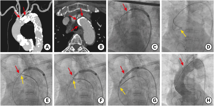
  • Figure 1 Snare-assisted delivery of a self-expanding valve.(A, B) Baseline computed tomography reveals a severely calcified aortic arch with dense calcified plaques protruding into the lumen. (C) The protruding calcification hampers the passage of a delivery catheter. (D) First, we insert a standard wire inside the snare introduced from the contralateral femoral access. Second, the standard wire inserted in the left ventricle is exchanged with the stiff wire. (E-G) Next, the snare is tightened and the stiff wire and nose cone of the delivery catheter are retained. Simultaneous pulling of the snare results in successful passage of the delivery catheter. (H) Final angiography shows an optimal valve position without aortic complications. Red arrows indicate the dense calcified plaque of the aortic arch, and yellow arrows indicate the snare position.


Reference

1. Szlapka M, Michel E, Ricciardi MJ, Malaisrie SC. Valve-in-valve-prosthesis embolization and aortic dissection: single procedure, double complication. Eur J Cardiothorac Surg. 2019; 56:204–205. PMID: 30561574.
Article
2. Kaneko U, Hachinohe D, Kobayashi K, et al. Evolut self-expanding transcatheter aortic valve replacement in patients with extremely horizontal aorta (aortic root angle ≥ 70°). Int Heart J. 2020; 61:1059–1069. PMID: 32921666.
Article
Full Text Links
  • KCJ
Actions
Cited
CITED
export Copy
Close
Share
  • Twitter
  • Facebook
Similar articles
Copyright © 2024 by Korean Association of Medical Journal Editors. All rights reserved.     E-mail: koreamed@kamje.or.kr