Pontic site development with an implant submergence technique for unaesthetic implant in the anterior maxilla
- Affiliations
-
- 1Department of Periodontology, Pusan National University Dental Hospital, Yangsan, Republic of Korea
- 2Department of Periodontology, School of Dentistry, Pusan National University, Yangsan, Republic of Korea
- KMID: 2512124
- DOI: http://doi.org/10.14368/jdras.2020.36.4.289
Abstract
- Improving implant esthetics is very difficult, especially in cases where unaesthetic problems are related to implants in the maxillary anterior dentition. A 69-year old male patient was referred by a prosthodontist for periodic pus discharge and an unaesthetic implant prosthesis (maxillary right lateral incisor). The implant was placed too deeply and showed soft tissue volume deficiency and a long clinical crown. After a clinical and radiographic examination, implant submergence and alveolar ridge augmentation were performed to enhance the aesthetics instead of an explantation. The treatment plan was as follows: extraction the adjacent teeth with tooth mobility, secondary caries, and poor prognosis; placement an additional dental implant with hard and soft tissue grafting; fabrication a fixed bridge using implant abutments. A fixed esthetic prosthesis using implants was fabricated, and the patient was satisfied with the prosthesis. A ridge augmentation with implant submergence may be an alternative for solving the problems of unaesthetic implant restorations in the esthetic zone.
Figure
-
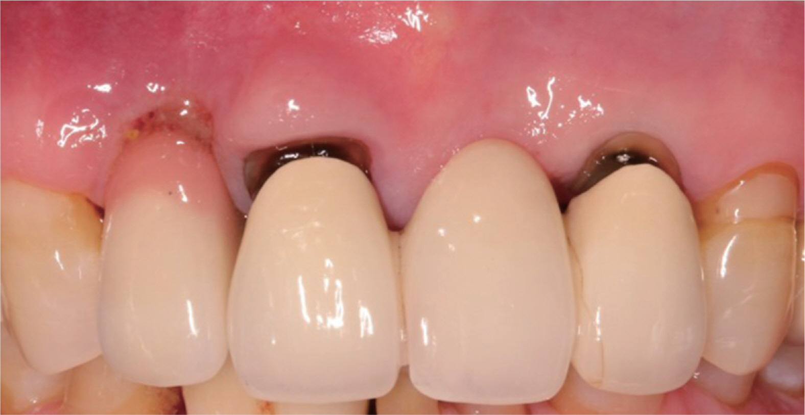
Fig. 1 Initial clinical presentation. Note the long clinical crown with pink ceramic and pus discharge around the maxillary right lateral incisor (#12 dental implant). The marginal soft-tissue interface around the cervical portion of the implant had a recession and lack of ridge volume. Maxillary right central incisor and left lateral incisor had poor esthetics and deep secondary caries.

Fig. 2 Initial periapical view (A), Cone beam computed tomography (B) and panoramic view (C). The preexisting implant fixture was placed reasonably in the mesiodistal and buccolingual direction. Note the deep positioning of the implant in the apico-coronal direction, mild marginal bone loss, and too wide fixture relative to the buccal bone thickness.

Fig. 3 Clinical presentation at each steps of the surgical phase of treatment. Two months after the extraction of the hopeless teeth (#11 and #22). Note shallow circular intrabony defect at implant site 12 (A). After implantation of #11 and #22, the defect around # 12 and buccal defect over #11 and #22 were augmented with bovine bone minerals (Bio-Oss, Geistlich) and non-cross linked bioresorbable membrane (Bio-Gide, Geistlich) (B). Suturing after palatal pedicle graft at implant site 12 (C).

Fig. 4 Preoperative clinical situation (A). Note the unaesthetic gingival margin and the deficiency of buccal ridge volume of the maxillary right lateral incisor after removing the implant prosthesis. After the surgical procedure, the ridge volume was restored and the gingival margin was corrected (B). Final restoration after implant surgery with ridge augmentation and implant submergence (C and D). Gingival volume was restored, and gingival level coincided with the neighboring dentition.

Fig. 5 Periapical view after completion of the final restoration. Deep positioning of the existing implant (#12) was remarkable.
Reference
-
References
1. Mesquita de Carvalho PF, Joly JC, Carvalho da Silva R, González-Martín O. 2019; Therapeutic alternatives for addressing pink esthetic complications in singletooth implants: A proposal for a clinical decision tree. J Esthet Restor Dent. 31:403–14. DOI: 10.1111/jerd.12487. PMID: 31095876.2. Cooper LF, De Kok IJ, Thalji G, Bryington MS. 2019; Prosthodontic Management of Implant Therapy: Esthetic Complications. Dent Clin North Am. 63:199–216. DOI: 10.1016/j.cden.2018.11.003. PMID: 30825986.3. Buser D, Dula K, Belser UC, Hirt HP, Berthold H. 1995; Localized ridge augmentation using guided bone regeneration. II. Surgical procedure in the mandible. Int J Periodontics Restorative Dent. 15:10–29. PMID: 7591520.4. Grunder U. 2011; Crestal Ridge Width Changes When Placing Implants at the Time of Tooth Extraction With and Without Soft Tissue Augmentation After a Healing Period of 6 Months: Report of 24 Consecutive Cases. Int J Periodontics Rest. 31:9–17. PMID: 21365022.5. Comut A, Mehra M, Saito H. 2013; Pontic site development with a root submergence technique for a screw-retained prosthesis in the anterior maxilla. J Prosthet Dent. 110:337–43. DOI: 10.1016/j.prosdent.2013.06.009. PMID: 24035254.6. Papaspyridakos P, Chen CJ, Singh M, Weber HP, Gallucci GO. 2012; Success criteria in implant dentistry: a systematic review. J Dent Res. 91:242–8. DOI: 10.1177/0022034511431252. PMID: 22157097.7. Joo JY, Son S, Lee JY. 2016; Implant Site Development for Enhancing Esthetics of Soft and Hard Tissue and Simplification of Implant Surgery Using a Forced Eruption. Int J Periodontics Restorative Dent. 36:583–9. DOI: 10.11607/prd.2291. PMID: 27333017.8. Kan JY, Rungcharassaeng K, Lozada JL, Zimmerman G. 2011; Facial gingival tissue stability following immediate placement and provisionalization of maxillary anterior single implants: a 2- to 8-year followup. Int J Oral Maxillofac Implants. 26:179–87. PMID: 21365054.9. Buser D, Martin W, Belser UC. 2004; Optimizing esthetics for implant restorations in the anterior maxilla: anatomic and surgical considerations. Int J Oral Maxillofac Implants. 19 Suppl:43–61. PMID: 15635945.10. Evans CD, Chen ST. 2008; Esthetic outcomes of immediate implant placements. Clin Oral Implants Res. 19:73–80. DOI: 10.1111/j.1600-0501.2007.01413.x. PMID: 17956569.11. Funato A, Salama MA, Ishikawa T, Garber DA, Salama H. 2007; Timing, positioning, and sequential staging in esthetic implant therapy: a four-dimensional perspective. Int J Periodontics Restorative Dent. 27:313–23. PMID: 17726987.12. Chackartchi T, Romanos GE, Sculean A. 2019; Soft tissue-related complications and management around dental implants. Periodontol 2000. 81:124–38. DOI: 10.1111/prd.12287. PMID: 31407443.13. Mombelli A, Lang NP. 1998; The diagnosis and treatment of peri-implantitis. Periodontol 2000. 17:63–76. DOI: 10.1111/j.1600-0757.1998.tb00124.x. PMID: 10337314.14. Januário AL, Barriviera M, Duarte WR. 2008; Soft tissue cone-beam computed tomography: a novel method for the measurement of gingival tissue and the dimensions of the dentogingival unit. J Esthet Restor Dent. 20:366–73. DOI: 10.1111/j.1708-8240.2008.00210.x. PMID: 19120781.15. González-Martín O, Oteo C, Ortega R, Alandez J, Sanz M, Veltri M. 2016; Evaluation of peri-implant buccal bone by computed tomography: an experimental study. Clin Oral Implants Res. 27:950–5. DOI: 10.1111/clr.12663. PMID: 26178780.16. Singh G, O'Neal RB, Brennan WA, Strong SL, Horner JA, Van Dyke TE. 1993; Surgical treatment of induced peri-implantitis in the micro pig: clinical and histological analysis. J Periodontol. 64:984–9. DOI: 10.1902/jop.1993.64.10.984. PMID: 8277409.17. Kreisler M, Kohnen W, Christoffers AB, Götz H, Jansen B, Duschner H, d'Hoedt B. 2005; In vitro evaluation of the biocompatibility of contaminated implant surfaces treated with an Er : YAG laser and an air powder system. Clin Oral Implants Res. 16:36–43. DOI: 10.1111/j.1600-0501.2004.01056.x. PMID: 15642029.18. Wetzel AC, Vlassis J, Caffesse RG, Hämmerle CH, Lang NP. 1999; Attempts to obtain re-osseointegration following experimental peri-implantitis in dogs. Clin Oral Implants Res. 10:111–9. DOI: 10.1034/j.1600-0501.1999.100205.x. PMID: 10219130.19. Salama M, Ishikawa T, Salama H, Funato A, Garber D. 2007; Advantages of the root submergence technique for pontic site development in esthetic implant therapy. Int J Periodontics Restorative Dent. 27:521–7. PMID: 18092446.
- Full Text Links
-
- Actions
-
Cited
- CITED
-
- Close
- Share
-
- Similar articles
-
- A root submergence technique for pontic site development in fixed dental prostheses in the maxillary anterior esthetic zone
- Cases of screw-retained implant prosthesis in the anterior maxilla through multidisciplinary approach, including orthodontic teeth alignment
- Outcome Evaluation of an Immediately Placed Maxillary Anterior Single-Tooth Implant Using Objective Esthetic Criteria: Case Report
- The impact of the alveolar bone sites on early implant failure: a systematic review with meta-analysis
- Clinical study on survival rate of osseointegrated implants
