Assessment of the radiant emittance of damaged/contaminated dental lightcuring tips by spectrophotometric methods
- Affiliations
-
- 1Dental Biomedical Sciences PhD Program, University of Maryland School of Dentistry, Baltimore, MD, USA
- 2Department of Restorative Dental Sciences, Imam Abdulrahman Bin Faisal University, College of Dentistry, Dammam, Saudi Arabia
- 3Dental Materials Laboratory, School of Dentistry, Federal University of Rio Grande do Sul, Porto Alegre, RS, Brazil
- 4BlueLight Analytics Company, Halifax, NS, Canada
- 5Department of Dental Clinical Sciences, Dalhousie University, Halifax, NS, Canada
- 6Division of Operative Dentistry, Department of General Dentistry, University of Maryland School of Dentistry, Baltimore, MD, USA
- KMID: 2512046
- DOI: http://doi.org/10.5395/rde.2020.45.e55
Abstract
Objectives
This study investigated the effects of physically damaged and resin-contaminated tips on radiant emittance, comparing them with new undamaged, non-contaminated tips using 3 pieces of spectrophotometric laboratory equipment.
Materials and Methods
Nine tips with damage and/or resin contaminants from actual clinical situations were compared with a new tip without damage or contamination (control group). The radiant emittance was recorded using 3 spectrophotometric methods: a laboratory-grade thermopile, a laboratory-grade integrating sphere, and a portable light collector (checkMARC).
Results
A significant difference between the laboratory-grade thermopile and the laboratory-grade integrating sphere was found when the radiant emittance values of the control or damaged/contaminated tips were investigated (p < 0.05), but both methods were comparable to checkMARC (p > 0.05). Regardless of the method used to quantify the light output, the mean radiant emittance values of the damaged/contaminated tips were significantly lower than those of the control (p < 0.05). The beam profile of the damaged/ contaminated tips was less homogeneous than that of the control.
Conclusions
Damaged/contaminated tips can reduce the radiant emittance output and the homogeneity of the beam, which may affect the energy delivered to composite restorations. The checkMARC spectrophotometer device can be used in dental offices, as it provided values close to those produced by a laboratory-grade integrated sphere spectrophotometer. Dentists should assess the radiant emittance of their light-curing units to ensure optimal curing in photoactivated, resin-based materials.
Keyword
Figure
-
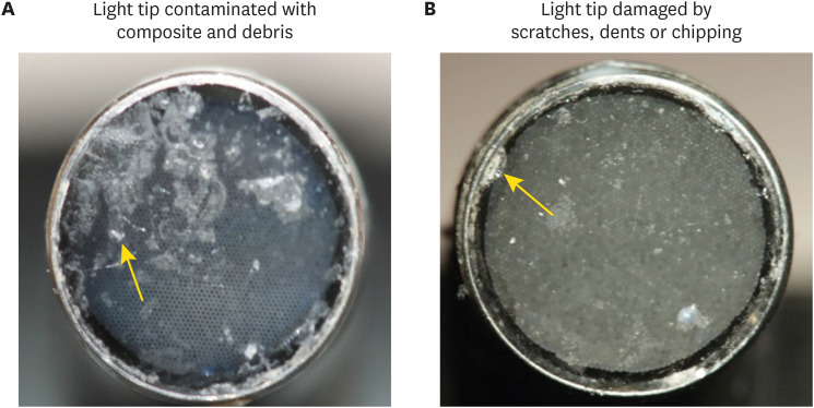
Figure 1 Contaminated and damaged light tips. (A) Light tip contaminated with resin composite and debris. (B) Damaged light tip by scratches, dents, and chipping.

Figure 2 The radiant emittance values were recorded via spectrophotometric methods. (A) A laboratory-grade thermopile system. (B) A laboratory-grade integrating sphere spectrophotometer. (C) An in-office checkMARC portable spectrophotometer.
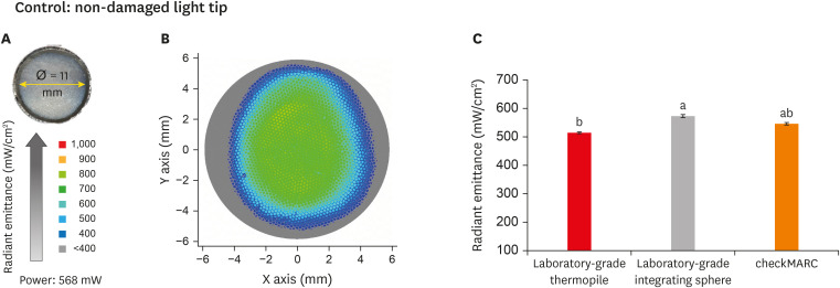
Figure 3 Radiant emittance levels of the control tip. (A) An appearance of the control tip and the measured active tip diameter. (B) Representative image of the light beam radiant emittance profile for the control tip. The color bar on the right shows the visual representation of radiant emittance expressed in mW per cm2 corresponding to the colors seen. The profile presents an abundance of the green color representative of the range of 600–700 mW/cm2 with scattered dots in a hot color (yellow-orange) representing radiance with emission in the range of 800–900 mW/cm2. The profile beam image also shows peripheral areas in blue and gray colors, representative of radiance at 400 and below 400 mW/cm2, respectively. (C) Radiant emittance assessment of non-damaged and non-contaminated tips by the 3 spectrophotometric methods. No significant difference was found between the checkMARC spectrophotometer and the other methods (p < 0.05). Values with different letters are significantly different (p < 0.05).
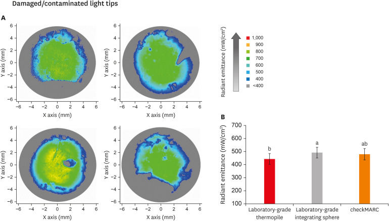
Figure 4 Radiant emittance levels of the damaged/contaminated tips. (A) Representative images of the beam profile of damaged/contaminated light tips. The colors in the beam profile represent the amount of radiant emittance captured at each area across the light-curing unit tip. The higher the normalized radiant emittance value, the better the performance of the light-curing unit (high-value areas: light green). The color bar on the right shows the visual representation of radiant emittance expressed in mW per cm2 corresponding to the colors seen. The profile beam images for damaged/contaminated tips show an increased abundance of peripheral and central areas in blue and gray colors representative of radiance at 400 and below 400 mW/cm2, respectively. In B, the radiant emittance assessments of damaged/contaminated tips (n = 9) by the 3 spectrophotometric methods are shown. The radiant emittance assessed by the thermopile was significantly (p < 0.05) lower than the values for the laboratory-grade sphere. No significant differences were found between the checkMARC spectrophotometer and the other methods (p < 0.05). Values with different letters are significantly different (p < 0.05).
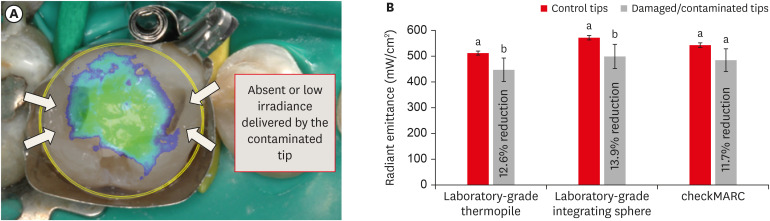
Figure 5 Impact of damage/contamination on radiant emittance. (A) Representative illustration of a clinical restorative scenario where a mesio-distal occlusal composite restoration is placed and cured by a damaged tip. The overlapping image of the non-uniform beam profile over the composite restoration should be noted. The lack of homogeneity in the produced radiant emittance profile could compromise the optimal polymerization of the restoration. The black arrows indicate areas that may not receive the radiant emittance appropriately. (B) Radiant emittance results and percentage reduction of radiance relative to the control. On the first y-axis, the bar plot shows radiant emittance results expressed in mW/cm2. Each method was used to capture the radiant emittance values from damaged/contaminated tips and compared to those with no damage or contamination (control). The difference in the power output between the damaged/contaminated and control tips was significant when the thermopile and the laboratory-grade sphere spectrophotometer were used. Damaged/contaminated tips significantly demonstrated lower radiant emittance values than the control (p < 0.05). In this plot, values with different letters are significantly different (p < 0.05).
Reference
-
1. Chan KHS, Mai Y, Kim H, Tong KCT, Ng D, Hsiao JCM. Review: resin composite filling. Materials (Basel). 2010; 3:1228–1243.
Article2. Balhaddad AA, Kansara AA, Hidan D, Weir MD, Xu HHK, Melo MAS. Toward dental caries: exploring nanoparticle-based platforms and calcium phosphate compounds for dental restorative materials. Bioact Mater. 2018; 4:43–55. PMID: 30582079.
Article3. World Health Organization. Oral health surveys: basic methods. 3rd ed. Geneva: World Health Organization;1987.4. Gordan VV, Riley JL 3rd, Rindal DB, Qvist V, Fellows JL, Dilbone DA, Brotman SG, Gilbert GH. National Dental Practice-Based Research Network Collaborative Group. Repair or replacement of restorations: a prospective cohort study by dentists in the National Dental Practice-Based Research Network. J Am Dent Assoc. 2015; 146:895–903. PMID: 26610834.5. David JR, Gomes OM, Gomes JC, Loguercio AD, Reis A. Effect of exposure time on curing efficiency of polymerizing units equipped with light-emitting diodes. J Oral Sci. 2007; 49:19–24. PMID: 17429178.
Article6. Santini A, Gallegos IT, Felix CM. Photoinitiators in dentistry: a review. Prim Dent J. 2013; 2:30–33.
Article7. Samaha S, Bhatt S, Finkelman M, Papathanasiou A, Perry R, Strassler H, Kugel G, Garcia-Godoy F, Price R. Effect of instruction, light curing unit, and location in the mouth on the energy delivered to simulated restorations. Am J Dent. 2017; 30:343–349. PMID: 29251458.8. Price RB, Ferracane JL, Shortall AC. Light-curing units: a review of what we need to know. J Dent Res. 2015; 94:1179–1186. PMID: 26156516.9. Maktabi H, Balhaddad AA, Alkhubaizi Q, Strassler H, Melo MAS. Factors influencing success of radiant exposure in light-curing posterior dental composite in the clinical setting. Am J Dent. 2018; 31:320–328. PMID: 30658380.10. AlShaafi MM. Factors affecting polymerization of resin-based composites: a literature review. Saudi Dent J. 2017; 29:48–58. PMID: 28490843.
Article11. Rueggeberg FA, Giannini M, Arrais CAG, Price RBT. Light curing in dentistry and clinical implications: a literature review. Braz Oral Res. 2017; 31:e61. PMID: 28902241.
Article12. Maktabi H, Ibrahim M, Alkhubaizi Q, Weir M, Xu H, Strassler H, Fugolin APP, Pfeifer CS, Melo MAS. Underperforming light curing procedures trigger detrimental irradiance-dependent biofilm response on incrementally placed dental composites. J Dent. 2019; 88:103110. PMID: 31022421.
Article13. Alshaafi MM. Evaluation of light-curing units in rural and urban areas. Saudi Dent J. 2012; 24:163–167. PMID: 23960546.
Article14. Price RBT, Ehrnford L, Andreou P, Felix CA. Comparison of quartz-tungsten-halogen, light-emitting diode, and plasma arc curing lights. J Adhes Dent. 2003; 5:193–207. PMID: 14621241.15. Vandenbulcke JDE, Marks LAM, Martens LC, Verbeeck RMH. Comparison of curing depth of a colored polyacid-modified composite resin with different light-curing units. Quintessence Int. 2010; 41:787–794. PMID: 20806104.16. Maghaireh GA, Alzraikat H, Taha NA. Assessing the irradiance delivered from light-curing units in private dental offices in Jordan. J Am Dent Assoc. 2013; 144:922–927. PMID: 23904579.
Article17. Barghi N, Fischer DE, Pham T. Revisiting the intensity output of curing lights in private dental offices. Compend Contin Educ Dent. 2007; 28:380–384. PMID: 17687900.18. Santos GC Jr, Santos MJMC, El-Mowafy O, El-Badrawy W. Intensity of quartz-tungsten-halogen light polymerization units used in dental offices in Brazil. Int J Prosthodont. 2005; 18:434–435. PMID: 16220811.19. Nassar HM, Ajaj R, Hasanain F. Efficiency of light curing units in a government dental school. J Oral Sci. 2018; 60:142–146. PMID: 29576574.
Article20. Al Shaafi M, Maawadh A, Al Qahtani M. Evaluation of light intensity output of QTH and LED curing devices in various governmental health institutions. Oper Dent. 2011; 36:356–361. PMID: 21834709.
Article21. El-Mowafy O, El-Badrawy W, Lewis DW, Shokati B, Kermalli J, Soliman O, Encioiu A, Zawi R, Rajwani F, Rajwani F. Intensity of quartz-tungsten-halogen light-curing units used in private practice in Toronto. J Am Dent Assoc. 2005; 136:766–773. PMID: 16022041.
Article22. Jandt KD, Mills RW, Blackwell GB, Ashworth SH. Depth of cure and compressive strength of dental composites cured with blue light emitting diodes (LEDs). Dent Mater. 2000; 16:41–47. PMID: 11203522.
Article23. Shortall AC, Price RB, MacKenzie L, Burke FJT. Guidelines for the selection, use, and maintenance of LED light-curing units - part II. Br Dent J. 2016; 221:551–554. PMID: 27811902.
Article24. International Organization for Standardization. ISO 10650-2. Dentistry—powered polymerization activators—part 2: light-emitting diode (LED) lamps. Geneva: International Organization for Standardization;2007.25. Shortall AC, Felix CJ, Watts DC. Robust spectrometer-based methods for characterizing radiant exitance of dental LED light curing units. Dent Mater. 2015; 31:339–350. PMID: 25773187.
Article26. Marović D, Matić S, Kelić K, Klarić E, Rakić M, Tarle Z. Time dependent accuracy of dental radiometers. Acta Clin Croat. 2013; 52:173–180. PMID: 24053077.27. Kameyama A, Haruyama A, Asami M, Takahashi T. Effect of emitted wavelength and light guide type on irradiance discrepancies in hand-held dental curing radiometers. ScientificWorldJournal. 2013; 2013:647941. PMID: 24222744.
Article28. Price RB, Labrie D, Kazmi S, Fahey J, Felix CM. Intra- and inter-brand accuracy of four dental radiometers. Clin Oral Investig. 2012; 16:707–717.
Article29. Michaud PL, Price RBT, Labrie D, Rueggeberg FA, Sullivan B. Localised irradiance distribution found in dental light curing units. J Dent. 2014; 42:129–139. PMID: 24287255.
Article30. Harlow JE, Sullivan B, Shortall AC, Labrie D, Price RB. Characterizing the output settings of dental curing lights. J Dent. 2016; 44:20–26. PMID: 26546716.
Article31. Shimokawa CAK, Harlow JE, Turbino ML, Price RB. Ability of four dental radiometers to measure the light output from nine curing lights. J Dent. 2016; 54:48–55. PMID: 27597433.
Article32. Konerding KL, Heyder M, Kranz S, Guellmar A, Voelpel A, Watts DC, Jandt KD, Sigusch BW. Study of energy transfer by different light curing units into a class III restoration as a function of tilt angle and distance, using a MARC patient simulator (PS). Dent Mater. 2016; 32:676–686. PMID: 27017156.
Article33. Soares CJ, Rodrigues MP, Oliveira LRS, Braga SSL, Barcelos LM, Silva GR, Giannini M, Price RB. An evaluation of the light output from 22 contemporary light curing units. Braz Dent J. 2017; 28:362–371. PMID: 29297558.
Article34. Price RBT, Labrie D, Rueggeberg FA, Felix CM. Irradiance differences in the violet (405 nm) and blue (460 nm) spectral ranges among dental light-curing units. J Esthet Restor Dent. 2010; 22:363–377. PMID: 21126292.
Article35. Price RBT, Rueggeberg FA, Labrie D, Felix CM. Irradiance uniformity and distribution from dental light curing units. J Esthet Restor Dent. 2010; 22:86–101. PMID: 20433559.
Article36. Rueggeberg FA, Caughman WF, Curtis JW Jr. Effect of light intensity and exposure duration on cure of resin composite. Oper Dent. 1994; 19:26–32. PMID: 8183730.37. Al-Zain AO, Eckert GJ, Lukic H, Megremis S, Platt JA. Polymerization pattern characterization within a resin-based composite cured using different curing units at two distances. Clin Oral Investig. 2019; 23:3995–4010.
Article38. Eshmawi YT, Al-Zain AO, Eckert GJ, Platt JA. Variation in composite degree of conversion and microflexural strength for different curing lights and surface locations. J Am Dent Assoc. 2018; 149:893–902. PMID: 30149887.
Article39. Yeh CL, Miyagawa Y, Powers JM. Optical properties of composites of selected shades. J Dent Res. 1982; 61:797–801. PMID: 6953116.
Article40. International Organization for Standardization. ISO 10650-1. Dentistry—powered polymerization activators—part 1: quartz tungsten halogen lamps. Geneva: International Organization for Standardization;2004.41. Arikawa H, Kanie T, Fujii K, Takahashi H, Ban S. Effect of inhomogeneity of light from light curing units on the surface hardness of composite resin. Dent Mater J. 2008; 27:21–28. PMID: 18309608.
Article42. Rueggeberg FA. State-of-the-art: dental photocuring--a review. Dent Mater. 2011; 27:39–52. PMID: 21122903.
Article
- Full Text Links
-
- Actions
-
Cited
- CITED
-
- Close
- Share
-
- Similar articles
-
- Shade comparative analysis of natural tooth measured by visual and spectrophotometric methods
- Comparison of instrumental methods for color change assessment of Giomer resins
- Electron Microscopy of Needle Tips Following Intravitreal Injections Using 30-Gauge Needles
- The efficacy of different implant surface decontamination methods using spectrophotometric analysis: an in vitro study
- The Effectiveness Of Decontamination Methods In Dental Instruments
