Restor Dent Endod.
2020 Aug;45(3):e38. 10.5395/rde.2020.45.e38.
Apical root canal cleaning after preparation with endodontic instruments: a randomized trial in vivo analysis
- Affiliations
-
- 1Endodontic Division, Department of Restorative Dentistry, Piracicaba Dental School, Campinas State University (UNICAMP), Piracicaba, SP, Brazil
- 2Meridional Dental Studies Center (CEOM), Passo Fundo, RS, Brazil
- 3Pathology Institute of Passo Fundo, Passo Fundo, RS, Brazil
- KMID: 2512029
- DOI: http://doi.org/10.5395/rde.2020.45.e38
Abstract
Objectives
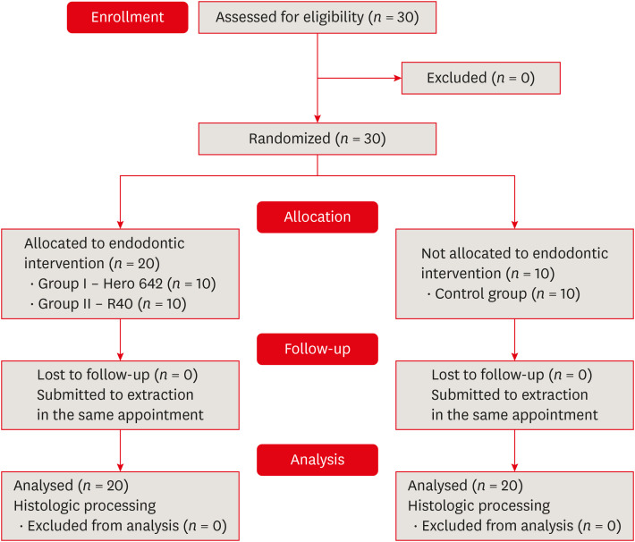
This study aimed to evaluate vital pulp tissue removal from different endodontic instrumentation systems from root canal apical third in vivo
Materials and Methods
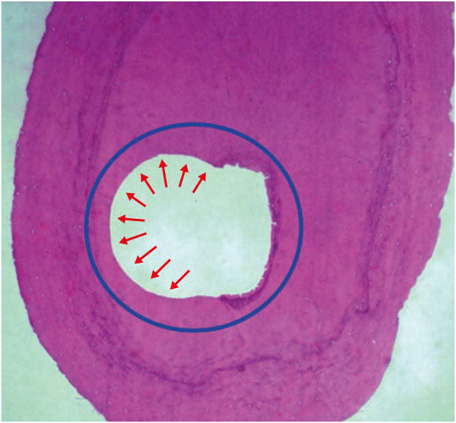
Thirty mandibular molars were selected and randomly divided into 2 test groups and one control group. Inclusion criteria were a positive response to cold sensibility test, curvature angle between 10 and 20 degrees, and curvature radius lower than 10 mm. Root canals prepared with Hero 642 system (size 45/0.02) (n = 10) and Reciproc R40 (size 40/0.06) (n = 10) and control (n = 10) without instrumentation. Canals were irrigated only with saline solution during root canal preparation. The apical third was evaluated considering the touched/untouched perimeter and area to evaluate the efficacy of root canal wall debridement. Statistical analysis used t-test for comparisons.
Results
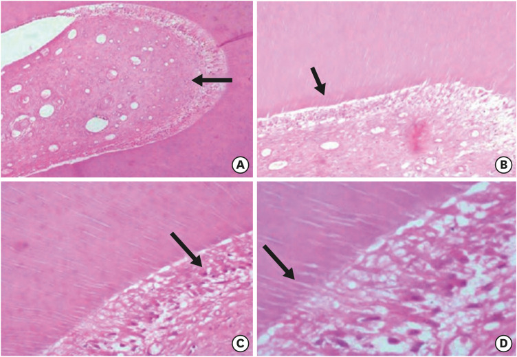
Untouched root canal at cross-section perimeter, the Hero 642 system showed 41.44% ± 5.62% and Reciproc R40 58.67% ± 12.39% without contact with instruments. Regarding the untouched area, Hero 642 system showed 22.78% ± 6.42% and Reciproc R40 34.35% ± 8.52%. Neither instrument achieved complete cross-sectional root canal debridement. Hero 642 system rotary taper 0.02 instruments achieved significant greater wall contact perimeter and area compared to reciprocate the Reciproc R40 taper 0.06 instrument.
Conclusions
Hero 642 achieved higher wall contact perimeter and area but, regardless of instrument size and taper, vital pulp during in vivo instrumentation is not entirely removed.
Keyword
Figure
Reference
-
1. Hülsmann M, Schade M, Schäfers F. A comparative study of root canal preparation with Hero 642 and Quantec SC rotary Ni-Ti instruments. Int Endod J. 2001; 34:538–546. PMID: 11601772.
Article2. De-Deus G, Barino B, Zamolyi RQ, Souza E, Fonseca A Jr, Fidel S, Fidel RA. Suboptimal debridement quality produced by the single-file F2 ProTaper technique in oval-shaped canals. J Endod. 2010; 36:1897–1900. PMID: 20951309.
Article3. Bürklein S, Benten S, Schäfer E. Shaping ability of different single-file systems in severely curved root canals of extracted teeth. Int Endod J. 2013; 46:590–597. PMID: 23240965.
Article4. Borges MF, Miranda CE, Silva SR, Marchesan M. Influence of apical enlargement in cleaning and extrusion in canals with mild and moderate curvatures. Braz Dent J. 2011; 22:212–217. PMID: 21915518.
Article5. Pagliosa A, Sousa-Neto MD, Versiani MA, Raucci-Neto W, Silva-Sousa YT, Alfredo E. Computed tomography evaluation of rotary systems on the root canal transportation and centering ability. Braz Oral Res. 2015; 29:1–7.
Article6. Fornari VJ, Silva-Sousa YT, Vanni JR, Pécora JD, Versiani MA, Sousa-Neto MD. Histological evaluation of the effectiveness of increased apical enlargement for cleaning the apical third of curved canals. Int Endod J. 2010; 43:988–994. PMID: 20722756.
Article7. Peters OA, Arias A, Paqué F. A micro-computed tomographic assessment of root canal preparation with a novel instrument, TRUShape, in mesial roots of mandibular molars. J Endod. 2015; 41:1545–1550. PMID: 26238528.
Article8. Marceliano-Alves MF, Sousa-Neto MD, Fidel SR, Steier L, Robinson JP, Pécora JD, Versiani MA. Shaping ability of single-file reciprocating and heat-treated multifile rotary systems: a micro-CT study. Int Endod J. 2015; 48:1129–1136. PMID: 25400256.
Article9. Lacerda MF, Marceliano-Alves MF, Pérez AR, Provenzano JC, Neves MA, Pires FR, Gonçalves LS, Rôças IN, Siqueira JF Jr. Cleaning and shaping oval canals with 3 instrumentation systems: a correlative micro-computed tomographic and histologic study. J Endod. 2017; 43:1878–1884. PMID: 28951035.10. Siqueira JF Jr, Pérez AR, Marceliano-Alves MF, Provenzano JC, Silva SG, Pires FR, Vieira GC, Rôças IN, Alves FR. What happens to unprepared root canal walls: a correlative analysis using micro-computed tomography and histology/scanning electron microscopy. Int Endod J. 2018; 51:501–508. PMID: 28196289.
Article11. Wu MK, Barkis D, Roris A, Wesselink PR. Does the first file to bind correspond to the diameter of the canal in the apical region? Int Endod J. 2002; 35:264–267. PMID: 11985678.
Article12. Hartmann RC, Fensterseifer M, Peters OA, de Figueiredo JA, Gomes MS, Rossi-Fedele G. Methods for measurement of root canal curvature: a systematic and critical review. Int Endod J. 2019; 52:169–180. PMID: 30099748.
Article13. Wu MK, R'oris A, Barkis D, Wesselink PR. Prevalence and extent of long oval canals in the apical third. Oral Surg Oral Med Oral Pathol Oral Radiol Endod. 2000; 89:739–743. PMID: 10846130.
Article14. Schilder H. Cleaning and shaping the root canal. Dent Clin North Am. 1974; 18:269–296. PMID: 4522570.15. Paqué F, Zehnder M, De-Deus G. Microtomography-based comparison of reciprocating single-file F2 ProTaper technique versus rotary full sequence. J Endod. 2011; 37:1394–1397. PMID: 21924189.
Article16. Bürklein S, Schäfer E. Apically extruded debris with reciprocating single-file and full-sequence rotary instrumentation systems. J Endod. 2012; 38:850–852. PMID: 22595125.
Article17. Robinson JP, Lumley PJ, Cooper PR, Grover LM, Walmsley AD. Reciprocating root canal technique induces greater debris accumulation than a continuous rotary technique as assessed by 3-dimensional micro-computed tomography. J Endod. 2013; 39:1067–1070. PMID: 23880279.
Article18. Shemesh H, Lindtner T, Portoles CA, Zaslansky P. Dehydration induces cracking in root dentin irrespective of instrumentation: a two-dimensional and three-dimensional study. J Endod. 2018; 44:120–125. PMID: 29079053.
Article19. Brunson M, Heilborn C, Johnson DJ, Cohenca N. Effect of apical preparation size and preparation taper on irrigant volume delivered by using negative pressure irrigation system. J Endod. 2010; 36:721–724. PMID: 20307751.
Article20. Card SJ, Sigurdsson A, Orstavik D, Trope M. The effectiveness of increased apical enlargement in reducing intracanal bacteria. J Endod. 2002; 28:779–783. PMID: 12470024.
Article21. Coldero LG, McHugh S, MacKenzie D, Saunders WP. Reduction in intracanal bacteria during root canal preparation with and without apical enlargement. Int Endod J. 2002; 35:437–446. PMID: 12059915.
Article22. Bürklein S, Hinschitza K, Dammaschke T, Schäfer E. Shaping ability and cleaning effectiveness of two single-file systems in severely curved root canals of extracted teeth: Reciproc and WaveOne versus Mtwo and ProTaper. Int Endod J. 2012; 45:449–461. PMID: 22188401.
Article
- Full Text Links
- Actions
-
Cited
- CITED
-
- Close
- Share
- Similar articles
-
- Effects of the endodontic access cavity on apical debris extrusion during root canal preparation using different single-file systems
- Success and failure of endodontic microsurgery
- Prognosis of the Apical Fragment of Root Fractures after Root Canal Treatment of Both Fragments in Immature Permanent Teeth
- Safe root canal preparation using reciprocating nickel-titanium instruments
- Apical prepration size in infected root canals


