J Korean Assoc Oral Maxillofac Surg.
2020 Dec;46(6):385-392. 10.5125/jkaoms.2020.46.6.385.
Study of soft tissue changes in the upper lip and nose after backward movement of the maxilla in orthognathic surgery
- Affiliations
-
- 1Department of Oral and Maxillofacial Surgery, School of Dentistry, Kyung Hee University, Seoul, Korea
- KMID: 2510008
- DOI: http://doi.org/10.5125/jkaoms.2020.46.6.385
Abstract
Objectives
This study evaluates soft tissue changes of the upper lip and nose after maxillary setback with orthognathic surgery such as Le Fort I or anterior segmental osteotomy.
Materials and Methods
All 50 patients with bimaxillary protrusion and skeletal Class II malocclusion underwent Le Fort I or anterior segmental osteotomy with backward movement. Soft and hard tissue changes were analyzed using cephalograms collected preoperatively and 6 months postoperatively.
Results
Cluster analysis on the ratios shows that 2 lines intersected at 4 mm point. Based on this point, we divided the subjects into 2 groups: Group A (less than 4 mm, 27 subjects) and Group B (more than 4 mm, 23 subjects). Also, each group was divided according to changes of upper incisor angle (≥4°=A1, B1 or <4°=A2, B2). The correlation between A and B groups for Aʼ/ANS and Ls/Is (P<0.001) was significant; Aʼ/A (P=0.002), PRN/A (P=0.043), PRN/ANS (P=0.032), and St/Is (P=0.010). Variation of nasolabial angle between the two groups was not significant. There was no significant correlation of vertical movement and angle variation.
Conclusion
The ratio of soft tissue to hard tissue movement depends on the amount of posterior movement in the maxilla, showing approximately two times higher rates in most of the midface when posterior movement was greater than 4 mm. The soft tissue changes caused by posterior movement of the maxilla were little affected by angular changes of upper incisors. Interestingly, nasolabial angle showed a different tendency between A and B groups and was more affected by incisal angular changes when horizontal posterior movement was less than 4 mm.
Figure
-

Fig. 1 Cephalometric hard tissue landmarks and reference planes. (HRL: horizontal reference line, S: sella, N: nasion, ANS: anterior nasal spine, A: A point, Ia: incisor anterius, Is: incisor superius, Upper incisor angle: upper incisor to HRL, VRL: vertical reference line)
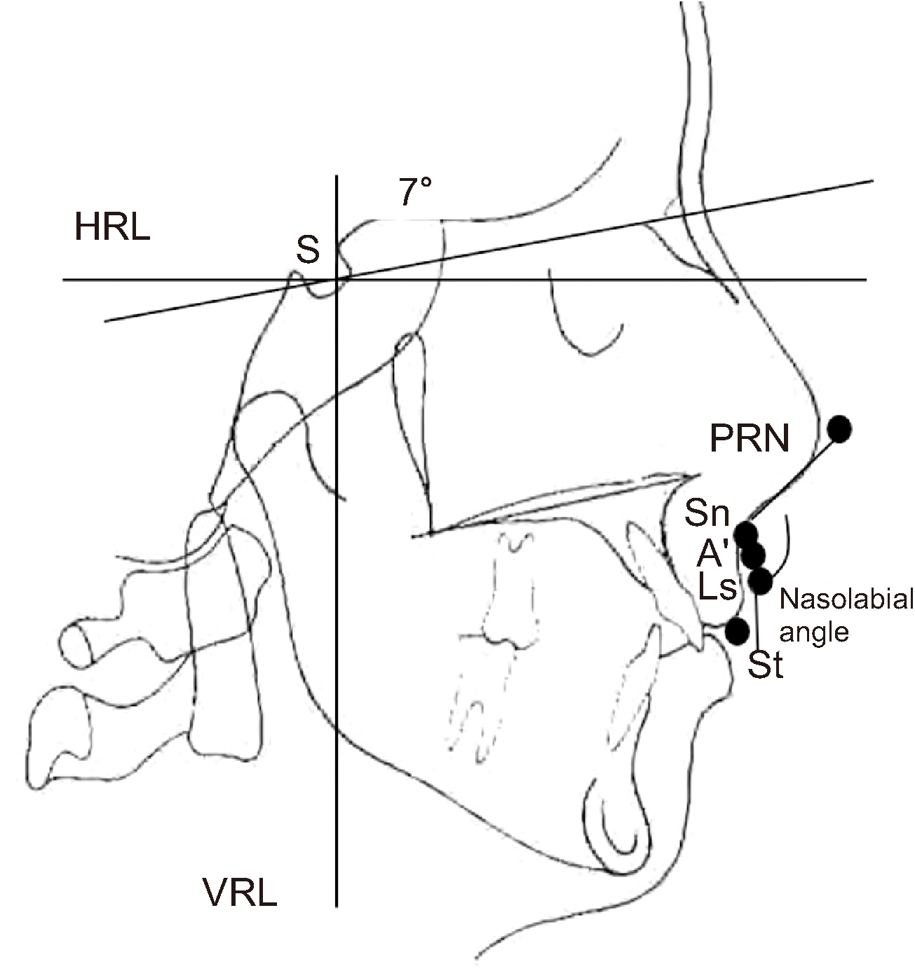
Fig. 2 Cephalometric soft tissue landmarks and reference planes. (HRL: horizontal reference line, S: sella, PRN: pronasale, Sn: subnasale, A’: soft tissue A point, Ls: labrale superius, St: stomion superius, VRL: vertical reference line)
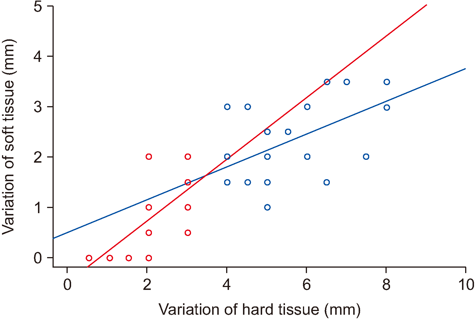
Fig. 3 Relaptionship between the movement of hard tissue and soft tissue. Ratio of soft tissue on the hard tissue. Two different gradient lines crossing near the 4 mm (x-axis) point.
Reference
-
References
1. O'Ryan F, Schendel S. 1989; Nasal anatomy and maxillary surgery. I. esthetic and anatomic principles. Int J Adult Orthodon Orthognath Surg. 4:27–37. PMID: 2689536.2. Betts NJ, Vig KW, Vig P, Spalding P, Fonseca RJ. 1993; Changes in the nasal and labial soft tissues after surgical repositioning of the maxilla. Int J Adult Orthodon Orthognath Surg. 8:7–23. PMID: 8509667.3. Hack GA, Nanda R. de Mol van Otterloo JJ. 1993; Long-term stability and prediction of soft tissue changes after LeFort I surgery. Am J Orthod Dentofacial Orthop. 104:544–55. https://doi.org/10.1016/s0889-5406(05)80438-x . DOI: 10.1016/s0889-5406(05)80438-x. PMID: 8249930.
Article4. Schendel SA, Eisenfeld JH, Bell WH, Epker BN. 1976; Superior repositioning of the maxilla: stability and soft tissue osseous relations. Am J Orthod. 70:663–74. https://doi.org/10.1016/0002-9416(76)90226-8 . DOI: 10.1016/0002-9416(76)90226-8. PMID: 1069482.
Article5. Chew MT. 2005; Soft and hard tissue changes after bimaxillary surgery in Chinese Class III patients. Angle Orthod. 75:959–63. https://doi.org/10.1043/0003-3219(2005)75[959:SAHTCA]2.0.CO;2 . DOI: 10.1043/0003-3219(2005)75[959:SAHTCA]2.0.CO;2. PMID: 16448238.
Article6. Dann JJ 3rd, Fonseca RJ, Bell WH. 1976; Soft tissue changes associated with total maxillary advancement: a preliminary study. J Oral Surg. 34:19–23. PMID: 1059747.7. Ewing M, Ross RB. 1993; Soft tissue response to orthognathic surgery in persons with unilateral cleft lip and palate. Cleft Palate Craniofac J. 30:320–7. https://doi.org/10.1597/1545-1569_1993_030_0320_strtos_2.3.co_2 . DOI: 10.1597/1545-1569_1993_030_0320_strtos_2.3.co_2. PMID: 8338863.
Article8. Mansour S, Burstone C, Legan H. 1983; An evaluation of soft-tissue changes resulting from Le Fort I maxillary surgery. Am J Orthod. 84:37–47. https://doi.org/10.1016/0002-9416(83)90146-x . DOI: 10.1016/0002-9416(83)90146-x. PMID: 6575616.
Article9. Schouman T, Baralle MM, Ferri J. 2010; Facial morphology changes after total maxillary setback osteotomy. J Oral Maxillofac Surg. 68:1504–11. https://doi.org/10.1016/j.joms.2009.09.095 . DOI: 10.1016/j.joms.2009.09.095. PMID: 20466475.
Article10. Radney LJ, Jacobs JD. 1981; Soft-tissue changes associated with surgical total maxillary intrusion. Am J Orthod. 80:191–212. https://doi.org/10.1016/0002-9416(81)90218-9 . DOI: 10.1016/0002-9416(81)90218-9. PMID: 6943938.
Article11. Stella JP, Streater MR, Epker BN, Sinn DP. 1989; Predictability of upper lip soft tissue changes with maxillary advancement. J Oral Maxillofac Surg. 47:697–703. https://doi.org/10.1016/s0278-2391(89)80008-4 . DOI: 10.1016/s0278-2391(89)80008-4. PMID: 2732829.
Article12. Eggensperger NM, Lieger O, Thüer U, Iizuka T. 2007; Soft tissue profile changes following mandibular advancement and setback surgery an average of 12 years postoperatively. J Oral Maxillofac Surg. 65:2301–10. https://doi.org/10.1016/j.joms.2007.06.644 . DOI: 10.1016/j.joms.2007.06.644. PMID: 17954329.
Article13. Yitschaky O, Redlich M, Abed Y, Faerman M, Casap N, Hiller N. 2011; Comparison of common hard tissue cephalometric measurements between computed tomography 3D reconstruction and conventional 2D cephalometric images. Angle Orthod. 81:11–6. https://doi.org/10.2319/031710-157.1 . DOI: 10.2319/031710-157.1. PMID: 20936949.
Article14. Burstone CJ, James RB, Legan H, Murphy GA, Norton LA. 1978; Cephalometrics for orthognathic surgery. J Oral Surg. 36:269–77. PMID: 273073.15. Moore JW. 1976; Variation of the sella-nasion plane and its effect on SNA and SNB. J Oral Surg. 34:24–6. PMID: 1059748.16. Quast DC, Biggerstaff RH, Haley JV. 1983; The short-term and long-term soft-tissue profile changes accompanying mandibular advancement surgery. Am J Orthod. 84:29–36. https://doi.org/10.1016/0002-9416(83)90145-8 . DOI: 10.1016/0002-9416(83)90145-8. PMID: 6575615.
Article17. Koh CH, Chew MT. 2004; Predictability of soft tissue profile changes following bimaxillary surgery in skeletal class III Chinese patients. J Oral Maxillofac Surg. 62:1505–9. https://doi.org/10.1016/j.joms.2004.04.022 . DOI: 10.1016/j.joms.2004.04.022. PMID: 15573350.
Article18. Mommaerts MY, Lippens F, Abeloos JV, Neyt LF. 2000; Nasal profile changes after maxillary impaction and advancement surgery. J Oral Maxillofac Surg. 58:470–5. discussion 475–6. https://doi.org/10.1016/s0278-2391(00)90002-8 . DOI: 10.1016/s0278-2391(00)90002-8. PMID: 10800900.
Article19. Brooks BW, Buschang PH, Bates JD, Adams TB, English JD. 2001; Predicting upper lip response to 4-piece maxillary LeFort I osteotomy. Am J Orthod Dentofacial Orthop. 120:124–33. https://doi.org/10.1067/mod.2001.115614 . DOI: 10.1067/mod.2001.115614. PMID: 11500653.
Article20. Gassmann CJ, Nishioka GJ, Van Sickels JE, Thrash WJ. 1989; A lateral cephalometric analysis of nasal morphology following Le Fort I osteotomy applying photometric analysis techniques. J Oral Maxillofac Surg. 47:926–30. https://doi.org/10.1016/0278-2391(89)90375-3 . DOI: 10.1016/0278-2391(89)90375-3. PMID: 2760729.
Article21. Rosen HM. 1988; Lip-nasal aesthetics following Le Fort I osteotomy. Plast Reconstr Surg. 81:171–82. https://doi.org/10.1097/00006534-198802000-00005 . DOI: 10.1097/00006534-198802000-00005. PMID: 3336648.
Article22. Westermark AH, Bystedt H, Von Konow L, Sällström KO. 1991; Nasolabial morphology after Le Fort I osteotomies. Effect of alar base suture. Int J Oral Maxillofac Surg. 20:25–30. https://doi.org/10.1016/s0901-5027(05)80690-3 . DOI: 10.1016/s0901-5027(05)80690-3. PMID: 2019779.
Article23. Jayaratne YS, Zwahlen RA, Lo J, Cheung LK. 2010; Facial soft tissue response to anterior segmental osteotomies: a systematic review. Int J Oral Maxillofac Surg. 39:1050–8. https://doi.org/10.1016/j.ijom.2010.07.002 . DOI: 10.1016/j.ijom.2010.07.002. PMID: 20705431.
Article24. Okudaira M, Kawamoto T, Ono T, Moriyama K. 2008; Soft-tissue changes in association with anterior maxillary osteotomy: a pilot study. Oral Maxillofac Surg. 12:131–8. https://doi.org/10.1007/s10006-008-0121-9 . DOI: 10.1007/s10006-008-0121-9. PMID: 18629553.
Article25. Jacobson R, Sarver DM. 2002; The predictability of maxillary repositioning in LeFort I orthognathic surgery. Am J Orthod Dentofacial Orthop. 122:142–54. https://doi.org/10.1067/mod.2002.125576 . DOI: 10.1067/mod.2002.125576. PMID: 12165768.
Article26. Park JU, Hwang YS. 2008; Evaluation of the soft and hard tissue changes after anterior segmental osteotomy on the maxilla and mandible. J Oral Maxillofac Surg. 66:98–103. https://doi.org/10.1016/j.joms.2005.09.007 . DOI: 10.1016/j.joms.2005.09.007. PMID: 18083422.
Article27. Lew K. 1989; Profile changes following orthodontic treatment of bimaxillary protrusion in adults with the Begg appliance. Eur J Orthod. 11:375–81. https://doi.org/10.1093/oxfordjournals.ejo.a036009 . DOI: 10.1093/oxfordjournals.ejo.a036009. PMID: 2591485.
Article28. Nadkarni PG. 1986; Soft tissue profile changes associated with orthognathic surgery for bimaxillary protrusion. J Oral Maxillofac Surg. 44:851–4. https://doi.org/10.1016/0278-2391(86)90220-x . DOI: 10.1016/0278-2391(86)90220-x. PMID: 3464710.
Article29. Jensen AC, Sinclair PM, Wolford LM. 1992; Soft tissue changes associated with double jaw surgery. Am J Orthod Dentofacial Orthop. 101:266–75. https://doi.org/10.1016/0889-5406(92)70096-S . DOI: 10.1016/0889-5406(92)70096-S. PMID: 1539554.
Article30. Muradin MS, Rosenberg A, van der Bilt A, Stoelinga PJ, Koole R. 2009; The effect of alar cinch sutures and V-Y closure on soft tissue dynamics after Le Fort I intrusion osteotomies. J Craniomaxillofac Surg. 37:334–40. https://doi.org/10.1016/j.jcms.2009.03.004 . DOI: 10.1016/j.jcms.2009.03.004. PMID: 19477655.
Article31. Peled M, Ardekian L, Krausz AA, Aizenbud D. 2004; Comparing the effects of V-Y advancement versus simple closure on upper lip aesthetics after Le Fort I advancement. J Oral Maxillofac Surg. 62:315–9. https://doi.org/10.1016/j.joms.2003.08.015 . DOI: 10.1016/j.joms.2003.08.015. PMID: 15015164.
Article32. Khamashta-Ledezma L, Naini FB. 2014; Systematic review of changes in maxillary incisor exposure and upper lip position with Le Fort I type osteotomies with or without cinch sutures and/or VY closures. Int J Oral Maxillofac Surg. 43:46–61. https://doi.org/10.1016/j.ijom.2013.08.005 . DOI: 10.1016/j.ijom.2013.08.005. PMID: 24103543.
Article33. O'Ryan F, Carlotti A. 1989; Nasal anatomy and maxillary surgery. III. surgical techniques for correction of nasal deformities in patients undergoing maxillary surgery. Int J Adult Orthodon Orthognath Surg. 4:157–74. PMID: 2639924.34. Vasudavan S, Jayaratne YS, Padwa BL. 2012; Nasolabial soft tissue changes after Le Fort I advancement. J Oral Maxillofac Surg. 70:e270–7. https://doi.org/10.1016/j.joms.2011.11.022 . DOI: 10.1016/j.joms.2011.11.022. PMID: 22285331.
Article
- Full Text Links
-
- Actions
-
Cited
- CITED
-
- Close
- Share
-
- Similar articles
-
- A study on long-term soft tissue changes after superior repositioning of the maxilla
- A cephalometric study on the changes of soft tissue profile (upper lip and nose) following two-jaw surgery
- A Clinical Study of Soft Tissue Changes of the Midface after Mandibular Setback Surgery
- A clinico-statistical study of soft tissue changes of upper lip & nose following Le Fort I maxillary movement
- Changes of cephalometric data in secondary cleft deformities after orthognathic surgery and clinical consideration
