An unusual anatomical variation of the inferior alveolar nerve
- Maekawa S
 1,2,3
1,2,3 - Nagata M4
- Matsushita Y4,5
- Tubbs RS6,9,10,11
- Iwanaga J6,7,8
- Affiliations
-
- 1Department of Periodontics and Oral Medicine, University of Michigan School of Dentistry, Ann Arbor, MI, USA
- 2Biointerfaces Institute, North Campus Research Complex, University of Michigan, Ann Arbor, MI, USA
- 3Department of Periodontology, Graduate School of Medical and Dental Sciences, Tokyo Medical and Dental University, Tokyo, Japan
- 4Department of Orthodontics and Pediatric Dentistry, University of Michigan School of Dentistry, Ann Arbor, MI, USA
- 5Department of Clinical Oral Oncology, Nagasaki University Graduate School of Biomedical Sciences, Nagasaki, Japan
- 6Department of Neurosurgery, Tulane Center for Clinical Neurosciences, Tulane University School of Medicine, New Orleans, LA, USA
- 7Dental and Oral Medical Center, Kurume University School of Medicine, Fukuoka, Japan
- 8Division of Gross and Clinical Anatomy, Department of Anatomy, Kurume University School of Medicine, Fukuoka, Japan
- 9Department of Neurosurgery and Ochsner Neuroscience Institute, Ochsner Health System, New Orleans, LA, USA
- 10Department of Anatomical Sciences, St. George’s University, St. George’s, Grenada
- 11Department of Structural & Cellular Biology, Tulane University School of Medicine, New Orleans, LA , USA
- KMID: 2509701
- DOI: http://doi.org/10.5115/acb.20.145
Abstract
- A number of studies have previously shown variations of inferior alveolar, however, only a few reports focused on nearby the foramen ovale. In a formalin fixed cadaver, we identified three minor branches (anterior, middle, and posterior branches) arising from the main trunk of the mandibular nerve adjacent to the foramen ovale, passing lateral to the maxillary artery (MA), and joining the inferior alveolar nerve. The diameter of the branches was 0.68 mm, 1.43 mm, and 0.40 mm, respectively. The branches traveled inside the lateral pterygoid muscle (LPM) or between the LPM and tensor/levator veli palatini. Moreover, all of the branches were superficial to MA. Knowledge of such a variation might be helpful to dentists during, for example, anesthetic blockade and various oral surgeries.
Figure
-
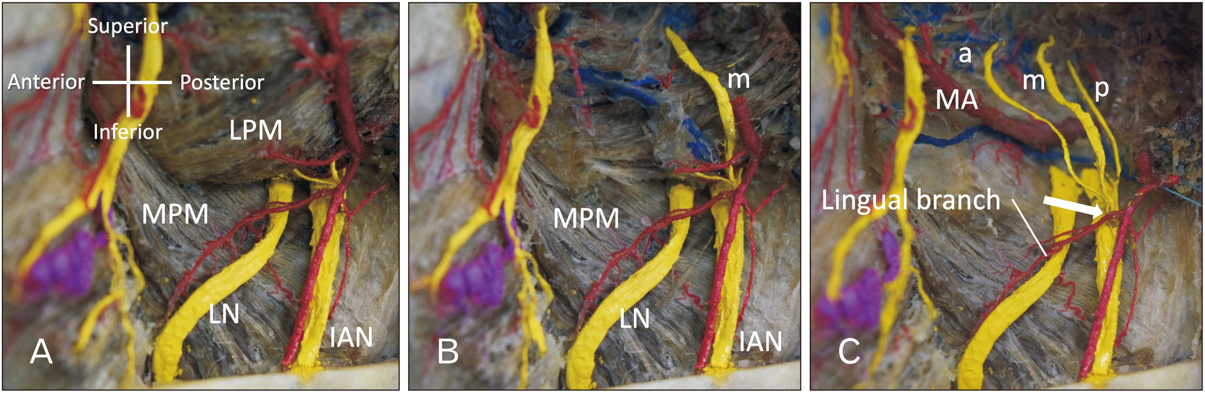
Fig. 1 Lateral view of the three variant branches arising from mandibular nerve in the left infratemporal fossa. The upper half of the mandibular ramus has been removed. (A) The three minor branches are not shown. (B) The middle branch (m) running within the lateral pterygoid muscle is shown. (C) The a, m, and p branches are shown after removal of the lateral pterygoid muscle. Note the three branches forming a common trunk to join the IAN at the root of the lingual branch of the inferior alveolar artery (arrow). a, anterior; IAN, inferior alveolar nerve; LPM, lateral pterygoid muscle; m, middle; MA, maxillary artery; MPM medial pterygoid muscle; p, posterior.
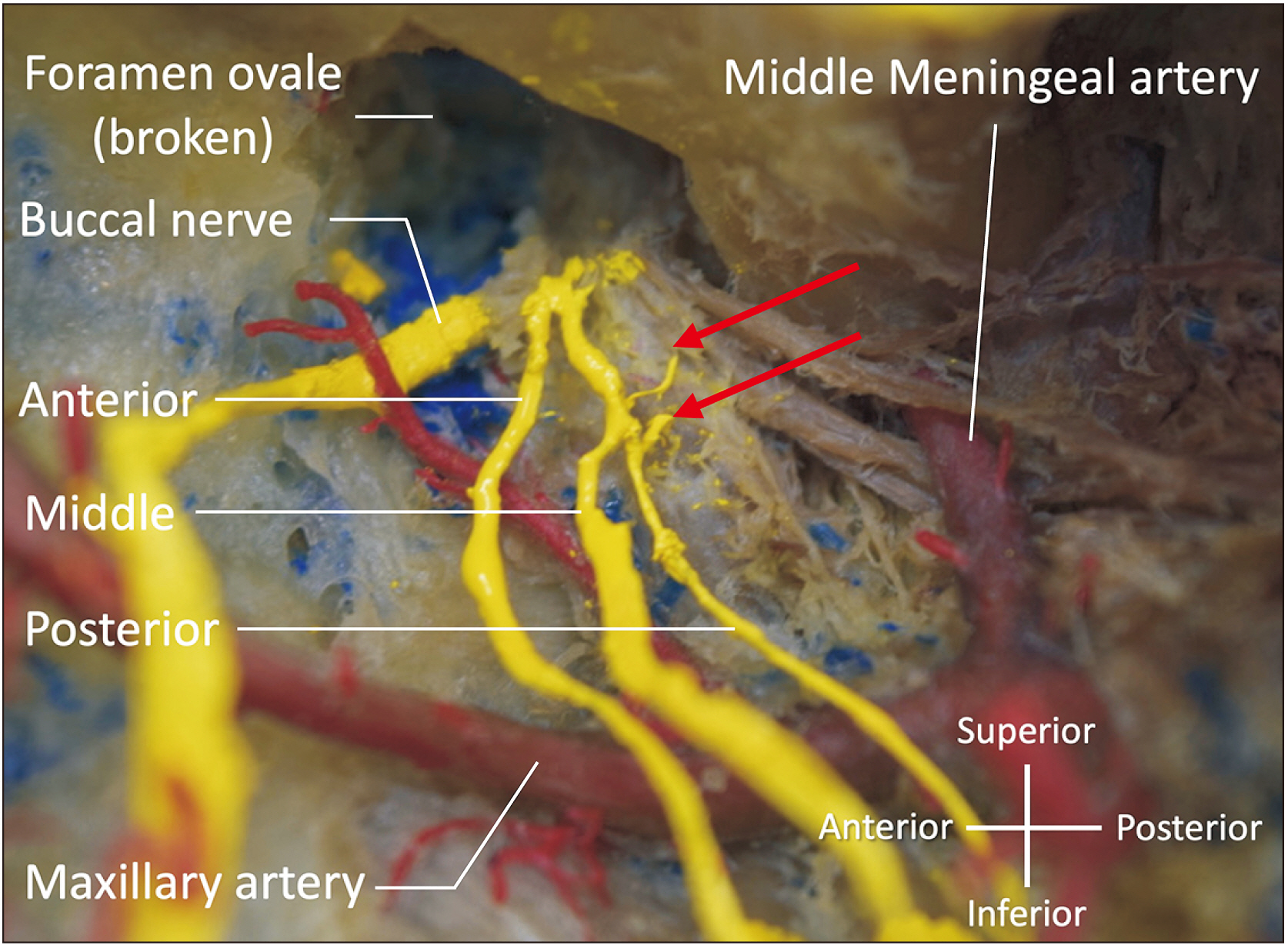
Fig. 2 A magnified image adjacent to the foramen ovale. Note the two small branches from the ganglion-like structure (arrows).
Reference
-
References
1. Kim SY, Hu KS, Chung IH, Lee EW, Kim HJ. 2004; Topographic anatomy of the lingual nerve and variations in communication pattern of the mandibular nerve branches. Surg Radiol Anat. 26:128–35. DOI: 10.1007/s00276-003-0179-x. PMID: 14586562.
Article2. Iamsaard S, Singsorn J, Boonruangsri P. 2015; An unusual communication between the trunk of the mandibular nerve and the lingual nerve in a female cadaver. Acta Med Acad. 44:201–2. DOI: 10.5644/ama2006-124.148. PMID: 26702916.
Article3. Muraleedharan A, Veeramani R, Chand P. 2014; Variations in the branching pattern of posterior division of mandibular nerve: a case report. Surg Radiol Anat. 36:947–50. DOI: 10.1007/s00276-014-1329-z. PMID: 24972574.
Article4. Carter RB, Keen EN. 1971; The intramandibular course of the inferior alveolar nerve. J Anat. 108(Pt 3):433–40. PMID: 5575310. PMCID: PMC1234179.5. Shimotakahara R, Lee H, Mine K, Ogata S, Tamatsu Y. 2019; Anatomy of the lingual nerve: application to oral surgery. Clin Anat. 32:635–41. DOI: 10.1002/ca.23361. PMID: 30815909.
Article6. Khoury J, Mihailidis S, Ghabriel M, Townsend G. 2010; Anatomical relationships within the human pterygomandibular space: relevance to local anesthesia. Clin Anat. 23:936–44. DOI: 10.1002/ca.21047. PMID: 20949494.
Article7. Loughner BA, Larkin LH, Mahan PE. 1990; Nerve entrapment in the lateral pterygoid muscle. Oral Surg Oral Med Oral Pathol. 69:299–306. DOI: 10.1016/0030-4220(90)90290-9. PMID: 2314856.
Article8. Anil A, Peker T, Turgut HB, Gülekon IN, Liman F. 2003; Variations in the anatomy of the inferior alveolar nerve. Br J Oral Maxillofac Surg. 41:236–9. DOI: 10.1016/S0266-4356(03)00113-X. PMID: 12946665.9. Wolf KT, Brokaw EJ, Bell A, Joy A. 2016; Variant inferior alveolar nerves and implications for local anesthesia. Anesth Prog. 63:84–90. DOI: 10.2344/0003-3006-63.2.84. PMID: 27269666. PMCID: PMC4896047.
Article10. Khan MM, Darwish HH, Zaher WA. 2010; Perforation of the inferior alveolar nerve by the maxillary artery: an anatomical study. Br J Oral Maxillofac Surg. 48:645–7. DOI: 10.1016/j.bjoms.2009.11.002. PMID: 20018415.
Article11. Harn SD, Durham TM. 2003; Anatomical variations and clinical implications of the artery to the lingual nerve. Clin Anat. 16:294–9. DOI: 10.1002/ca.10110. PMID: 12794911.
Article12. Gow-Gates GA. 1973; Mandibular conduction anesthesia: a new technique using extraoral landmarks. Oral Surg Oral Med Oral Pathol. 36:321–8. DOI: 10.1016/0030-4220(73)90208-9. PMID: 4516460.
Article13. Haas DA. 2011; Alternative mandibular nerve block techniques: a review of the Gow-Gates and Akinosi-Vazirani closed-mouth mandibular nerve block techniques. J Am Dent Assoc. 142 Suppl 3:8S–12S. DOI: 10.14219/jada.archive.2011.0341. PMID: 21881056.14. Iwanaga J, Saga T, Tabira Y, Nakamura M, Kitashima S, Watanabe K, Kusukawa J, Yamaki K. 2015; The clinical anatomy of accessory mental nerves and foramina. Clin Anat. 28:848–56. DOI: 10.1002/ca.22597. PMID: 26201838.
Article
- Full Text Links
-
- Actions
-
Cited
- CITED
-
- Close
- Share
-
- Similar articles
-
- Updates on the Inferior Alveolar Nerve Block Anesthesia
- Mylohyoid foramen of mandible: a rare exit point of intra-mandibular origin of nerve to mylohyoid
- A case of inferior alveolar nerve encircling the arteria maxillaris
- Facial blanching after inferior alveolar nerve block anesthesia: an unusual complication
- Diplopia after Inferior Alveolar Nerve Block Anesthesia: A Case Report
