Acromial morphology and morphometry associated with subacromial impingement syndrome
- Affiliations
-
- 1Department of anatomy Faculty of Medicine, Chiang Mai University, Chiang Mai, Thailand
- 2Forensic Osteology Research Center, Faculty of Medicine, Chiang Mai University, Chiang Mai, Thailand
- 3Excellence Center in Osteology Research and Training Center (ORTC), Chiang Mai University, Chiang Mai, Thailand
- KMID: 2509689
- DOI: http://doi.org/10.5115/acb.20.166
Abstract
- Acromion is a major associated structure of subacromial impingement syndrome, one of the most common diagnoses in chronic shoulder pain world-wide. The aims of this study are to study morphometry of acromion and to find risk group using acromial morphometry. Total samples were 392 scapulae. The samples were of both sexes, ranging from the age of 31 to 90. Acromion type and osteophytes were observed. Acromial parameters were measured. The relationships were analyzed among acromion type, acromial osteophyte, acromial parameters, age group, sex and side. Curved acromion had the highest prevalence in all age groups. Hooked acromion had the second highest prevalence since the age of 41. Hooked acromion prevalence was higher in male than in female. The highest prevalence of acromial osteophytes was on anteroinferior surface in all age group. The prevalence of acromial osteophytes on anteroinferior surface and acromial facet increased with age. In addition, acromion type was associated with only osteophytes on anteroinferior surface of acromion. Anterior one-third acromial thickness in the age of 31 to 50 was different from those of 51 to 90. There are differences between all parameters and sexes, but not side. General population with age above 50 and concerned male group with age above 40 who have chronic shoulder pain should be investigated for subacromial impingement syndrome. Surgical treatment is recommended because hooked acromion and osteophytes are mostly the root of problem.
Keyword
Figure
-
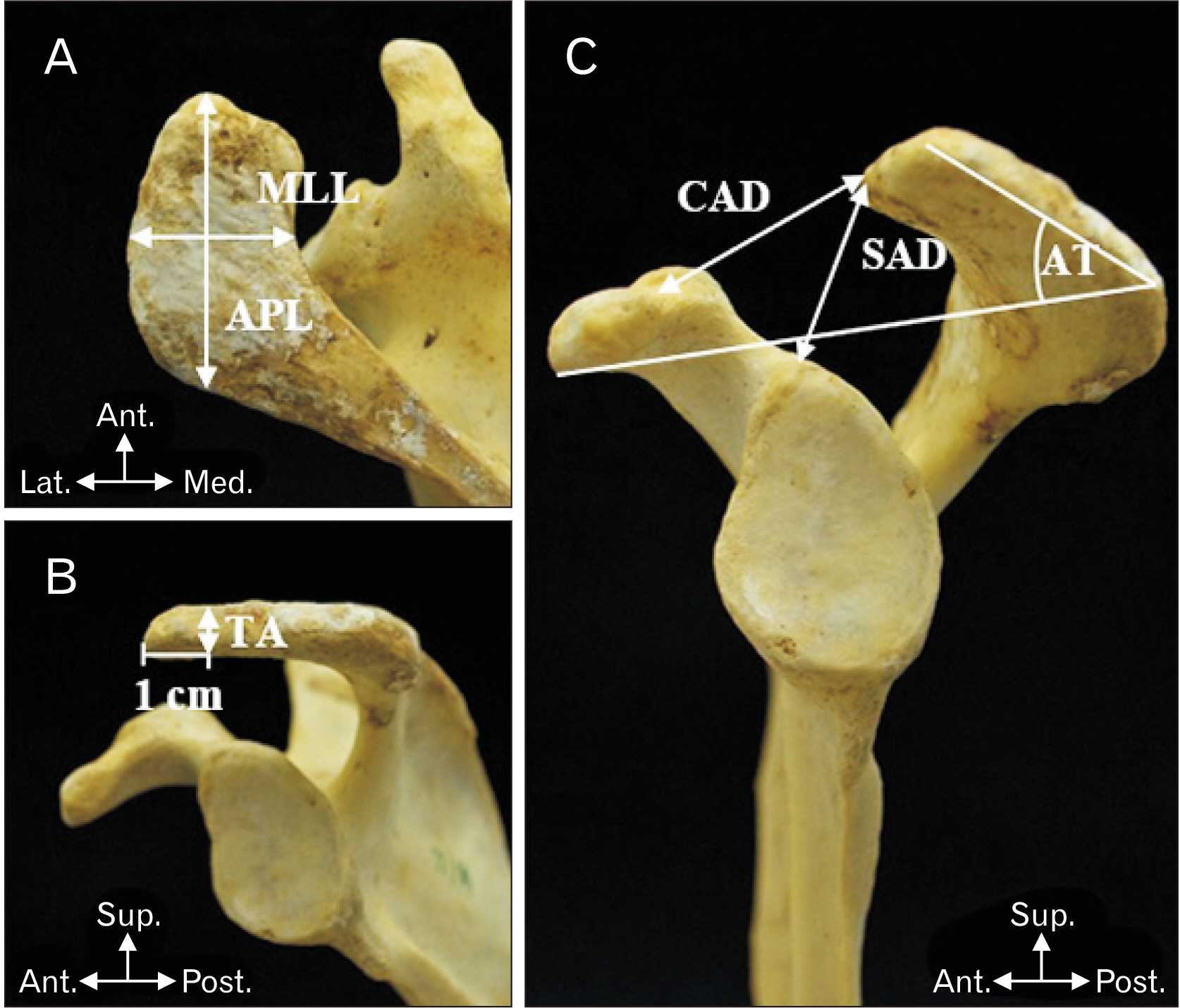
Fig. 1 (A) showed APL and MLL. (B) showed TA. (C) showed CAD, SAD, and AT. Ant., anterior; APL, maximum acromial length; AT, acromial tilt.; CAD, coracoacromial distance; Lat., lateral; Med., medial; MLL, maximum acromial width; Post., posterior; SAD, supraglenoacromial distanc; Sup., superior; TA, anterior one-third thickness.
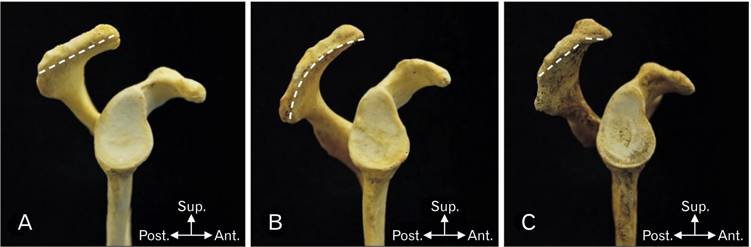
Fig. 2 Classification of acromion type; (A) Type I or Flat type, (B) Type II or curved type, (C) Type III or hooked type. Ant., anterior; Lat., lateral; Med., medial; Post., posterior; Sup., superior.
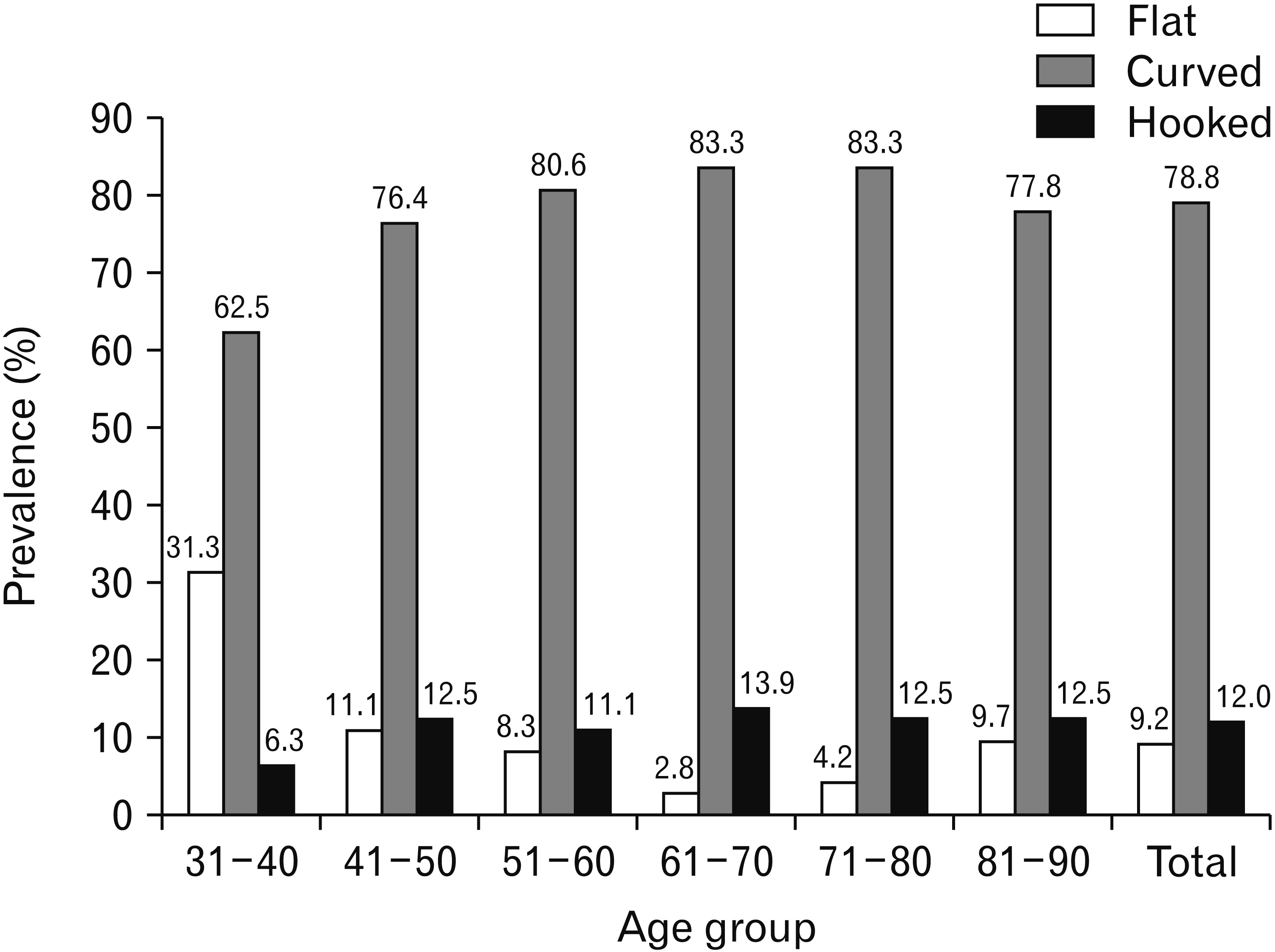
Fig. 3 Prevalence of acromion type in each age group.

Fig. 4 Osteophyte location; (A) Anteroinferior surface, (B) Acromial facet, (C) Lateral surface. Ac, acromion; Ant., anterior; Co, coracoid; F, acromial facet; G, glenoid; Lat., lateral; Med., medial; Post., posterior; Sup., superior.

Fig. 5 Prevalence of acromial osteophyte in each age group.
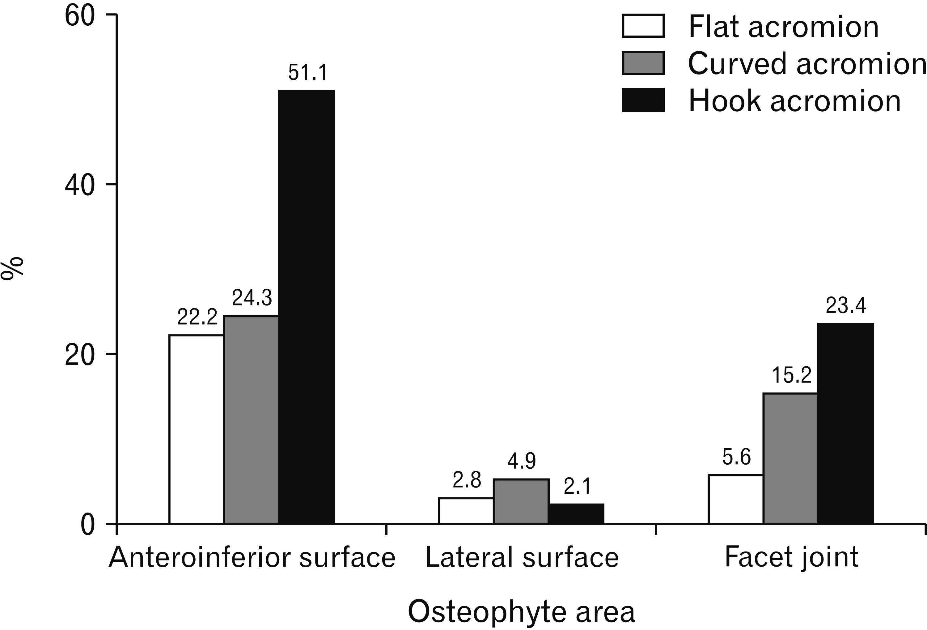
Fig. 6 Percentage of acromion type having osteophyte in each area.
Reference
-
References
1. Garving C, Jakob S, Bauer I, Nadjar R, Brunner UH. 2017; Impingement syndrome of the shoulder. Dtsch Arztebl Int. 114:765–76. DOI: 10.3238/arztebl.2017.0765. PMID: 29202926. PMCID: PMC5729225.
Article2. De Yang Tien J, Tan AHC. 2014; Shoulder impingement syndrome, a common affliction of the shoulder: a comprehensive review. Proc Singapore Healthc. 23:297–305. DOI: 10.1177/201010581402300406.
Article3. Juel NG, Natvig B. 2014; Shoulder diagnoses in secondary care, a one year cohort. BMC Musculoskelet Disord. 15:89. DOI: 10.1186/1471-2474-15-89. PMID: 24642168. PMCID: PMC3995190.
Article4. van der Windt DA, Koes BW, de Jong BA, Bouter LM. 1995; Shoulder disorders in general practice: incidence, patient characteristics, and management. Ann Rheum Dis. 54:959–64. DOI: 10.1136/ard.54.12.959. PMID: 8546527. PMCID: PMC1010060.
Article5. Blom AW, Warwick D, Whitehouse MR. 2018. Apley and Solomon's system of orthopaedics and trauma. 10th ed. Taylor & Francis;Boca Raton:6. Lazaro R. 2005; Shoulder impingement syndromes: implications on physical therapy examination and intervention. J Jpn Phys Ther Assoc. 8:1–7. DOI: 10.1298/jjpta.8.1. PMID: 25792938. PMCID: PMC4316502.
Article7. Dhillon KS. 2019; Subacromial impingement syndrome of the shoulder: a musculoskeletal disorder or a medical myth? Malays Orthop J. 13:1–7. DOI: 10.5704/MOJ.1911.001. PMID: 31890103. PMCID: PMC6915323.
Article8. Neer CS 2nd. 1972; Anterior acromioplasty for the chronic impingement syndrome in the shoulder: a preliminary report. J Bone Joint Surg Am. 54:41–50. DOI: 10.2106/00004623-197254010-00003. PMID: 5054450.9. Edwards SL, Bell JE, Bigliani LU. Wilk KE, Reinold MM, Andrews JR, editors. 2009. Subacromial impingement. The Athlete's Shoulder. 2nd ed. Churchill Livingstone;Philadelphia: p. 115–22. DOI: 10.1016/B978-044306701-3.50013-X.
Article10. Morrison DS, Frogameni AD, Woodworth P. 1997; Non-operative treatment of subacromial impingement syndrome. J Bone Joint Surg Am. 79:732–7. DOI: 10.2106/00004623-199705000-00013. PMID: 9160946.
Article11. Nyffeler RW, Meyer DC. 2017; Acromion and glenoid shape: why are they important predictive factors for the future of our shoulders? EFORT Open Rev. 2:141–50. DOI: 10.1302/2058-5241.2.160076. PMID: 28630752. PMCID: PMC5467673.
Article12. Natsis K, Tsikaras P, Totlis T, Gigis I, Skandalakis P, Appell HJ, Koebke J. 2007; Correlation between the four types of acromion and the existence of enthesophytes: a study on 423 dried scapulas and review of the literature. Clin Anat. 20:267–72. DOI: 10.1002/ca.20320. PMID: 16683236.
Article13. Paraskevas G, Tzaveas A, Papaziogas B, Kitsoulis P, Natsis K, Spanidou S. 2008; Morphological parameters of the acromion. Folia Morphol (Warsz). 67:255–60. PMID: 19085865.14. Mansur DI, Khanal K, Haque MK, Sharma K. 2012; Morphometry of acromion process of human scapulae and its clinical importance amongst Nepalese population. Kathmandu Univ Med J (KUMJ). 10:33–6. DOI: 10.3126/kumj.v10i2.7340. PMID: 23132472.
Article15. Guo X, Ou M, Yi G, Qin B, Wang G, Fu S, Zhang L. 2018; Correction between the morphology of acromion and acromial angle in Chinese population: a study on 292 scapulas. Biomed Res Int. 2018:3125715. DOI: 10.1155/2018/3125715. PMID: 30534558. PMCID: PMC6252189.
Article16. Alraddadi A, Alashkham A, Lamb C, Soames R. 2019; Examining changes in acromial morphology in relation to spurs at the anterior edge of acromion. Surg Radiol Anat. 41:409–14. DOI: 10.1007/s00276-018-2141-y. PMID: 30483867.
Article17. Mahakkanukrauh P, Surin P. 2003; Prevalence of osteophytes associated with the acromion and acromioclavicular joint. Clin Anat. 16:506–10. DOI: 10.1002/ca.10182. PMID: 14566897.
Article18. Umer M, Qadir I, Azam M. 2012; Subacromial impingement syndrome. Orthop Rev (Pavia). 4:e18. DOI: 10.4081/or.2012.e18. PMID: 22802986. PMCID: PMC3395987.
Article19. Nicholson GP, Goodman DA, Flatow EL, Bigliani LU. 1996; The acromion: morphologic condition and age-related changes. A study of 420 scapulas. J Shoulder Elbow Surg. 5:1–11. DOI: 10.1016/S1058-2746(96)80024-3. PMID: 8919436.
Article20. Sangiampong A, Chompoopong S, Sangvichien S, Thongtong P, Wongjittraporn S. 2007; The acromial morphology of Thais in relation to gender and age: study in scapular dried bone. J Med Assoc Thai. 90:502–7. PMID: 17427527.21. Kadavkolan AS, Lehmann LJ, Reichert M, Lattka K, Moursy M. 2017; Does acromion morphology depend on the extremity or on gender in the population? J Comput Assist Tomogr. 41:121–4. DOI: 10.1097/RCT.0000000000000474. PMID: 27680413.
Article22. Pesquer L, Borghol S, Meyer P, Ropars M, Dallaudière B, Abadie P. 2018; Multimodality imaging of subacromial impingement syndrome. Skeletal Radiol. 47:923–37. DOI: 10.1007/s00256-018-2875-y. PMID: 29445933.
Article23. Li X, Xu W, Hu N, Liang X, Huang W, Jiang D, Chen H. 2017; Relationship between acromial morphological variation and subacromial impingement: a three-dimensional analysis. PLoS One. 12:e0176193. DOI: 10.1371/journal.pone.0176193. PMID: 28441418. PMCID: PMC5404845.
Article24. Roidis NT, Motamed S, Vaishnav S, Ebramzadeh E, Karachalios TS, Itamura JM. 2009; The influence of the acromioclavicular joint degeneration on supraspinatus outlet impingement and the acromion shape. J Orthop Surg (Hong Kong). 17:331–4. DOI: 10.1177/230949900901700318. PMID: 20065375.
Article25. Klasan A, Malcherczyk D, Neri T, Saul J, Schofer MD, Heyse TJ, El-Zayat BF. 2020; Reporting on acromion morphology: classifications demonstrate significant correlation. J ISAKOS. 5:83–7. DOI: 10.1136/jisakos-2019-000403.
Article26. Maalouly J, Tawk A, Aouad D, Abdallah A, Darwiche M, Abboud G, El Rassi G. 2020; Association of acromial morphological parameters and rotator cuff tears, and evaluation of the influence of age and gender on the parameters and impact on cuff tears: a study on a Middle Eastern population. Asia Pac J Sports Med Arthrosc Rehabil Technol. 20:17–23. DOI: 10.1016/j.asmart.2020.02.002. PMID: 32161714. PMCID: PMC7058845.
Article27. El-Din WA, Ali MH. 2015; A morphometric study of the patterns and variations of the acromion and glenoid cavity of the scapulae in Egyptian population. J Clin Diagn Res. 9:AC08–11. DOI: 10.7860/JCDR/2015/14362.6386. PMID: 26435934. PMCID: PMC4576525.
Article28. Bigliani LU, Morrison DS, April EW. 1986; The morphology of the acromion and its relationship to rotator cuff tears. Orthop Trans. 10:228.29. Banas MP, Miller RJ, Totterman S. 1995; Relationship between the lateral acromion angle and rotator cuff disease. J Shoulder Elbow Surg. 4:454–61. DOI: 10.1016/S1058-2746(05)80038-2. PMID: 8665291.
Article30. Tangtrakulwanich B, Kapkird A. 2012; Analyses of possible risk factors for subacromial impingement syndrome. World J Orthop. 3:5–9. DOI: 10.5312/wjo.v3.i1.5. PMID: 22470844. PMCID: PMC3302047.
Article31. Saha S, Vasudeva N. 2017; Morphometric evaluation of adult acromion process in North Indian population. J Clin Diagn Res. 11:AC08–11. DOI: 10.7860/JCDR/2017/21060.9312. PMID: 28273959. PMCID: PMC5324404.32. Cabezas AF, Krebes K, Hussey MM, Santoni BG, Kim HS, Frankle MA, Oh JH. 2016; Morphologic variability of the shoulder between the populations of North American and East Asian. Clin Orthop Surg. 8:280–7. DOI: 10.4055/cios.2016.8.3.280. PMID: 27583111. PMCID: PMC4987312.
Article33. Adamopoulos IE. 2018; Inflammation in bone physiology and pathology. Curr Opin Rheumatol. 30:59–64. DOI: 10.1097/BOR.0000000000000449. PMID: 29016371. PMCID: PMC5963529.
Article34. Wang JC, Hatch JD, Shapiro MS. 2000; Comparison of MRI and radiographs in the evaluation of acromial morphology. Orthopedics. 23:1269–71. PMID: 11144495.
Article35. Aragão JA, Silva LP, Reis FP, Dos Santos Menezes CS. 2014; Analysis on the acromial curvature and its relationships with the subacromial space and types of acromion. Rev Bras Ortop. 49:636–41. DOI: 10.1016/j.rbo.2013.10.018. PMID: 26229874. PMCID: PMC4487451.
Article36. Haizlip KM, Harrison BC, Leinwand LA. 2015; Sex-based differences in skeletal muscle kinetics and fiber-type composition. Physiology (Bethesda). 30:30–9. DOI: 10.1152/physiol.00024.2014. PMID: 25559153. PMCID: PMC4285578.
Article37. Tekavec E, Jöud A, Rittner R, Mikoczy Z, Nordander C, Petersson IF, Englund M. 2012; Population-based consultation patterns in patients with shoulder pain diagnoses. BMC Musculoskelet Disord. 13:238. DOI: 10.1186/1471-2474-13-238. PMID: 23190941. PMCID: PMC3552995.
Article
- Full Text Links
-
- Actions
-
Cited
- CITED
-
- Close
- Share
-
- Similar articles
-
- Study for Acromial Type, Acromial Tilt and Subacromial Distances in Subacromial Impingement Syndrome
- A Study of Acromial Shape, Acromial Angle, Subacromial Distance in Normal Shoulder
- Acromial Downslping and Subacromial Interval in Shoulder Impingement Syndrome
- Simple Radiographic Finding of Subacromial Impingement Syndrome
- Comparative Analysis of Acromial Morphology in Normal and Impingement Syndrome
