J Korean Foot Ankle Soc.
2020 Dec;24(4):161-164. 10.14193/jkfas.2020.24.4.161.
Operative Treatment for Bilateral Chronic Recurrent Dislocation of the Peroneal Tendon: A Case Report
- Affiliations
-
- 1Department of Orthopedic Surgery, Bundang Jesaeng Hospital, Seongnam, Korea
- KMID: 2509538
- DOI: http://doi.org/10.14193/jkfas.2020.24.4.161
Abstract
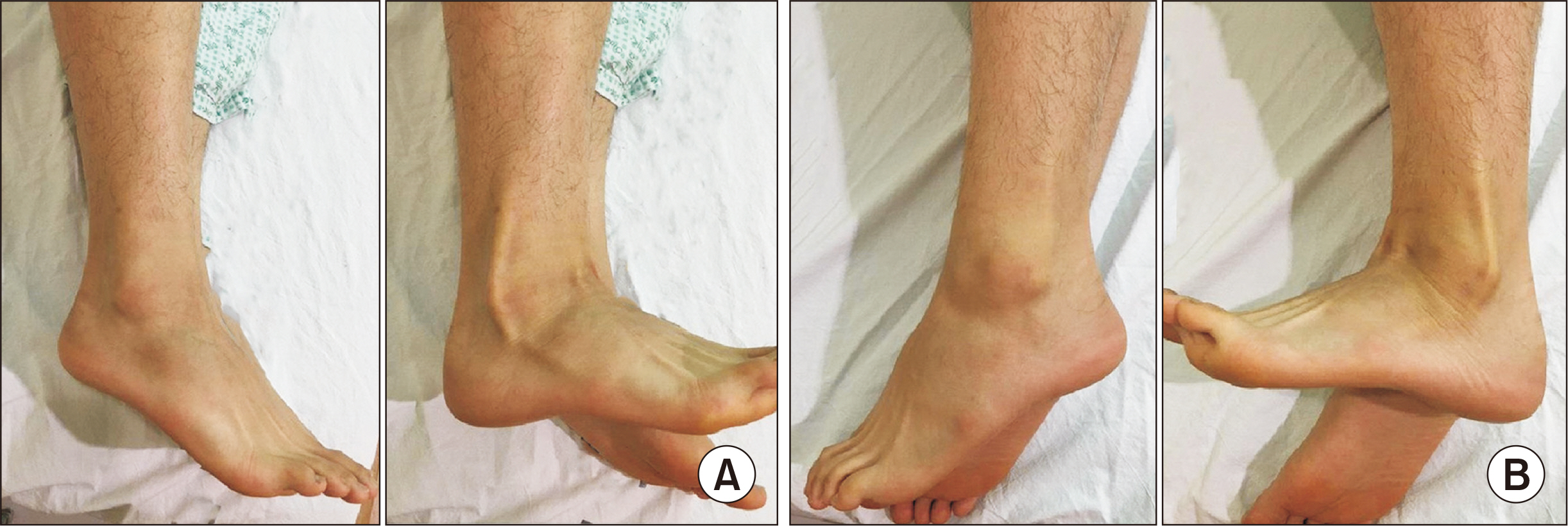
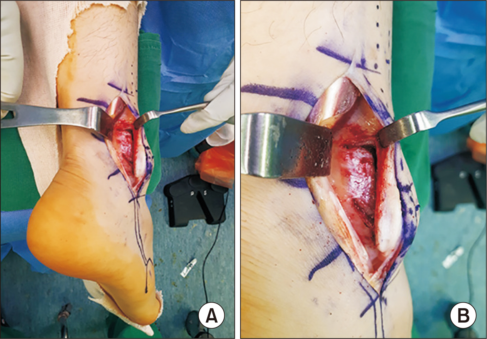
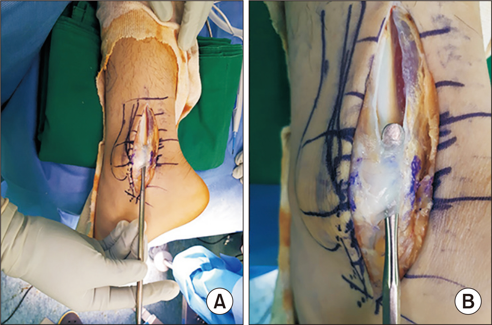
- A peroneal dislocation is a rare disease that is often misdiagnosed as a simple sprain and can be treated inadequately in the acute phase. For this reason, it is important to have an appropriate diagnosis in the early stages because it can progress to chronic and recurrent conditions. Surgical treatment is considered mainly when progressing to chronic recurrent dislocation. Recently, patients with an acute peroneal dislocation tend to prefer surgical treatment, so accurate initial diagnosis and management are very important. This paper reports a case of chronic recurrent peroneal tendon dislocation in both ankle joints, which was treated by a superior peroneal retinaculum reconstruction and a groove deepening procedure.
Keyword
Figure
Reference
-
1. Eckert WR, Davis EA Jr. 1976; Acute rupture of the peroneal retinaculum. J Bone Joint Surg Am. 58:670–2. DOI: 10.2106/00004623-197658050-00016.
Article2. Slätis P, Santavirta S, Sandelin J. 1988; Surgical treatment of chronic dislocation of the peroneal tendons. Br J Sports Med. 22:16–8. doi: 10.1136/bjsm.22.1.16.DOI: 10.1136/bjsm.22.1.16. PMID: 3370396. PMCID: PMC1478500.3. Kitaoka HB, Alexander IJ, Adelaar RS, Nunley JA, Myerson MS, Sanders M. 1994; Clinical rating systems for the ankle-hindfoot, midfoot, hallux, and lesser toes. Foot Ankle Int. 15:349–53. doi: 10.1177/107110079401500701.DOI: 10.1177/107110079401500701. PMID: 7951968.
Article4. Mendicino RW, Orsini RC, Whitman SE, Catanzariti AR. 2001; Fibular groove deepening for recurrent peroneal subluxation. J Foot Ankle Surg. 40:252–63. doi: 10.1016/s1067-2516(01)80026-0.DOI: 10.1016/S1067-2516(01)80026-0.
Article5. Ogawa BK, Thordarson DB. 2007; Current concepts review: peroneal tendon subluxation and dislocation. Foot Ankle Int. 28:1034–40. doi: 10.3113/FAI.2007.1034.DOI: 10.3113/FAI.2007.1034. PMID: 17880883.6. Monteggia GB. 1803. [Istituzioni chirurgiche]. Giuseppe Maspero;Milan: Itanlian.7. Edwards ME. 1928; The relations of the peroneal tendons to the fibula, calcaneus, and cuboideum. Am J Anat. 42:213–53. doi: 10.1002/aja.1000420109.DOI: 10.1002/aja.1000420109.
Article8. Marti R. 1977; Dislocation of the peroneal tendons. Am J Sports Med. 5:19–22. doi: 10.1177/036354657700500104.DOI: 10.1177/036354657700500104. PMID: 403818.
Article9. Pozo JL, Jackson AM. 1984; A rerouting operation for dislocation of peroneal tendons: operative technique and case report. Foot Ankle. 5:42–4. doi: 10.1177/107110078400500106.DOI: 10.1177/107110078400500106. PMID: 6479763.
Article10. Lee EW, Kim YS, Jeon JM. 1985; Dislocation of peroneal tendons two cases report. J Korean Orthop Assoc. 20:527–30. doi: 10.4055/jkoa.1985.20.3.527.DOI: 10.4055/jkoa.1985.20.3.527.
Article
- Full Text Links
- Actions
-
Cited
- CITED
-
- Close
- Share
- Similar articles
-
- Operative Treatment of Chronic Recurrent Dislocation of Peroneal Tendon: A Case Report
- Bilateral Recurrent Dislocation of the Peroneal Tendon: A Case Report
- Distal Fibular Rotational Plasty for Chronic Peroneal Tendon Recurrent Dislocation: A Technical Report
- Operative Treatment of Acute Peroneal Tendon Subluxation in Athletes: A Case Report - 2 Cases
- Dislocation of Peroneal Tendons Two Cases Report


