J Pathol Transl Med.
2020 Nov;54(6):508-512. 10.4132/jptm.2020.05.21.
Intraoperative frozen cytology of intraosseous cystic meningioma in the sphenoid bone
- Affiliations
-
- 1Department of Pathology, Gil Medical Center, Gachon University College of Medicine, Incheon, Korea
- 2Department of Neurosurgery, Gil Medical Center, Gachon University College of Medicine, Incheon, Korea
- KMID: 2509505
- DOI: http://doi.org/10.4132/jptm.2020.05.21
Abstract
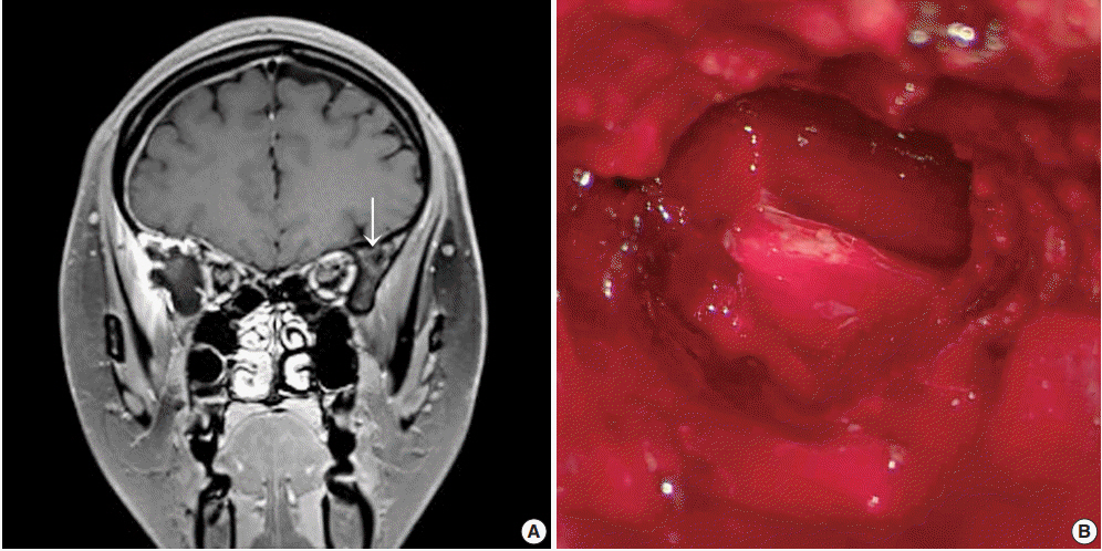
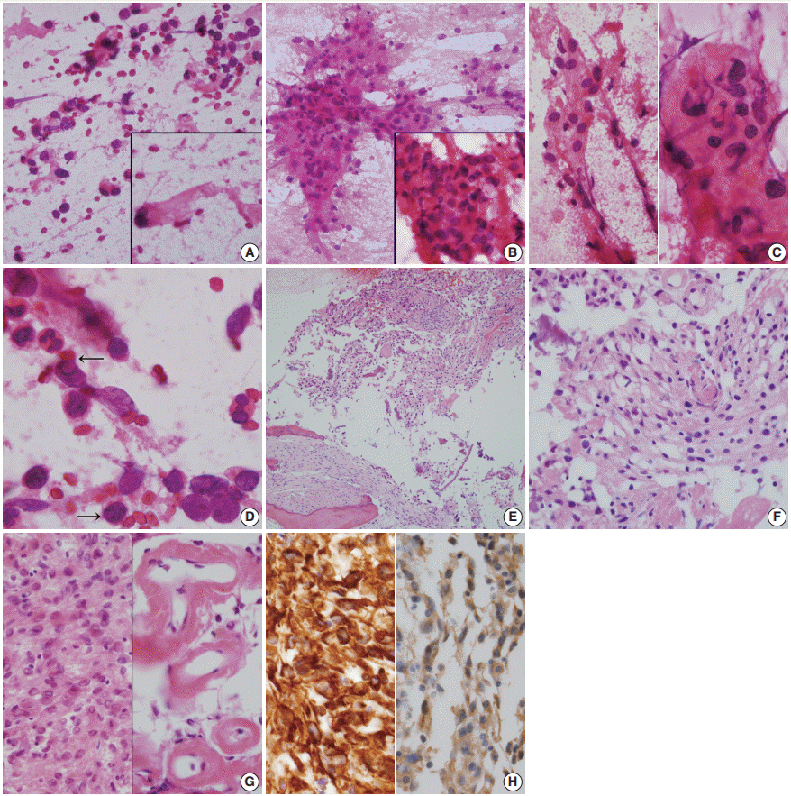
- Meningiomas in bone are rarely subjected to fine-needle aspiration diagnosis, and those arising in the skull bone with a cystic presentation are rare. A 24-year-old woman presented with subdural hemorrhage, and subsequent radiology depicted an osteolytic mass-like lesion in the sphenoid bone. Intraoperatively, a solid and cystic hemorrhagic lesion mimicking an aneurysmal bone cyst was observed in the sphenoid bone with dural tearing. Frozen cytology showed singly scattered or epithelioid clusters of round to elongated cells intermixed with many neutrophils. Tumor cells had bland-looking round nuclei with rare prominent nucleoli and nuclear inclusions and eosinophilic granular to globoid cytoplasm in capillary-rich fragments. Histology revealed intraosseous meningothelial and microcystic meningioma (World Health Organization grade 1) in right lesser wing of the sphenoid bone. Considering its unusual location and cytologic findings, differential diagnoses included chordoma, chondroma, chondrosarcoma, and aneurysmal bone cyst. The present case posed a diagnostic challenge due to possible confusion with these entities.
Keyword
Figure
Reference
-
References
1. Lang FF, Macdonald OK, Fuller GN, DeMonte F. Primary extradural meningiomas: a report on nine cases and review of the literature from the era of computerized tomography scanning. J Neurosurg. 2000; 93:940–50.2. Agrawal V, Ludwig N, Agrawal A, Bulsara KR. Intraosseous intracranial meningioma. AJNR Am J Neuroradiol. 2007; 28:314–5.3. Rosahl SK, Mirzayan MJ, Samii M. Osteolytic intra-osseous meningiomas: illustrated review. Acta Neurochir (Wien). 2004; 146:1245–9.
Article4. Butscheidt S, Ernst M, Rolvien T, et al. Primary intraosseous meningioma: clinical, histological, and differential diagnostic aspects. J Neurosurg. 2019 Jun 21 [Epub]. https://doi.org/10.3171/2019.3.JNS182968.
Article5. Caruso R, Fini G, Pesce A, et al. A primary intraosseous cystic meningioma: case report. Int J Surg Case Rep. 2017; 37:189–92.
Article6. Aghaghazvini L, Sedighi N, Karami P, Yeganeh O. Skull base aneurysmal bone cyst presented with foramen jugular syndrome and multi-osseous involvement. Iran J Radiol. 2012; 9:157–60.
Article7. Ilica AT, Mossa-Basha M, Zan E, et al. Cranial intraosseous meningioma: spectrum of neuroimaging findings with respect to histopathological grades in 65 patients. Clin Imaging. 2014; 38:599–604.
Article8. Turner OA, Laird AT. Meningioma with traumatic etiology: report of a case. J Neurosurg. 1966; 24:96–8.9. Tokgoz N, Oner YA, Kaymaz M, Ucar M, Yilmaz G, Tali TE. Primary intraosseous meningioma: CT and MRI appearance. AJNR Am J Neuroradiol. 2005; 26:2053–6.10. Kee TP, Liauw L, Sathiyamoorthy S, Lee HY, Tan GSL, Yu WY. Large solitary lytic skull vault lesions in adults: radiological review with pathological correlation. Clin Imaging. 2020; 59:129–43.
Article11. Ustabasioglu FE, Samanci C, Asik M, et al. Aneurysmal bone cyst of sphenoid bone and clivus misdiagnosed as chordoma: a case report. Brain Tumor Res Treat. 2015; 3:115–7.
Article12. Kang M, Chung DH, Kim NR, et al. Intraoperative frozen cytology of central nervous system neoplasms: an ancillary tool for frozen diagnosis. J Pathol Transl Med. 2019; 53:104–11.
Article13. Seok JY, Kim NR, Cho HY, Chung DH, Yee GT, Kim EY. Crush cytology of microcystic meningioma with extensive sclerosis. Korean J Pathol. 2014; 48:77–80.
Article14. Pinton L, Solito S, Masetto E, et al. Immunosuppressive activity of tumor-infiltrating myeloid cells in patients with meningioma. Oncoimmunology. 2018; 7:e1440931.
Article15. Lee S, Kim NR, Chung DH, Yee GT, Cho HY. Squash cytology of a dural-based high-grade chondrosarcoma may mimic that of glioblastoma in the central nervous system. Acta Cytol. 2015; 59:219–24.
Article16. Nijhawan VS, Rajwanshi A, Das A, Jayaram N, Gupta SK. Fineneedle aspiration cytology of sacrococcygeal chordoma. Diagn Cytopathol. 1989; 5:404–7.
Article17. Creager AJ, Madden CR, Bergman S, Geisinger KR. Aneurysmal bone cyst: fine-needle aspiration findings in 23 patients with clinical and radiologic correlation. Am J Clin Pathol. 2007; 128:740–5.18. Kang M, Kim NR, Chung DH, Yie GT. Frozen cytology of meningeal malignant solitary fibrous tumor/hemangiopericytoma. J Pathol Transl Med. 2019; 53:192–7.
Article19. Fritchie K, Jensch K, Moskalev EA, et al. The impact of histopathology and NAB2-STAT6 fusion subtype in classification and grading of meningeal solitary fibrous tumor/hemangiopericytoma. Acta Neuropathol. 2019; 137:307–19.
Article20. Ligon KL, Alberta JA, Kho AT, et al. The oligodendroglial lineage marker OLIG2 is universally expressed in diffuse gliomas. J Neuropathol Exp Neurol. 2004; 63:499–509.
Article
- Full Text Links
- Actions
-
Cited
- CITED
-
- Close
- Share
- Similar articles
-
- Primary Intraosseous Calvarial Meningioma: A Case Report
- Primary Intraosseous Meningioma in the Orbital Bony Wall: A Case Report and Review of the Literature Review
- Primary Intraosseous Meningioma
- Extracranial Extension of A Pterional Meningioma: Case Report
- Primary Benign Intraosseous Meningioma on 18F-FDG PET/CT Mimicking Malignancy


