J Cardiovasc Imaging.
2020 Apr;28(2):158-160. 10.4250/jcvi.2019.0095.
Non-Obstructive Accessory Mitral Valve Tissue in the Left Ventricular Outflow Tract with PerimembranousVentricular Septal Defect: A Rare Entity
- Affiliations
-
- 1Department of Preventive and Non-Invasive Cardiology, Jaipur Heart Institute, Jaipur, India
- 2Department of Cardiology, Jaipur Heart Institute, Jaipur, India
- KMID: 2509462
- DOI: http://doi.org/10.4250/jcvi.2019.0095
Figure
-
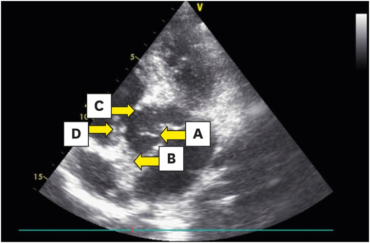
Figure 1 Apical five-chamber view; arrows point to filamentous accessory mitral valve tissue (A), aortic valve (B), perimembranous ventricular septal defect (C), and septal leaflet of the tricuspid valve (D).
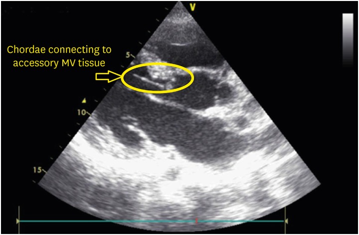
Figure 2 Parasternal long-axis view showing accessory mitral valve tissue attached to the chordae.
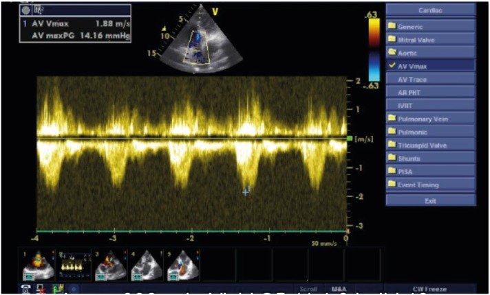
Figure 3 Apical five-chamber view with continuous width Doppler showing a maximum pressure gradient of 14.16 mmHg through the left ventricular outflow tract.
- Full Text Links
-
- Actions
-
Cited
- CITED
-
- Close
- Share
-
- Similar articles
-
- Left Ventricular Outflow Tract Obstruction Caused by Accessory Mitral Valve Tissue in a Child: A case report
- Relationship between Systolic Anterior Motion of the Mitral Valve and the Left Ventricular Outflow Pressure Gradient in Patients with Hypertrophic Obstructive Cardiomyopathy
- Two Cases of Double-Orifice Mitral Valve Detected by Echocardiography
- Echocardiographic features of accessory mitral valve tissue presenting left ventricular outflow tract obstruction in a dog
- A Case of Left Ventricular Outflow Obstruction Caused by Mitral Valve Replacement
