J Cardiovasc Imaging.
2020 Apr;28(2):156-157. 10.4250/jcvi.2019.0070.
A Bone Cement Mass in the Pulmonary Artery
- Affiliations
-
- 1Division of Cardiology, Department of Internal Medicine, Gyeongsang National University School of Medicine and Gyeongsang National University Hospital, Jinju, Korea
- KMID: 2509461
- DOI: http://doi.org/10.4250/jcvi.2019.0070
Figure
-
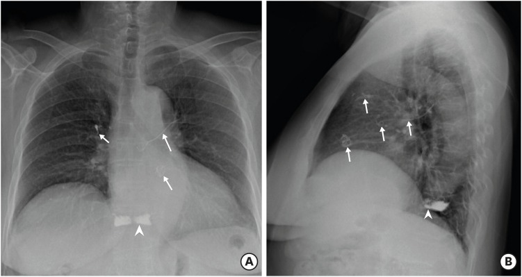
Figure 1 Simple X-ray images of chest PA (A) and chest left-lateral (B) show a round radio-opaque mass made of coiled lines in the pulmonary artery and linear materials in the left and right pulmonary artery (arrow). The vertebroplasty was seen in a simple X-ray (A and B, arrow head).
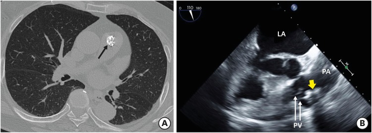
Figure 2 (A) Computed tomography shows a lobulated mass with high attenuation (arrow) in the pulmonary artery. (B) The mass of bone cement was attached at pulmonary valve by transesophageal echocardiogram. LA: left atrium, PA: pulmonary artery, PV: pulmonary valve.
Reference
-
1. Fadili Hassani S, Cormier E, Shotar E, et al. Intracardiac cement embolism during percutaneous vertebroplasty: incidence, risk factors and clinical management. Eur Radiol. 2019; 29:663–673. PMID: 30054794.2. Kim YJ, Lee JW, Park KW, et al. Pulmonary cement embolism after percutaneous vertebroplasty in osteoporotic vertebral compression fractures: incidence, characteristics, and risk factors. Radiology. 2009; 251:250–259. PMID: 19332856.
- Full Text Links
-
- Actions
-
Cited
- CITED
-
- Close
- Share
-
- Similar articles
-
- Pulmonary Bone Cement Embolism: CT Angiographic Evaluation with Material Decomposition Using Gemstone Spectral Imaging
- Transcatheter Removal of Bone Cement Embolism in the Right Atrium after Percutaneous Vertebroplasty: The Embolus Broke in Half and Migrated to the Right Pulmonary Artery Intraoperatively
- Asymptomatic Bone Cement Pulmonary Embolism after Percutaneous Vertebroplasty: A Case Report
- Pulmonary Bone Cement Embolism Following Percutaneous Vertebroplasty
- Changes in Cardiopulmonary Variables during Cemented Hip Arthroplasty in the Elderly Patients
