Heart Failure with Masked Atrial Contraction Detected on Echocardiography
- Affiliations
-
- 1Department of Cardiology, The Sakakibara Heart Institute of Okayama, Okayama, Japan
- 2Clinical Laboratory, The Sakakibara Heart Institute of Okayama, Okayama, Japan
- KMID: 2509460
- DOI: http://doi.org/10.4250/jcvi.2019.0098
Figure
-
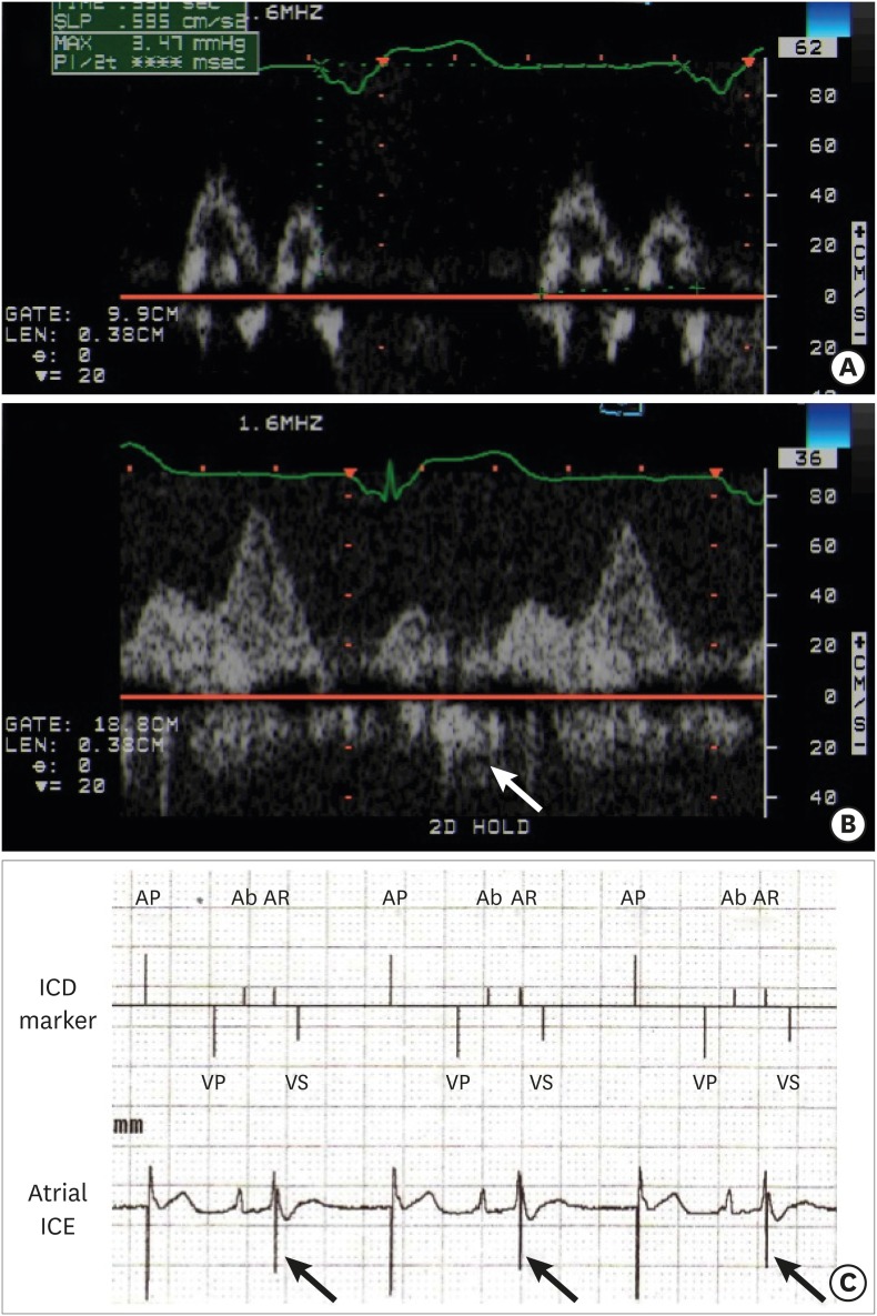
Figure 1 Echocardiography and intracardiac electrocardiography during atrioventricular delay of 350 ms. (A) The patient exhibited heart failure symptoms; however, transmitral flow was not a restrictive pattern. (B) By pulmonary venous flow, systolic reversal flow (arrow) due to ventriculoatrial conduction is noted. (C) Constant ventriculoatrial conduction (arrow) is shown using intracardiac electrocardiography. AR represents successfully masked ventriculoatrial conduction by prolonged postventricular atrial refractory period. Ab: atrial blanking period, AP: atrial pacing, AR: atrial refractory period, ICD: implantable cardioverter-defibrillator, ICE: intracardiac electrocardiogram, VP: ventricular pacing, VS: ventricular sensing.
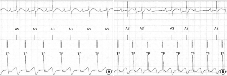
Figure 2 Electrophysiological study via ICD. (A) Ventricular pacing of 100 bpm demonstrated constant VA conduction. (B) Ventricular pacing of 120 bpm demonstrated refractory period of VA conduction. No decrement represented VA conduction via accessory pathway. AS: atrial sensing, ICD: implantable cardioverter-defibrillator, TP: tachypacing, VA: ventriculoatrial.
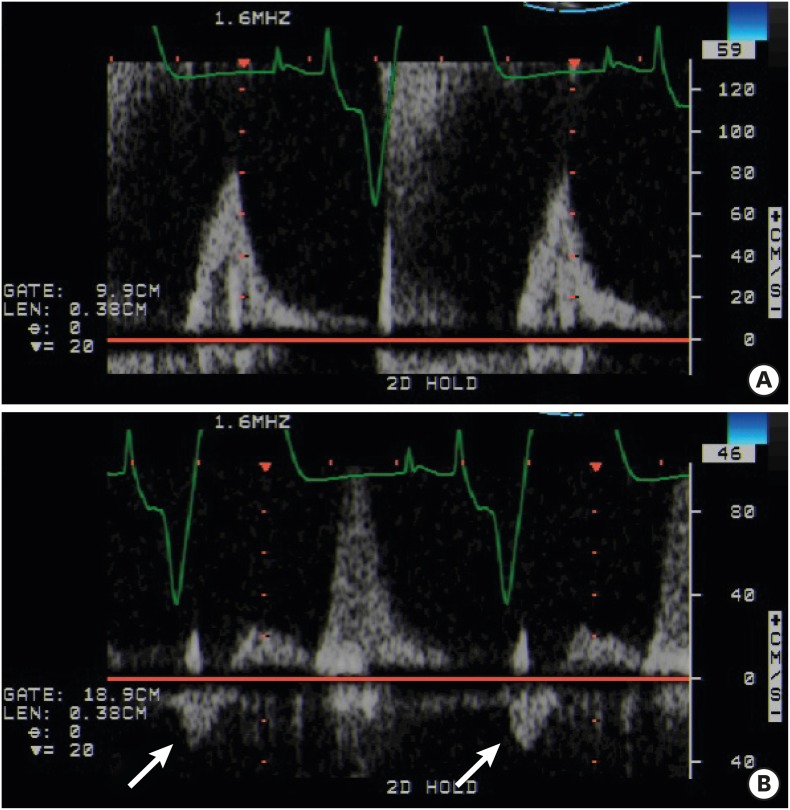
Figure 3 Echocardiography during atrioventricular delay of 150 ms. (A) Atrial contraction disappeared by transmitral flow. (B) By pulmonary venous flow, systolic reversal flow (arrow) due to delayed atrial conduction is noted.
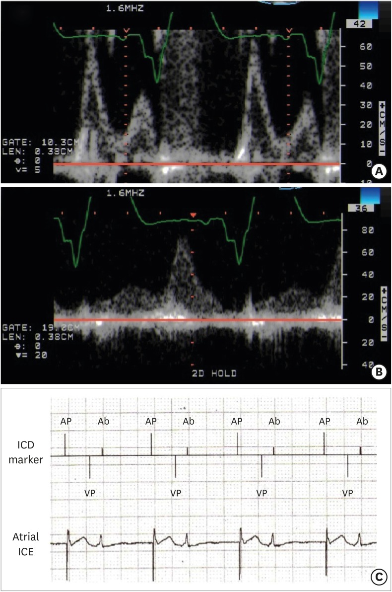
Figure 4 Echocardiography and intracardiac electrocardiography during atrioventricular delay of 280 ms. (A) Atrial contraction appeared by transmitral flow. (B) By pulmonary venous flow, systolic reversal flow disappeared. (C) No constant ventriculoatrial conduction is detected using intracardiac electrocardiography. Atrial sensing during atrial blanking is detected due to far-field potential of ventricular pacing. Ab: atrial blanking period, AP: atrial pacing, ICD: implantable cardioverter-defibrillator, ICE: intracardiac electrocardiogram, VP: ventricular pacing.
Reference
-
1. Tabata T, Grimm RA, Bauer FJ, et al. Giant flow reversal in pulmonary venous flow as a possible mechanism for asynchronous pacing-induced heart failure. J Am Soc Echocardiogr. 2005; 18:722–728. PMID: 16003269.
