J Cardiovasc Imaging.
2020 Apr;28(2):123-133. 10.4250/jcvi.2019.0109.
Clinical and Echocardiographic Characterization of False-Positive Results from Stress Echocardiography
- Guerreiro RA
 1
1 - Fazendas P2
- Pereira AR2
- Marques A2
- Pais J1
- Alegria S2
- Congo KH1
- Gomes AC2
- Carvalho J1
- Morgado G2
- Cruz I2
- Almeida AR2
- João I2
- Pereira H2
- Affiliations
-
- 1Cardiology Department, Hospital do Espírito Santo, Évora, Portugal
- 2Cardiology Department, Hospital Garcia de Orta, Almada, Portugal
- KMID: 2509454
- DOI: http://doi.org/10.4250/jcvi.2019.0109
Abstract
- BACKGROUND
Stress echocardiography has a 72%–85% sensitivity and an 80%–95% specificity. In this study, we characterized patients who received a false-positive stress echocardiogram result.
METHODS
A total of 5,256 patients underwent a stress echocardiogram (induced by exercise, dobutamine, or dipyridamole) between 2009 to 2018, and 405 patients (7.7%) received a positive result. Among the positive patients, 300 underwent coronary angiography within 12 months, and these patients were included in this study (mean age = 64.9 ± 9.4 years, 230 men [76.7%]). Coronary artery disease was diagnosed by stenosis ≥50% in any epicardial coronary artery. Clinical and echocardiographic variables were compared between patients with true- and false-positive stress echocardiogram results.
RESULTS
Seventy-two patients (24%) had a false-positive stress echocardiogram, with similar rates across stressor types (p = 0.574). Patients with false positives were less frequently men (63.9% vs. 80.7%, p = 0.003), had lower diabetes mellitus prevalence (15.3% vs. 45.6%, p = 0.001), were similar to true positive patients with regard to body-mass index, arterial hypertension prevalence, hyperlipidemia and smoking, and had lower pre-test probability of coronary artery disease (23% vs. 32%, p = 0.016). The wall motion score index (WMSI) was higher in the true-positive stress group, and wall motion abnormalities were more frequent in the apical segments (70.5% vs. 56.7%, p = 0.034). In a multivariable predictive model, men (odds ratio [OR] = 2.994), diabetes (OR = 5.440), and peak WMSI (OR = 10.690) were associated with a true-positive result.
CONCLUSIONS
Twenty-four percent of our study population received a false-positive stress echocardiogram result, with similar rates across stressor types. Patients with true-positive stress echocardiogram results are more likely to be men, diabetic, and have a high peak WMSI.
Keyword
Figure
-
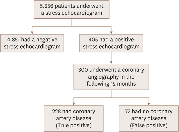
Figure 1 Study design.
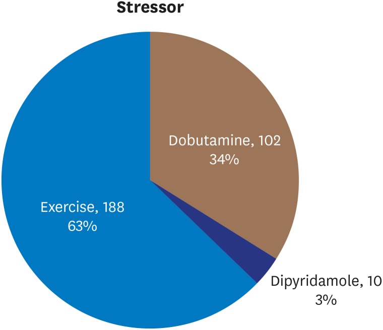
Figure 2 Distribution of stressors.
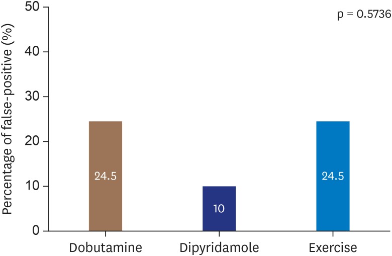
Figure 3 False-positive stress echocardiogram results by stressor.
Cited by 1 articles
-
The Role of Echocardiography in Evaluating Cardiovascular Diseases in Patients with Diabetes Mellitus
Sun Hwa Lee, Jae-Hyeong Park
Diabetes Metab J. 2023;47(4):470-483. doi: 10.4093/dmj.2023.0036.
Reference
-
1. Senior R, Monaghan M, Becher H, Mayet J, Nihoyannopoulos P. British Society of Echocardiography. Stress echocardiography for the diagnosis and risk stratification of patients with suspected or known coronary artery disease: a critical appraisal. Supported by the British Society of Echocardiography. Heart. 2005; 91:427–436. PMID: 15772187.2. Task Force Members. Montalescot G, Sechtem U, et al. 2013 ESC guidelines on the management of stable coronary artery disease: the Task Force on the management of stable coronary artery disease of the European Society of Cardiology. Eur Heart J. 2013; 34:2949–3003. PMID: 23996286.3. Heijenbrok-Kal MH, Fleischmann KE, Hunink MG. Stress echocardiography, stress single-photon-emission computed tomography and electron beam computed tomography for the assessment of coronary artery disease: a meta-analysis of diagnostic performance. Am Heart J. 2007; 154:415–423. PMID: 17719283.4. Pellikka PA, Nagueh SF, Elhendy AA, Kuehl CA, Sawada SG. American Society of Echocardiography. American Society of Echocardiography recommendations for performance, interpretation, and application of stress echocardiography. J Am Soc Echocardiogr. 2007; 20:1021–1041. PMID: 17765820.5. Qamruddin S. False-positive stress echocardiograms: a continuing challenge. Ochsner J. 2016; 16:277–279. PMID: 27660577.6. Lang RM, Badano LP, Mor-Avi V, et al. Recommendations for cardiac chamber quantification by echocardiography in adults: an update from the American Society of Echocardiography and the European Association of Cardiovascular Imaging. Eur Heart J Cardiovasc Imaging. 2015; 16:233–270. PMID: 25712077.7. Bach DS, Muller DW, Gros BJ, Armstrong WF. False positive dobutamine stress echocardiograms: characterization of clinical, echocardiographic and angiographic findings. J Am Coll Cardiol. 1994; 24:928–933. PMID: 7930226.8. Poldermans D, Fioretti PM, Boersma E, et al. Long-term prognostic value of dobutamine-atropine stress echocardiography in 1737 patients with known or suspected coronary artery disease: A single-center experience. Circulation. 1999; 99:757–762. PMID: 9989960.9. Elhendy A, Cornel JH, Roelandt JR, et al. Relation between contractile response of akinetic segments during dobutamine stress echocardiography and myocardial ischemia assessed by simultaneous thallium-201 single-photon emission computed tomography. Am J Cardiol. 1996; 77:955–959. PMID: 8644645.10. Fletcher GF, Ades PA, Kligfield P, et al. Exercise standards for testing and training: a scientific statement from the American Heart Association. Circulation. 2013; 128:873–934. PMID: 23877260.11. Knuuti J, Wijns W, Saraste A, et al. 2019 ESC Guidelines for the diagnosis and management of chronic coronary syndromes. Eur Heart J. 2020; 41:407–477. PMID: 31504439.12. Labovitz AJ. The “myth” of the false positive stress echo. J Am Soc Echocardiogr. 2010; 23:215–216. PMID: 20152704.13. Latcham AP, Orsinelli DA, Pearson AC. Recognition of the segmental tendency of false-positive dobutamine stress echocardiograms and its effects on test sensitivity and specificity. Am Heart J. 1995; 129:1047–1050. PMID: 7732970.14. From AM, Prasad A, Pellikka PA, McCully RB. Are some false-positive stress echocardiograms a forme fruste variety of apical ballooning syndrome? Am J Cardiol. 2009; 103:1434–1438. PMID: 19427442.15. From AM, Kane G, Bruce C, Pellikka PA, Scott C, McCully RB. Characteristics and outcomes of patients with abnormal stress echocardiograms and angiographically mild coronary artery disease (<50% stenoses) or normal coronary arteries. J Am Soc Echocardiogr. 2010; 23:207–214. PMID: 20152703.
- Full Text Links
-
- Actions
-
Cited
- CITED
-
- Close
- Share
-
- Similar articles
-
- Preoperative cardiac evaluation with transthoracic echocardiography before non-cardiac surgery
- Echocardiographic Findings of Heart Disease in Children
- Left Atrial Enlargement: Echocardiographic Assessment of Electrocardiographic Criteria
- False Tendon in Cardiovascular Diseases: Friend, Foe, or Bystander?
- Recent Advances in Echocardiography
