Korean J Gastroenterol.
2020 Nov;76(5):265-267. 10.4166/kjg.2020.131.
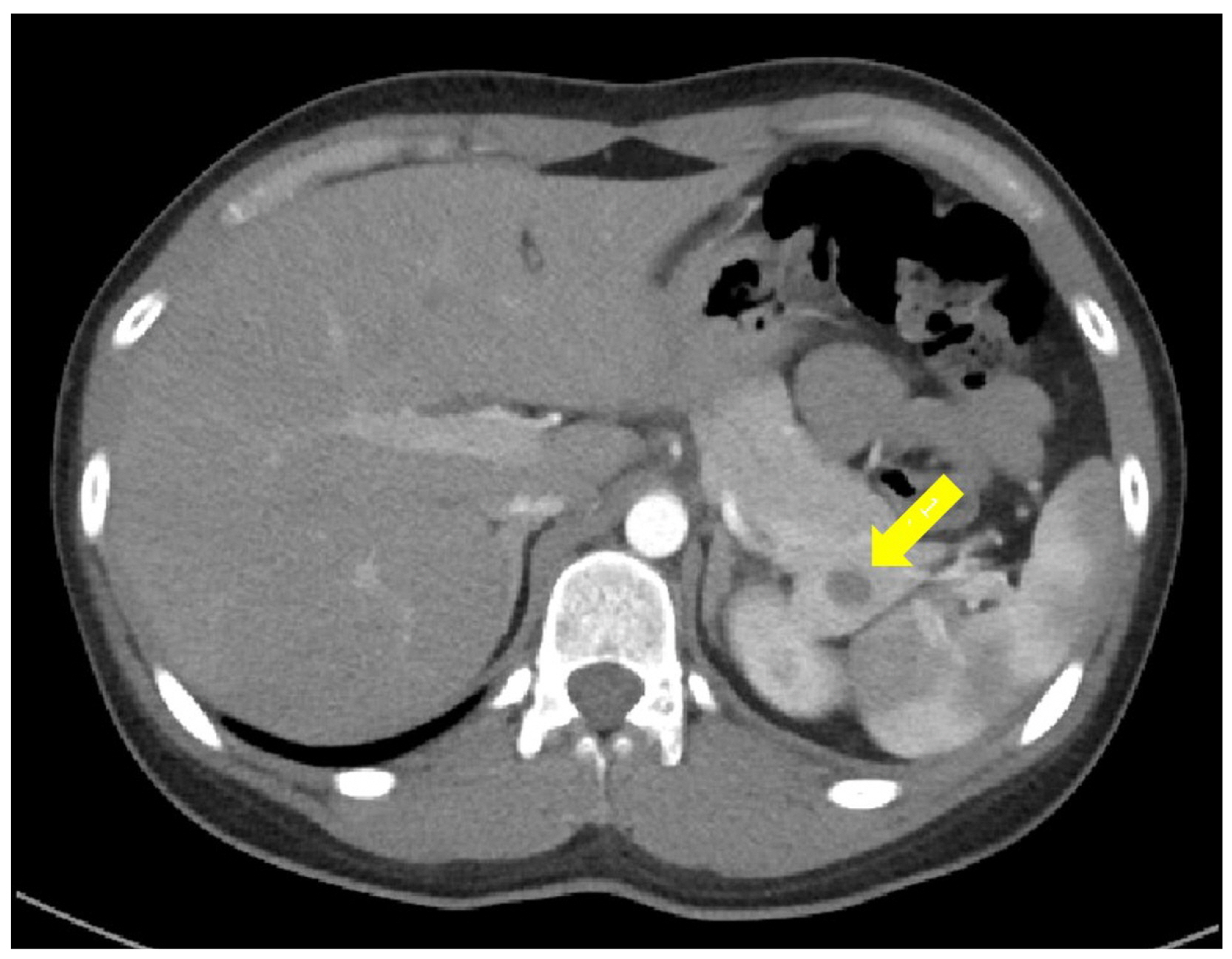
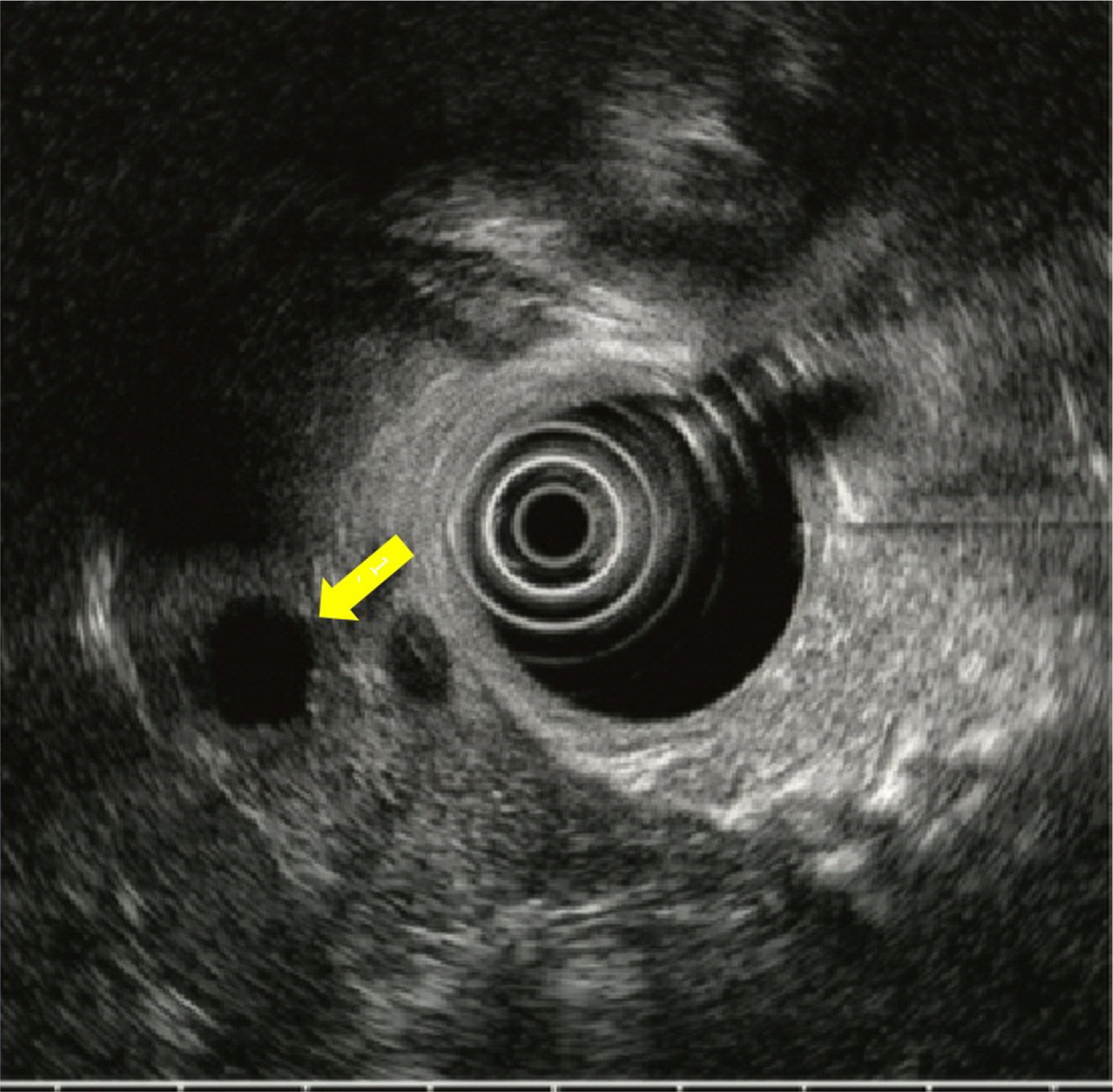
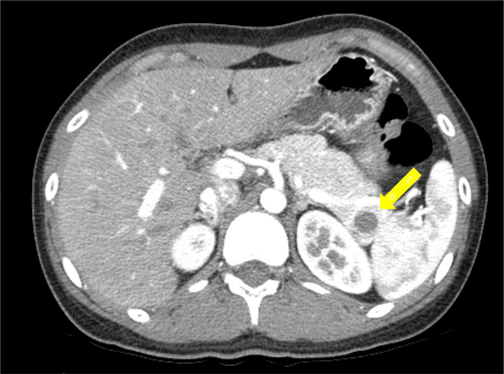
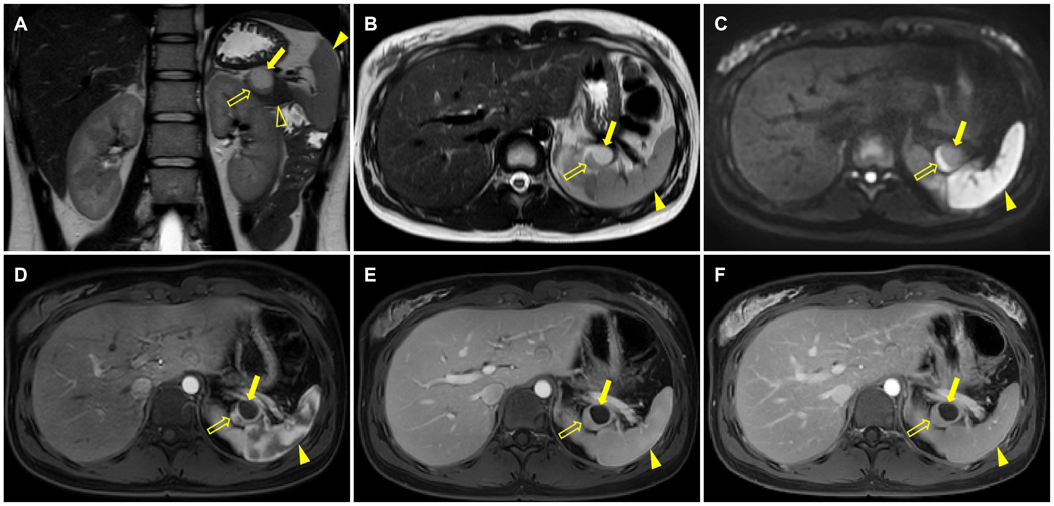
Epidermoid Cyst Arising from Intrapancreatic Accessory Spleen
- Affiliations
-
- 1Department of Radiology, Soonchunhyang University Cheonan Hospital, Soonchunhyang University College of Medicine, Cheonan, Korea
- KMID: 2508878
- DOI: http://doi.org/10.4166/kjg.2020.131
Figure
Reference
-
1. Halpert B, Alden ZA. 1964; Accessory spleens in or at the tail of the pancreas. A survey of 2,700 additional necropsies. Arch Pathol. 77:652–654. PMID: 14130052.2. Freeman JL, Jafri SZ, Roberts JL, Mezwa DG, Shirkhoda A. 1993; CT of congenital and acquired abnormalities of the spleen. Radiographics. 13:597–610. DOI: 10.1148/radiographics.13.3.8316667. PMID: 8316667.
Article3. Kato S, Mori H, Zakimi M, et al. 2016; Epidermoid cyst in an intrapancreatic accessory spleen: case report and literature review of the preoperative imaging findings. Intern Med. 55:3445–3452. DOI: 10.2169/internalmedicine.55.7140. PMID: 27904107. PMCID: PMC5216141.
Article4. Kim SH, Lee JM, Han JK, et al. 2008; Intrapancreatic accessory spleen: findings on MR Imaging, CT, US and scintigraphy, and the pathologic analysis. Korean J Radiol. 9:162–174. DOI: 10.3348/kjr.2008.9.2.162. PMID: 18385564. PMCID: PMC2627219.
Article5. Kim SS, Choi GC, Jou SS. 2018; Pancreas ductal adenocarcinoma and its mimics: review of cross-sectional imaging findings for differential diagnosis. J Belg Soc Radiol. 102:71. DOI: 10.5334/jbsr.1644. PMID: 30386851. PMCID: PMC6208287.
Article6. Horibe Y, Murakami M, Yamao K, Imaeda Y, Tashiro K, Kasahara M. 2001; Epithelial inclusion cyst (epidermoid cyst) formation with epithelioid cell granuloma in an intrapancreatic accessory spleen. Pathol Int. 51:50–54. DOI: 10.1046/j.1440-1827.2001.01155.x. PMID: 11148465.
Article7. Higaki K, Jimi A, Watanabe J, Kusaba A, Kojiro M. 1998; Epidermoid cyst of the spleen with CA19-9 or carcinoembryonic antigen productions: report of three cases. Am J Surg Pathol. 22:704–708. DOI: 10.1097/00000478-199806000-00007. PMID: 9630177.8. Zhang Z, Wang JC. 2009; An epithelial splenic cyst in an intrapancreatic accessory spleen. A case report. JOP. 10:664–666. PMID: 19890189.9. Sonomura T, Kataoka S, Chikugo T, et al. 2002; Epidermoid cyst originating from an intrapancreatic accessory spleen. Abdom Imaging. 27:560–562. DOI: 10.1007/s00261-001-0145-1. PMID: 12172998.
Article
- Full Text Links
- Actions
-
Cited
- CITED
-
- Close
- Share
- Similar articles
-
- Malignant Transformation of an Epidermoid Cyst in an Intrapancreatic Accessory Spleen: A Case Report
- An epidermoid cyst in an intrapancreatic accessory spleen
- Two Cases of Epidermoid Cysts in the Intrapancreatic Accessory Spleen Mimicking Pancreatic Cystic Neoplasm
- A Case of Epidermoid Cyst in the Intrapancreatic Accessory Spleen Mimicking Pancreas Mucinous Cystic Neoplasm
- Epithelial Cysts in the Intrapancreatic Accessory Spleen that Clinically Mimic Pancreatic Cystic Tumor: A Report of Two Cases


