Navigation-Assisted Balloon Eustachian Tuboplasty for Eustachian Tube Dilatory Dysfunction
- Affiliations
-
- 1Department of Otorhinolaryngology and Biomedical Research Institute, Pusan National University Hospital, Pusan National University School of Medicine, Busan, Korea
- KMID: 2508515
- DOI: http://doi.org/10.21053/ceo.2019.01305
Abstract
Objectives
. Balloon Eustachian tuboplasty (BET) is a novel treatment method for Eustachian tube dilatory dysfunction (ETD). However, surgeons cannot identify the insertion depth of the catheter during BET, resulting in potential risks such as internal carotid artery (ICA) injury. Therefore, we developed an image-guided navigation balloon catheter to identify the insertion depth of the catheter and to establish awareness of the proximity of the ICA. This study aimed to evaluate the technical feasibility of this image-guided navigation balloon catheter system in patients with ETD.
Methods
. Twenty-nine patients (38 ears; nine bilateral; 21 right ears, and 17 left ears) diagnosed with ETD were assessed. All patients who showed no improvement despite medical therapy with topical steroids, anti-reflux medication, and the Valsalva maneuver for a minimum of 6 weeks received image-guided navigation-assisted BET. The 7-item Eustachian Tube Dysfunction Questionnaire (ETDQ-7) score and Valsalva maneuver were used to evaluate patients’ symptoms preoperatively and at the postoperative follow-up.
Results
. Image-guided navigation-assisted BET was safely performed in all patients. The mean total ETDQ-7 score was 25.4±7.1 preoperatively, 17.5±6.2 at 1 month, and 15.2±7.0 at 6 months (P<0.001). In total, a Valsalva maneuver was possible for 28 of 38 ears (73.7%) at the time of the patient’s final visit at 6 months post-procedure.
Conclusion
. Image-guided navigation balloon catheters are a potentially valuable tool in patients with ETD. Their use is also technically feasible and safe when performing BET to treat ETD.
Keyword
Figure
-
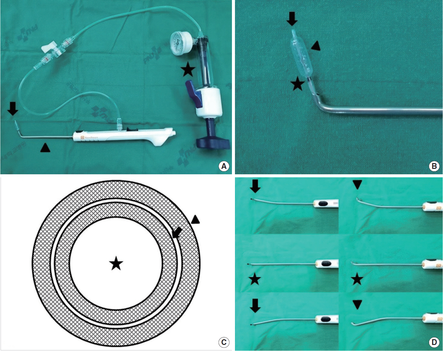
Fig. 1. (A) Photographs of the balloon catheter (arrow), guide catheter (arrowhead), and inflation device (asterisk). (B) Distal end of the balloon catheter, comprising an electromagnetic navigation sensor (arrow), a balloon (16 mm in length and 6 mm in diameter at 12 atm; arrowhead), and a black proximal balloon marker (asterisk). (C) Cross-sectional view of the balloon catheter consisting of an electromagnetic sensor part (0.9 mm in diameter; asterisk), inner tube (1.2 mm in diameter; arrow) and outer tube (1.6 mm in diameter; arrowhead). (D) Flexibility of the guide catheter. Neutral position (asterisks), right- and left-sided bending (arrows), and upward and downward bending (arrowheads).

Fig. 2. (A) Intraoperative view showing the insertion depth of the catheter tip as confirmed during a balloon Eustachian tuboplasty procedure. The catheter tip was advanced until its tip reached the isthmus. The on-screen crosshair provided real-time localization in three dimensions. Intraoperative view showing the internal carotid artery (asterisks). (B) Endoscopic photograph of catheter tip insertion over a black proximal endoscopic marker (denoting canalization of the cartilaginous segment of the Eustachian tube).
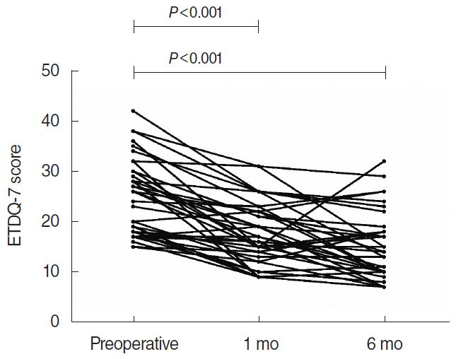
Fig. 3. Total 7-item Eustachian Tube Dysfunction Questionnaire (ETDQ-7) scores preoperatively, and at 1 month and 6 months post-procedure.
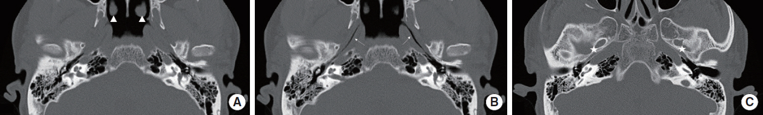
Fig. 4. (A) Preoperative computed tomography (CT) scans during the Valsalva maneuver, showing the nasopharyngeal end of the Eustachian tube (arrowheads). (B) Six-month postoperative CT scans during the Valsalva maneuver, showing patency of the cartilaginous Eustachian tube (arrows). (C) Six-month postoperative CT scans without the Valsalva maneuver. No micro-fracture was found along the bony canal of the internal carotid artery (asterisks).
Cited by 2 articles
-
Eustachian Tube Function Test
Jeon Mi Lee, Hyun Jin Lee
Korean J Otorhinolaryngol-Head Neck Surg. 2022;65(4):193-201. doi: 10.3342/kjorl-hns.2022.00220.Expanding Indications: Balloon Dilation of the Eustachian Tube for Patients Undergoing Surgery for Chronic Otitis Media
So Young Kim
Clin Exp Otorhinolaryngol. 2022;15(4):295-296. doi: 10.21053/ceo.2022.01410.
Reference
-
1. Martino E, Di Thaden R, Krombach GA, Westhofen M. Function tests for the Eustachian tube: current knowledge. HNO. 2004; Nov. 52(11):1029–39.2. Browning GG, Gatehouse S. The prevalence of middle ear disease in the adult British population. Clin Otolaryngol Allied Sci. 1992; Aug. 17(4):317–21.
Article3. Luukkainen V, Kivekas I, Hammaren-Malmi S, Rautiainen M, Poyhonen L, Aarnisalo AA, et al. Balloon Eustachian tuboplasty under local anesthesia: is it feasible? Laryngoscope. 2017; May. 127(5):1021–5.
Article4. Ockermann T, Reineke U, Upile T, Ebmeyer J, Sudhoff HH. Balloon dilation eustachian tuboplasty: a feasibility study. Otol Neurotol. 2010; Sep. 31(7):1100–3.5. Catalano PJ, Jonnalagadda S, Yu VM. Balloon catheter dilatation of Eustachian tube: a preliminary study. Otol Neurotol. 2012; Dec. 33(9):1549–52.6. Miller BJ, Elhassan HA. Balloon dilatation of the Eustachian tube: an evidence-based review of case series for those considering its use. Clin Otolaryngol. 2013; Dec. 38(6):525–32.
Article7. Randrup TS, Ovesen T. Balloon eustachian tuboplasty: a systematic review. Otolaryngol Head Neck Surg. 2015; Mar. 152(3):383–92.8. Poe DS, Hanna BM. Balloon dilation of the cartilaginous portion of the eustachian tube: initial safety and feasibility analysis in a cadaver model. Am J Otolaryngol. 2011; Mar-Apr. 32(2):115–23.
Article9. Ashry Y, Kawai K, Poe D. Utility of adjunctive procedures with balloon dilation of the Eustachian tube. Laryngoscope Investig Otolaryngol. 2017; Nov. 2(6):337–43.
Article10. Kepchar J, Acevedo J, Schroeder J, Littlefield P. Transtympanic balloon dilatation of eustachian tube: a human cadaver pilot study. J Laryngol Otol. 2012; Nov. 126(11):1102–7.
Article11. Busby DR, Slemmons DH, Miller TF Jr. Fatal epistaxis via carotid aneurysm and eustachian tube. Arch Otolaryngol. 1968; Mar. 87(3):295–8.
Article12. McCoul ED, Anand VK, Christos PJ. Validating the clinical assessment of eustachian tube dysfunction: The Eustachian Tube Dysfunction Questionnaire (ETDQ-7). Laryngoscope. 2012; May. 122(5):1137–41.
Article13. Tarabichi M, Najmi M. Visualization of the eustachian tube lumen with Valsalva computed tomography. Laryngoscope. 2015; Mar. 125(3):724–9.
Article14. Poe DS, Silvola J, Pyykko I. Balloon dilation of the cartilaginous eustachian tube. Otolaryngol Head Neck Surg. 2011; Apr. 144(4):563–9.
Article15. Hwang SY, Kok S, Walton J. Balloon dilation for eustachian tube dysfunction: systematic review. J Laryngol Otol. 2016; Jul. 130 Suppl 4:S2–6.
Article16. Williams B, Taylor BA, Clifton N, Bance M. Balloon dilation of the Eustachian tube: a tympanometric outcomes analysis. J Otolaryngol Head Neck Surg. 2016; Feb. 45:13.
Article17. Ockermann T, Reineke U, Upile T, Ebmeyer J, Sudhoff HH. Balloon dilatation eustachian tuboplasty: a clinical study. Laryngoscope. 2010; Jul. 120(7):1411–6.
Article18. Bowles PF, Agrawal S, Salam MA. Balloon tuboplasty in patients with Eustachian tube dysfunction: a prospective study in 39 patients (55 ears). Clin Otolaryngol. 2017; Oct. 42(5):1057–60.
Article19. Jufas N, Treble A, Newey A, Patel N. Endoscopically guided transtympanic balloon catheter dilatation of the eustachian tube: a cadaveric pilot study. Otol Neurotol. 2016; Apr. 37(4):350–5.20. Poe D, Anand V, Dean M, Roberts WH, Stolovitzky JP, Hoffmann K, et al. Balloon dilation of the eustachian tube for dilatory dysfunction: a randomized controlled trial. Laryngoscope. 2018; May. 128(5):1200–6.
Article21. Schroder S, Lehmann M, Ebmeyer J, Upile T, Sudhoff H. Balloon Eustachian tuboplasty: a retrospective cohort study. Clin Otolaryngol. 2015; Dec. 40(6):629–38.
Article22. Silvola J, Kivekas I, Poe DS. Balloon dilation of the cartilaginous portion of the Eustachian tube. Otolaryngol Head Neck Surg. 2014; Jul. 151(1):125–30.
Article23. Jufas N, Patel N. Transtympanic balloon dilatation of the eustachian tube: systematic review. J Laryngol Otol. 2016; May. 130(5):425–30.
Article24. McCoul ED, Anand VK. Eustachian tube balloon dilation surgery. Int Forum Allergy Rhinol. 2012; May-Jun. 2(3):191–8.
Article25. Huda W, Ogden KM, Khorasani MR. Converting dose-length product to effective dose at CT. Radiology. 2008; Sep. 248(3):995–1003.
Article26. Christner JA, Kofler JM, McCollough CH. Estimating effective dose for CT using dose-length product compared with using organ doses: consequences of adopting International Commission on Radiological Protection publication 103 or dual-energy scanning. AJR Am J Roentgenol. 2010; Apr. 194(4):881–9.
Article27. Schilder AG, Bhutta MF, Butler CC, Holy C, Levine LH, Kvaerner KJ, et al. Eustachian tube dysfunction: consensus statement on definition, types, clinical presentation and diagnosis. Clin Otolaryngol. 2015; Oct. 40(5):407–11.
Article
- Full Text Links
-
- Actions
-
Cited
- CITED
-
- Close
- Share
-
- Similar articles
-
- A Case of Subcutaneous Emphysema and Pneumomediastinum After Balloon Eustachian Tuboplasty
- Two Cases of Intra-Operative Cardiac Arrhythmia During Balloon Eustachian Tuboplasty: The Mechanism and Treatment
- Comparison of Eustachian Tube Function Before and After Septoplasty: A Systematic Review and Meta-Analysis
- Analysis of Eustachian Tube Dysfunction by Dynamic Slow Motion Video Endoscopy and Eustachian Tube Dysfunction Questionnaire in Chronic Otitis Media
- A Case of Collagen Graft for Patulous Eustachian Tube
