Anesth Pain Med.
2020 Oct;15(4):492-497. 10.17085/apm.19087.
Pneumocephalus following fluoroscopy-guided lumbar epidural injection in elderly patients: two cases report and a review of Korean literatures - Two cases report -
- Affiliations
-
- 1Department of Anesthesiology and Pain Medicine, Jeju National University Hospital, Jeju, Korea
- KMID: 2508413
- DOI: http://doi.org/10.17085/apm.19087
Abstract
- Background
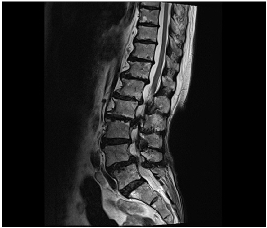
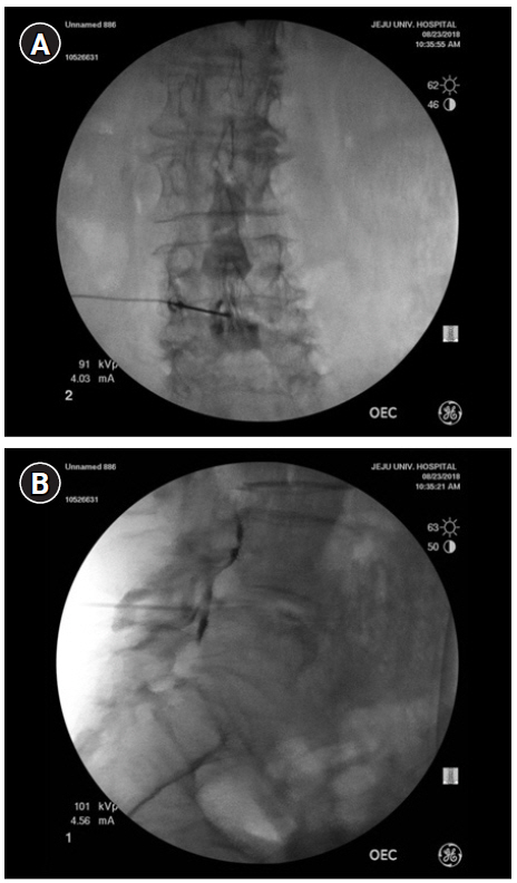
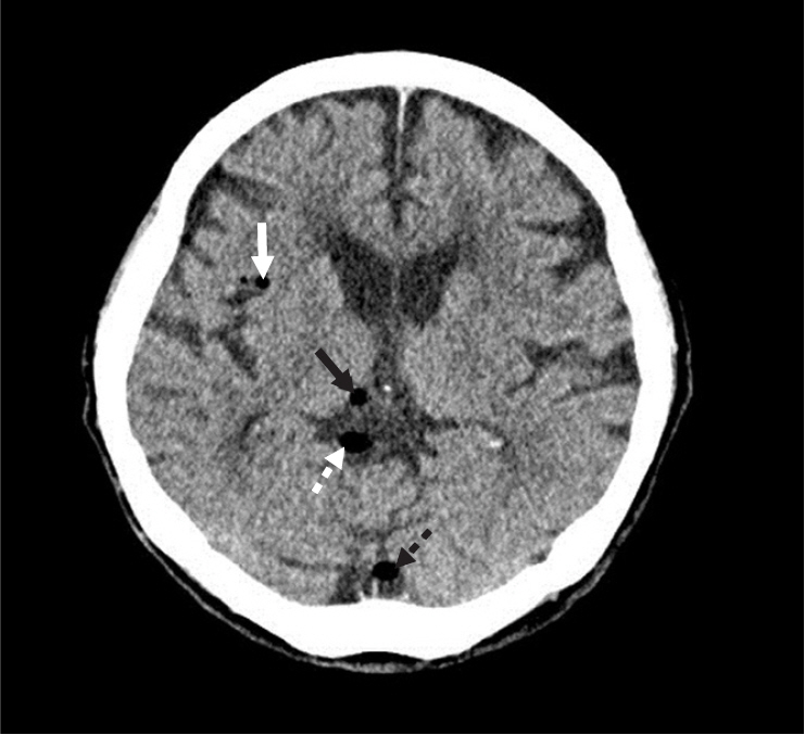
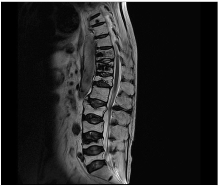
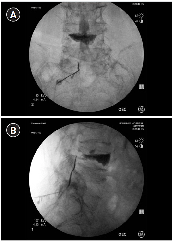
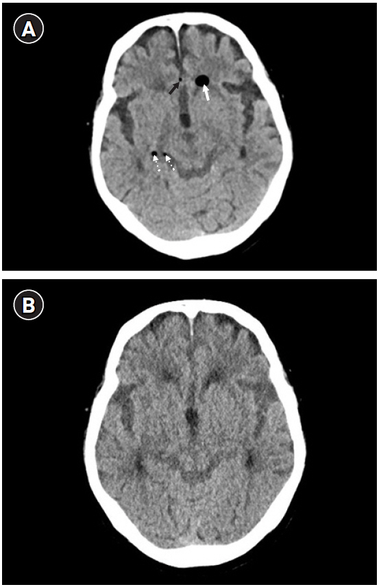
Pneumocephalus can originate from accidental dural puncture while performing epidural block using the loss-of-resistance (LOR) technique with an air-filled syringe. Case: We present two cases of pneumocephalus after lumbar epidural block under fluoroscopy for pain control in elderly patients.
Conclusions
Lumbar epidural block should be performed under fluoroscopic guidance in elderly patients with severe lesions. The physician should be aware of the increased possibility of a dural puncture occurring due to anatomical changes in older patients. The use of saline is recommended for the LOR technique. A contrast injection should be used together with the LOR technique to locate the epidural space. If a dural puncture occur, the patient should be carefully monitored to determine whether pneumocephalus has developed.
Keyword
Figure
Reference
-
1. Verdun AV, Cohen SP, Williams BS, Hurley RW. Pneumocephalus after lumbar epidural steroid injection: a case report and review of the literature. A A Case Rep. 2014; 3:9–13.2. Brogly N, Guasch E, Alsina E, García C, Puertas L, Dominguez A, et al. Epidural space identification with loss of resistance technique for epidural analgesia during labor: a randomized controlled study using air or saline-new arguments for an old controversy. Anesth Analg. 2018; 126:532–6.3. Schier R, Guerra D, Aguilar J, Pratt GF, Hernandez M, Boddu K, et al. Epidural space identification: a meta-analysis of complications after air versus liquid as the medium for loss of resistance. Anesth Analg. 2009; 109:2012–21.4. Aida S, Taga K, Yamakura T, Endoh H, Shimoji K. Headache after attempted epidural block: the role of intrathecal air. Anesthesiology. 1998; 88:76–81.5. Zaki SM. Study of the human ligamentum flavum in old age: a histological and morphometric study. Folia Morphol (Warsz). 2014; 73:492–9.6. Hogan QH. Epidural anatomy examined by cryomicrotome section. Influence of age, vertebral level, and disease. Reg Anesth. 1996; 21:395–406.7. Han CS, Yu JS, Kim IH, Kim YJ, Kim CS, Ahn KR. Headache and pneumocephalus after lumbar epidural block -a case report-. Korean J Pain. 1996; 9:251–5.8. Ahn B, Noh SM, Kim NH. Pneumocephalus after an epidural injection. J Korean Neurol Assoc. 2012; 30:148–50.9. Kim YD, Lee JH, Cheong YK. Pneumocephalus in a patient with no cerebrospinal fluid leakage after lumbar epidural block - a case report -. Korean J Pain. 2012; 25:262–6.10. Jung SK, Park KH. Pneumocephalus after epidural steroid injection -a case report-. Korean J Pain. 2001; 14:276–9.11. Kim YD, Ham HD, Moon HS, Kim SH. Delayed pneumocephalus following fluoroscopy guided cervical interlaminar epidural steroid injection: a rare complication and anatomical considerations. J Korean Neurosurg Soc. 2015; 57:376–8.12. Chung YW, Seo HY, Lee DH, Kim SK. Pneumocephalus after interlaminar lumbar epidural block. J Korean Orthop Assoc. 2017; 52:552–5.13. Landa J, Kim Y. Outcomes of interlaminar and transforminal spinal injections. Bull NYU Hosp Jt Dis. 2012; 70:6–10.14. Stojanovic MP, Vu TN, Caneris O, Slezak J, Cohen SP, Sang CN. The role of fluoroscopy in cervical epidural steroid injections: an analysis of contrast dispersal patterns. Spine (Phila Pa 1976). 2002; 27:509–14.15. Goodman BS, Posecion LW, Mallempati S, Bayazitoglu M. Complications and pitfalls of lumbar interlaminar and transforaminal epidural injections. Curr Rev Musculoskelet Med. 2008; 1:212–22.
- Full Text Links
- Actions
-
Cited
- CITED
-
- Close
- Share
- Similar articles
-
- Pneumocephalus after Epidural Steroid Injection: A case report
- Delayed Pneumocephalus Following Fluoroscopy Guided Cervical Interlaminar Epidural Steroid Injection: A Rare Complication and Anatomical Considerations
- Pneumocephalus after an Epidural Injection
- Pneumocephalus in a Patient with No Cerebrospinal Fluid Leakage after Lumbar Epidural Block: A Case Report
- Headache and Pneumocephalus after Lumbar Epidural Block: A case report

