J Korean Assoc Oral Maxillofac Surg.
2020 Oct;46(5):313-320. 10.5125/jkaoms.2020.46.5.313.
Correlation between mandibular morphology and masticatory muscle thickness in normal occlusion and mandibular prognathism
- Affiliations
-
- 1Department of Oral and Maxillofacial Surgery, College of Dentistry, Dankook University, Cheonan, Korea
- KMID: 2508255
- DOI: http://doi.org/10.5125/jkaoms.2020.46.5.313
Abstract
Objectives
The aim of this study was to evaluate the relationship between masticatory muscle thickness and mandibular morphology in young Korean adults with normal occlusion and mandibular prognathism.
Patients and Methods
Multidetector computed tomography (MDCT) was used to measure the masticatory muscle thickness on the right side in 100 Korean young adults (50 normal occlusion group, 50 mandibular prognathism group). Cephalometric analysis was done to measure mandibular morphology. Pearson correlation analysis was done to investigate the relationship between the masticatory muscle thickness and mandibular morphometry.
Results
The four masticatory muscles showed positive correlation with intergonial width in all subjects. All muscles, except temporalis, positively correlated with height of the ramus and mandibular length. Positive correlation was also observed in all muscles, except medial pterygoid, with thickness of the ramus. In the normal occlusion group, all four masticatory muscles showed positive correlation with intergonial width and ramus thickness. Positive correlation was also observed in all muscles (except lateral pterygoid) with mandibular length. Masseter and lateral pterygoid positively correlated with height of the ramus. In the mandibular prognathism group, all masticatory muscles, except lateral pterygoid, showed positive correlation with intergonial width. The masseter muscle showed negative correlation with ANB.
Conclusion
The results suggest a positive correlation of the thickness of masticatory muscles with both horizontal and vertical dimensions of the mandible. However, thickness of the masseter was found to decrease in patients with increasing severity of mandibular prognathism.
Keyword
Figure
-

Fig. 1 A. Thickness of the masseter muscle (m.) measured in axial view. B. Thickness of the medial pterygoid muscle (m.) measured in coronal view. C. Thickness of the lateral pterygoid muscle (m.) measured in axial view. D. Thickness of the temporalis muscle (m.) measured in axial view.
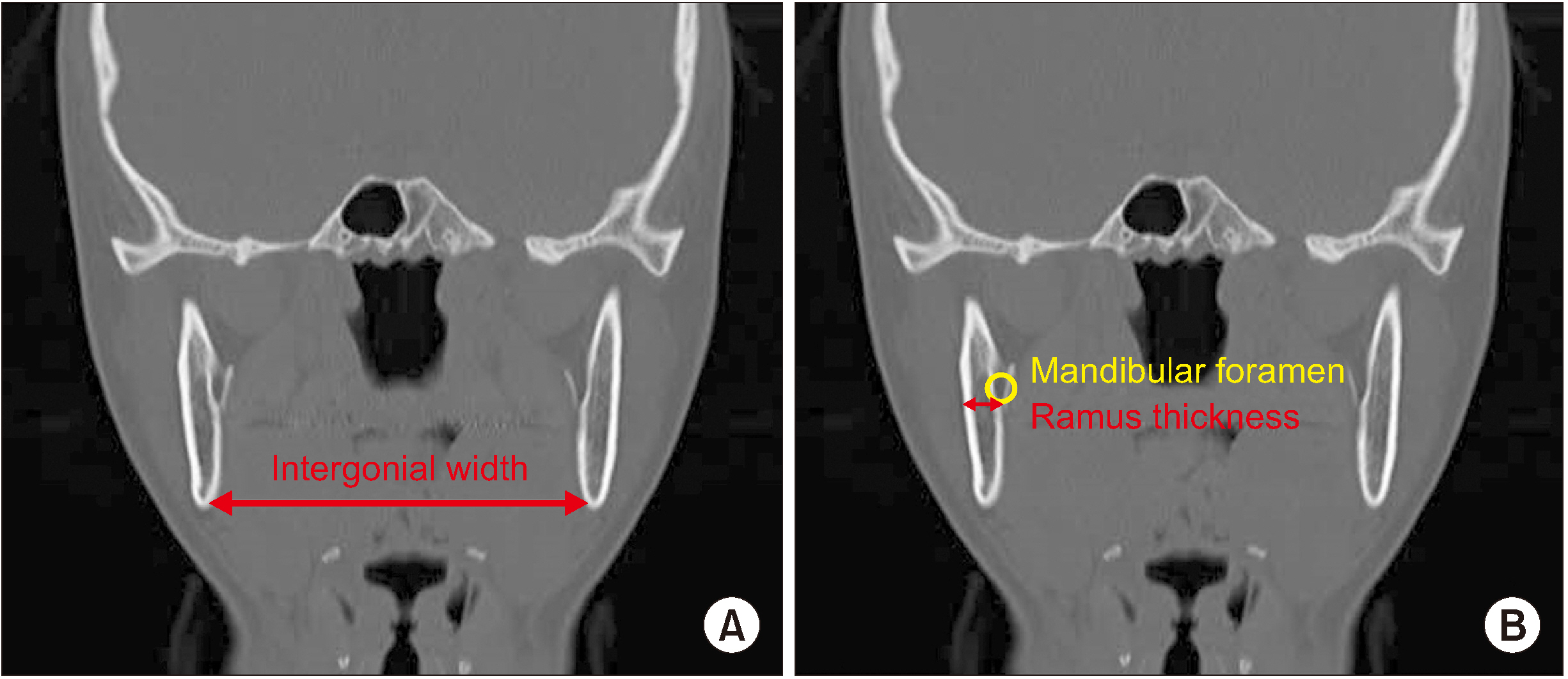
Fig. 2 Intergonial width (A) and ramus thickness (B) measured in coronal view.
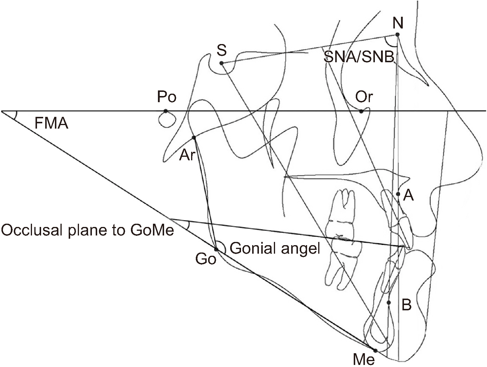
Fig. 3 Landmarks used for measurement: S (sella), N (nasion), A (point A), B (point B), Me (menton), Go (gonion), Ar (articulare), Or (orbitale), Po (porion). Linear measurements: ramus height (Go-Ar), mandibular length (Go-Me). Angular measurements: SNA, SNB, gonial angle (Ar-Go-Me), occlusal plane to GoMe, FMA (FH plane-GoMe).
Reference
-
References
1. Weijs WA, Hillen B. 1984; Relationships between masticatory muscle cross-section and skull shape. J Dent Res. 63:1154–7. https://doi.org/10.1177/00220345840630091201 . DOI: 10.1177/00220345840630091201. PMID: 6589280.
Article2. Proffit WR, Fields HW, Nixon WL. 1983; Occlusal forces in normal- and long-face adults. J Dent Res. 62:566–70. https://doi.org/10.1177/00220345830620051201 . DOI: 10.1177/00220345830620051201. PMID: 6573373.
Article3. Kitai N, Fujii Y, Murakami S, Furukawa S, Kreiborg S, Takada K. 2002; Human masticatory muscle volume and zygomatico-mandibular form in adults with mandibular prognathism. J Dent Res. 81:752–6. https://doi.org/10.1177/0810752 . DOI: 10.1177/0810752. PMID: 12407089.
Article4. Raadsheer MC, Kiliaridis S, Van Eijden TM, Van Ginkel FC, Prahl-Andersen B. 1996; Masseter muscle thickness in growing individuals and its relation to facial morphology. Arch Oral Biol. 41:323–32. https://doi.org/10.1016/0003-9969(95)00136-0 . DOI: 10.1016/0003-9969(95)00136-0.
Article5. Benington PC, Gardener JE, Hunt NP. 1999; Masseter muscle volume measured using ultrasonography and its relationship with facial morphology. Eur J Orthod. 21:659–70. https://doi.org/10.1093/ejo/21.6.659 . DOI: 10.1093/ejo/21.6.659. PMID: 10665195.
Article6. van Spronsen PH, Weijs WA, Valk J, Prahl-Andersen B, van Ginkel FC. 1991; Relationships between jaw muscle cross-sections and craniofacial morphology in normal adults, studied with magnetic resonance imaging. Eur J Orthod. 13:351–61. https://doi.org/10.1093/ejo/13.5.351 . DOI: 10.1093/ejo/13.5.351. PMID: 1748181.
Article7. Yang WS. 1995; The study on the orthodontic patients who visited Department of Orthodontics, Seoul National University Hospital during last 10 years (1985-1994). Korean J Orthod. 25:497–509.8. Ariji Y, Kawamata A, Yoshida K, Sakuma S, Nawa H, Fujishita M, et al. 2000; Three-dimensional morphology of the masseter muscle in patients with mandibular prognathism. Dentomaxillofac Radiol. 29:113–8. DOI: 10.1038/sj.dmfr.4600515. PMID: 10808226.
Article9. Sasaki K, Hannam AG, Wood WW. 1989; Relationships between the size, position, and angulation of human jaw muscles and unilateral first molar bite force. J Dent Res. 68:499–503. https://doi.org/10.1177/00220345890680031401 . DOI: 10.1177/00220345890680031401. PMID: 2921394.
Article10. Pepicelli A, Woods M, Briggs C. 2005; The mandibular muscles and their importance in orthodontics: a contemporary review. Am J Orthod Dentofacial Orthop. 128:774–80. https://doi.org/10.1016/j.ajodo.2004.09.023 . DOI: 10.1016/j.ajodo.2004.09.023. PMID: 16360920.
Article11. Boom HP, van Spronsen PH, van Ginkel FC, van Schijndel RA, Castelijns JA, Tuinzing DB. 2008; A comparison of human jaw muscle cross-sectional area and volume in long- and short-face subjects, using MRI. Arch Oral Biol. 53:273–81. https://doi.org/10.1016/j.archoralbio.2007.08.013 . DOI: 10.1016/j.archoralbio.2007.08.013. PMID: 18096133.
Article12. Maughan RJ, Watson JS, Weir J. 1983; Strength and cross-sectional area of human skeletal muscle. J Physiol. 338:37–49. https://doi.org/10.1113/jphysiol.1983.sp014658 . DOI: 10.1113/jphysiol.1983.sp014658. PMID: 6875963. PMCID: PMC1197179.
Article13. Ueki K, Takazakura D, Marukawa K, Shimada M, Nakagawa K, Yamamoto E. 2006; Relationship between the morphologies of the masseter muscle and the ramus and occlusal force in patients with mandibular prognathism. J Oral Maxillofac Surg. 64:1480–6. https://doi.org/10.1016/j.joms.2006.03.036 . DOI: 10.1016/j.joms.2006.03.036. PMID: 16982305.
Article14. Kiliaridis S, Kälebo P. 1991; Masseter muscle thickness measured by ultrasonography and its relation to facial morphology. J Dent Res. 70:1262–5. https://doi.org/10.1177/00220345910700090601 . DOI: 10.1177/00220345910700090601. PMID: 1918575.
Article15. Park KM, Choi E, Kwak EJ, Kim S, Park W, Jeong JS, et al. 2018; The relationship between masseter muscle thickness measured by ultrasonography and facial profile in young Korean adults. Imaging Sci Dent. 48:213–21. https://doi.org/10.5624/isd.2018.48.3.213 . DOI: 10.5624/isd.2018.48.3.213. PMID: 30276158. PMCID: PMC6148042.
Article16. Weijs WA, Hillen B. 1986; Correlations between the cross-sectional area of the jaw muscles and craniofacial size and shape. Am J Phys Anthropol. 70:423–31. https://doi.org/10.1002/ajpa.1330700403 . DOI: 10.1002/ajpa.1330700403. PMID: 3766712.
Article17. Moore WJ. 1973; An experimental study of the functional components of growth in the rat mandible. Acta Anat (Basel). 85:378–85. https://doi.org/10.1159/000144005 . DOI: 10.1159/000144005. PMID: 4723385.
Article18. Yonemitsu I, Muramoto T, Soma K. 2007; The influence of masseter activity on rat mandibular growth. Arch Oral Biol. 52:487–93. https://doi.org/10.1016/j.archoralbio.2006.10.019 . DOI: 10.1016/j.archoralbio.2006.10.019. PMID: 17126288.
Article19. Uchida Y, Motoyoshi M, Shigeeda T, Shinohara A, Igarashi Y, Sakaguchi M, et al. 2011; Relationship between masseter muscle size and maxillary morphology. Eur J Orthod. 33:654–9. https://doi.org/10.1093/ejo/cjq152 . DOI: 10.1093/ejo/cjq152. PMID: 21262936.
Article20. Iwase M, Ohashi M, Tachibana H, Toyoshima T, Nagumo M. 2006; Bite force, occlusal contact area and masticatory efficiency before and after orthognathic surgical correction of mandibular prognathism. Int J Oral Maxillofac Surg. 35:1102–7. https://doi.org/10.1016/j.ijom.2006.08.014 . DOI: 10.1016/j.ijom.2006.08.014. PMID: 17097270.
Article21. Ishida T, Yabushita T, Soma K. 2009; Functional changes of temporomandibular joint mechanoreceptors induced by reduced masseter muscle activity in growing rats. Angle Orthod. 79:978–83. https://doi.org/10.2319/081108-424.1 . DOI: 10.2319/081108-424.1. PMID: 19705933.
Article22. Islam I, Lim AAT, Wong RCW. 2017; Changes in bite force after orthognathic surgical correction of mandibular prognathism: a systematic review. Int J Oral Maxillofac Surg. 46:746–55. https://doi.org/10.1016/j.ijom.2017.01.012 . DOI: 10.1016/j.ijom.2017.01.012. PMID: 28209396.
Article
- Full Text Links
-
- Actions
-
Cited
- CITED
-
- Close
- Share
-
- Similar articles
-
- A Comparative Study On The Location Of The Mandibular Foramen In Panoramic Radiographs Of Normal Occlusion and Mandibular Prognathism
- Study on the Anatomical Position of Mandibular Canal Using Computed Tomography in Mandibular Prognathism Patients
- A radiological study on the morphology of labial alveolar bone in the mandibular incisor area of mandibular prognathism patients
- Ultrasonographic study on the masseter muscle thickness of adult Korean
- A roentgenocephalometric study on mandibular prognatnism
