Garbled and Incoherent Text Messages Are a Sign of Acute Ischemic Stroke: A New Sign of Aphasia in the Era of Chat Applications
- Affiliations
-
- 1Division of Interventional Neuroradiology, Department of Diagnostic and Therapeutic Radiology, Ramathibodi Hospital, Mahidol University, Bangkok, Thailand
- 2Division of Neurology, Department of Internal Medicine, Ramathibodi Hospital, Mahidol University, Bangkok, Thailand
- KMID: 2508069
- DOI: http://doi.org/10.5469/neuroint.2020.00290
Abstract
- We report a 68-year-old female was diagnosed acute ischemic stroke with an interesting clinical presentation. She was unable to send the messages in chat application normally and accurately. Neurological examination revealed global aphasia without weakness. Computed tomography angiography (CTA) showed the occlusion of the inferior branch of the left M2 of middle cerebral artery (MCA) but showed a good collateral score. Intravenous fibrinolysis and mechanical thrombectomy were not indicated. The patient showed spontaneous clinical improvement and almost fully recovered by the day of hospital discharge. Currently, chat applications have been widely adopted for communication and have replaced direct or telephone conversations in daily life. Dystextia and dystypia may serve as modern sign of aphasia on text conversation.
Keyword
Figure
-

Fig. 1. Conversation in the chat application revealed garbled and incoherent answers. (A) The husband asked, “Is the weather hot there?” The patient repeated after him, “Is the weather hot there?”. (B) The patient sent many pictures (stickers) to her husband instead of text conversation as she usual does. (C) Her husband asked, “Is it raining there?” The patient answered, “rainy?”.

Fig. 2. (A) and (B), Non-contrast computed tomography (NCCT) brain scan performed 9 hours after the onset of stroke demonstrated early acute infarction at the posterior part of the left temporal, insular, and parietal lobe (white arrows). (C) Hyperdense MCA sign (arrowhead) at the inferior branch of the left M2 of MCA. (D) The incidental finding of meningioma at the right anterior temporal pole convexity (black arrow).
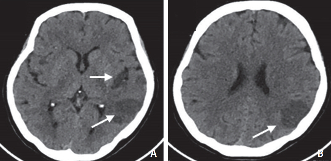
Fig. 3. (A) and (B), Non-contrast computed tomography (NCCT) brain done after 72 hours, obviously seen acute infarction at the posterior part of the left temporal, insular, and parietal lobe (white arrows).
Cited by 1 articles
-
Commentary to: Garbled and Incoherent Text Messages Are a Sign of Acute Ischemic Stroke: A New Sign of Aphasia in the Era of Chat Applications
Hyungjong Park
Neurointervention. 2021;16(1):88-89. doi: 10.5469/neuroint.2020.00430.
Reference
-
1. Azhar A, Maqbool S, Butt GA, Iftikhar S, Iftikhar G. Frequency of aphasia and its symptoms in stroke patients. J Speech Pathol Ther. 2017; 2:121.
Article2. Kim WJ, Paik NJ. Lesion localization of global aphasia without hemiparesis by overlapping of the brain magnetic resonance images. Neural Regen Res. 2014; 9:2081–2086.
Article3. Sharma AK, Fridman S, Gleichgerrcht E, Sposato LA. Dystextia and dystypia as modern stroke symptoms: a case series and literature review. Clin Neurol Neurosurg. 2019; 180:25–27.
Article4. Cabrera-Maqueda JM, Minhas JS. New horizons for stroke medicine: understanding the value of social media. Stroke. 2018; 49:e25–e27.
Article5. Kothari RU, Pancioli A, Liu T, Brott T, Broderick J. Cincinnati prehospital stroke scale: reproducibility and validity. Ann Emerg Med. 1999; 33:373–378.
Article6. Aroor S, Singh R, Goldstein LB. BE-FAST (balance, eyes, face, arm, speech, time): reducing the proportion of strokes missed using the FAST mnemonic. Stroke. 2017; 48:479–481.
- Full Text Links
-
- Actions
-
Cited
- CITED
-
- Close
- Share
-
- Similar articles
-
- Commentary to: Garbled and Incoherent Text Messages Are a Sign of Acute Ischemic Stroke: A New Sign of Aphasia in the Era of Chat Applications
- Analysis of Aphasia Patients Resulting from Acute Ischemic Stroke Using Quantitative Methods of Aphasia Test
- Prolonged Ictal Aphasia Presenting as Clinical-Diffusion Mismatch in a Patient with Acute Ischemic Stroke
- A Case of Ictal Aphasia Mimicking Transient Ischemic Attack
- Spot Sign on Initial Brain Computed Tomography Angiography Source Image to Predict Large Hemorrhagic Transformation after Middle Cerebral Artery Infarction
