Novel Endoscopic Criteria for Predicting Tumor Invasion Depth in Superficial Esophageal Squamous Carcinoma
- Affiliations
-
- 1Health Promotion Center, Asan Medical Center, University of Ulsan College of Medicine, Seoul, Korea
- 2Department of Gastroenterology, Asan Medical Center, University of Ulsan College of Medicine, Seoul, Korea
- 3Department of Pathology, Asan Medical Center, University of Ulsan College of Medicine, Seoul, Korea
- KMID: 2507824
- DOI: http://doi.org/10.3346/jkms.2020.35.e336
Abstract
- Background
Accurate prediction of tumor invasion depth in superficial esophageal squamous carcinoma (SESC) is essential for deciding the appropriate treatment strategy. We proposed novel endoscopic criteria to differentiate between mucosal and submucosal esophageal cancers and to evaluate the diagnostic accuracy and usefulness of the criteria.
Methods
A total of 352 patients who underwent endoscopic or surgical resection for SESC between 1991 and 2010 were included. First, the novel endoscopic criteria were created based on the endoscopic features of 60 randomly selected patients as follows: for T1m cancers, I. flat or slightly elevated or depressed lesion with smooth/even surface of any size, II. slightly elevated lesion of ≤ 1 cm with granular or uneven surface, III. hyperemic flat lesion of ≤ 3 cm with granular or uneven surface, IV. slightly depressed lesion of ≤ 2 cm with uneven surface and for T1sm cancers, I. irregularly (unevenly) nodular or protruded lesion of any size, II. slightly elevated lesion of > 1 cm with granular or uneven surface, III. hyperemic flat lesion of > 3 cm with granular or uneven surface, IV. irregularly (unevenly) depressed lesion of > 2 cm, and V. ulcerative lesion of any size. Next, the endoscopic findings of the remaining 292 patients were reviewed according to the criteria.
Results
The accuracy of novel endoscopic criteria was 79.5% (232/292). The sensitivity and specificity of mucosal cancers were 78.4% and 81.0%, respectively, whereas those for submucosal cancers were 81.0% and 78.4%, respectively. The accuracy for mucosal cancers was high (97.3%, 72/74) when the lesions were flat or slightly elevated/depressed with smooth/even surface regardless of size, whereas that for submucosal cancers was high (85.7%, 18/21) when the lesions were irregular/nodular protrusions regardless of size. In multivariate analysis, macroscopic type IIb lesion was identified as an independent factor affecting accuracy (P < 0.05). The difference in recurrence-free survival rates between endoscopically mucosal and submucosal cancers was significant (P = 0.026).
Conclusion
The novel endoscopic criteria appear to be accurate and useful in predicting invasion depth in SESC. Our criteria might help not only to decide the treatment strategy between surgery and endoscopic resection but also to predict the outcomes of SESC.
Keyword
Figure
-
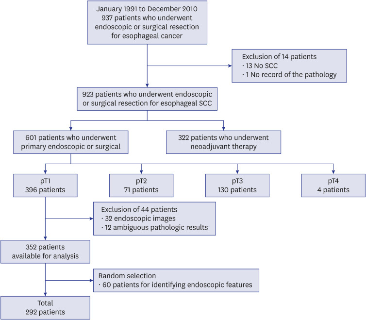
Fig. 1 Patient selection criteria.SCC = squamous cell carcinoma.
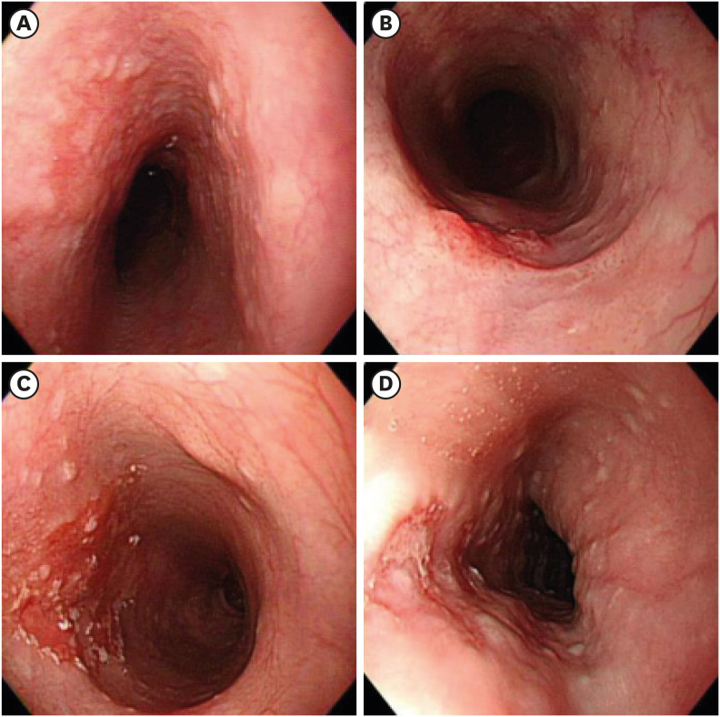
Fig. 2 Endoscopic criteria proposed for mucosal cancer. (A) flat or slightly elevated or depressed lesion with smooth/even surface of any size, (B) slightly elevated lesion of ≤ 1 cm with granular or uneven surface, (C) hyperemic flat lesion of ≤ 3 cm with granular or uneven surface, (D) slightly depressed lesion of ≤ 2 cm with uneven surface.
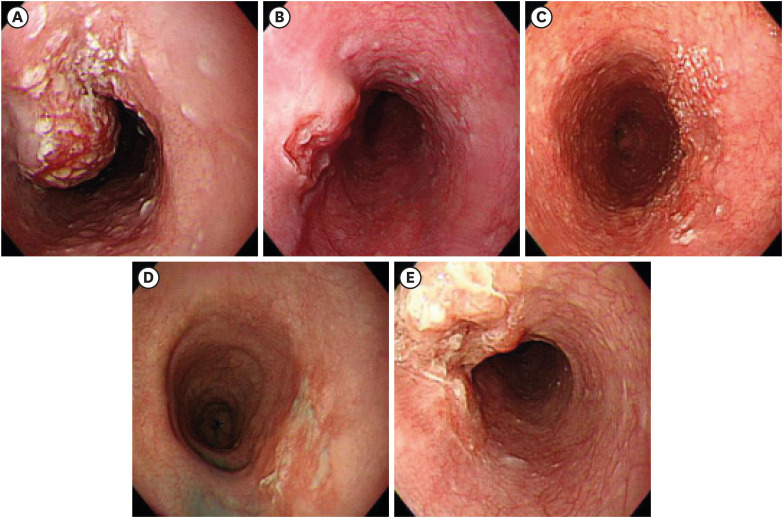
Fig. 3 Endoscopic criteria proposed for submucosal cancer. (A) irregularly (unevenly) nodular or protruded lesion of any size, (B) slightly elevated lesion of > 1 cm with granular or uneven surface, (C) hyperemic flat lesion of > 3 cm with granular or uneven surface, (D) irregularly (unevenly) depressed lesion of > 2 cm, (E) ulcerative lesion of any size.
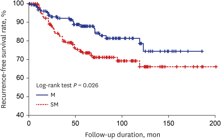
Fig. 4 Kaplan-Meier curve for recurrence-free survival in patients with endoscopically diagnosed M or SM. The difference between the M and SM groups was statistically significant (log-rank test, P = 0.026).M = mucosal cancer, SM = submucosal cancer.
Reference
-
1. Berger A, Rahmi G, Perrod G, Pioche M, Canard JM, Cesbron-Métivier E, et al. Long-term follow-up after endoscopic resection for superficial esophageal squamous cell carcinoma: a multicenter Western study. Endoscopy. 2019; 51(4):298–306. PMID: 30261535.
Article2. Min YW, Lee H, Song BG, Min BH, Kim HK, Choi YS, et al. Comparison of endoscopic submucosal dissection and surgery for superficial esophageal squamous cell carcinoma: a propensity score-matched analysis. Gastrointest Endosc. 2018; 88(4):624–633. PMID: 29750981.
Article3. Furue Y, Katada C, Tanabe S, Ishido K, Kondo Y, Kubota Y, et al. Effectiveness and safety of endoscopic aspiration mucosectomy and endoscopic submucosal dissection in patients with superficial esophageal squamous-cell carcinoma. Surg Endosc. 2019; 33(5):1433–1440. PMID: 30187201.
Article4. Song BG, Min YW, Lee JH, Lee H, Min BH, Rhee PL, et al. Efficacy and safety of endoscopic submucosal dissection in elderly patients with esophageal squamous cell carcinoma. Surg Endosc. 2017; 31(10):3905–3911. PMID: 28342128.
Article5. Choi JY, Park YS, Jung HY, Ahn JY, Kim MY, Lee JH, et al. Feasibility of endoscopic resection in superficial esophageal squamous carcinoma. Gastrointest Endosc. 2011; 73(5):881–889. 889.e1–882. PMID: 21392755.
Article6. Puli SR, Reddy JB, Bechtold ML, Antillon D, Ibdah JA, Antillon MR. Staging accuracy of esophageal cancer by endoscopic ultrasound: a meta-analysis and systematic review. World J Gastroenterol. 2008; 14(10):1479–1490. PMID: 18330935.
Article7. Thosani N, Singh H, Kapadia A, Ochi N, Lee JH, Ajani J, et al. Diagnostic accuracy of EUS in differentiating mucosal versus submucosal invasion of superficial esophageal cancers: a systematic review and meta-analysis. Gastrointest Endosc. 2012; 75(2):242–253. PMID: 22115605.
Article8. Lightdale CJ, Kulkarni KG. Role of endoscopic ultrasonography in the staging and follow-up of esophageal cancer. J Clin Oncol. 2005; 23(20):4483–4489. PMID: 16002838.
Article9. Oyama T, Inoue H, Arima M, Momma K, Omori T, Ishihara R, et al. Prediction of the invasion depth of superficial squamous cell carcinoma based on microvessel morphology: magnifying endoscopic classification of the Japan Esophageal Society. Esophagus. 2017; 14(2):105–112. PMID: 28386209.
Article10. Qu J, Zhang H, Wang Z, Zhang F, Liu H, Ding Z, et al. Comparison between free-breathing radial VIBE on 3-T MRI and endoscopic ultrasound for preoperative T staging of resectable oesophageal cancer, with histopathological correlation. Eur Radiol. 2018; 28(2):780–787. PMID: 28799124.
Article11. Young PE, Gentry AB, Acosta RD, Greenwald BD, Riddle M. Endoscopic ultrasound does not accurately stage early adenocarcinoma or high-grade dysplasia of the esophagus. Clin Gastroenterol Hepatol. 2010; 8(12):1037–1041. PMID: 20831900.
Article12. Goda K, Tajiri H, Ikegami M, Yoshida Y, Yoshimura N, Kato M, et al. Magnifying endoscopy with narrow band imaging for predicting the invasion depth of superficial esophageal squamous cell carcinoma. Dis Esophagus. 2009; 22(5):453–460. PMID: 19222533.
Article13. May A, Günter E, Roth F, Gossner L, Stolte M, Vieth M, et al. Accuracy of staging in early oesophageal cancer using high resolution endoscopy and high resolution endosonography: a comparative, prospective, and blinded trial. Gut. 2004; 53(5):634–640. PMID: 15082579.
Article14. Endoscopic Classification Review Group. Update on the paris classification of superficial neoplastic lesions in the digestive tract. Endoscopy. 2005; 37(6):570–578. PMID: 15933932.15. Japanese Gastric Cancer Association. Japanese classification of gastric carcinoma: 3rd English edition. Gastric Cancer. 2011; 14(2):101–112. PMID: 21573743.16. Brierley JD, Gospodarowicz MK, Wittekind C. TNM Classification of Malignant Tumours. 8th ed. Chichester: John Wiley & Sons;2017.17. Lokuhetty D, White VA, Watanabe R, Cree IA. World Health Organization. Digestive System Tumours. 5th ed. Lyon: International Agency for Research on Cancer;2019.18. Yoshida T, Inoue H, Usui S, Satodate H, Fukami N, Kudo SE. Narrow-band imaging system with magnifying endoscopy for superficial esophageal lesions. Gastrointest Endosc. 2004; 59(2):288–295. PMID: 14745410.
Article19. Lee MW, Kim GH, I H, Park DY, Baek DH, Lee BE, et al. Predicting the invasion depth of esophageal squamous cell carcinoma: comparison of endoscopic ultrasonography and magnifying endoscopy. Scand J Gastroenterol. 2014; 49(7):853–861. PMID: 24957951.
Article20. Nakagawa K, Ishihara R, Aoyama K, Ohmori M, Nakahira H, Matsuura N, et al. Classification for invasion depth of esophageal squamous cell carcinoma using a deep neural network compared with experienced endoscopists. Gastrointest Endosc. 2019; 90(3):407–414. PMID: 31077698.
Article21. Participants in the Paris Workshop. The Paris endoscopic classification of superficial neoplastic lesions: esophagus, stomach, and colon: November 30 to December 1, 2002. Gastrointest Endosc. 2003; 58(6):Suppl. S3–S43. PMID: 14652541.
- Full Text Links
-
- Actions
-
Cited
- CITED
-
- Close
- Share
-
- Similar articles
-
- Endoscopic Submucosal Dissection for Esophageal Squamous Cell Carcinoma
- Superficial Esophageal Cancer with Deep Submucosal Invasion Misdiagnosed as a Subepithelial Tumor
- Strategies for the Endoscopic Management and Post-Resection Treatment of Superficial Esophageal Squamous Cell Carcinoma
- Treatment of Superficial Esophageal Cancer: An Update
- Endoscopic Treatment for Esophageal Cancer
