Gastric Gastrointestinal Stromal Tumor with Repeated Recurrence at the Anastomosis Site in a Very Elderly Patient
- Affiliations
-
- 1Department of Internal Medicine, Pathology, Chung-Ang University College of Medicine, Seoul, Korea
- 2Department of Internal Medicine, Surgery, Chung-Ang University College of Medicine, Seoul, Korea
- KMID: 2507768
- DOI: http://doi.org/10.4166/kjg.2020.76.4.206
Abstract
- Although high-risk gastrointestinal stromal tumors (GISTs) frequently recur, even after a complete resection and imatinib therapy, local recurrence at the suture line after complete resection is rare. The present case was an 88-year-old woman who was initially diagnosed with high-risk GIST without a distant metastasis. She underwent a complete surgical resection of the lesion and received adjuvant imatinib therapy for 18 months, which was discontinued due to severe drug-induced anemia. During the follow-up, an endoscopic examination performed 40 months after the initial surgery revealed local recurrence at the anastomosis site. Although a complete surgical resection was performed, repeated local recurrence was detected 18 months later, which progressed rapidly to metastatic disease. This paper reports a case of a completely resected gastric GIST with repeated local recurrence, despite the complete surgical resections and adjuvant imatinib therapy.
Figure
-
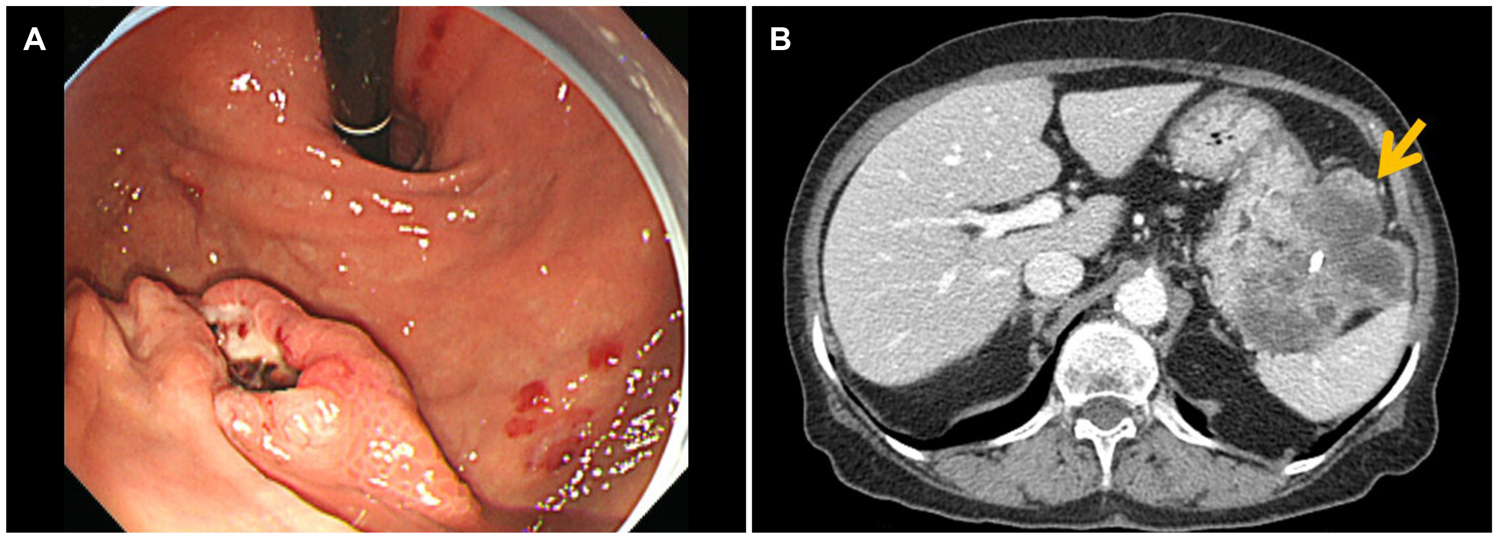
Fig. 1 Endoscopic images of the gastric lesion and abdominal computed tomography findings at the initial presentation. (A) A 2-cm sized, irregular shaped ulcerofungating mass on the greater curvature of the high body of the stomach. (B) An exophytic, heterogeneous enhancing mass with necrosis and central calcification, located adjacent to the gastric high body, closely abutting the splenic hilum (arrow).

Fig. 2 Histologic features of the gastric gastrointestinal stromal tumor at the first surgery. (A) Histopathological evaluation reveals blandly spindle cells with faintly eosinophilic cytoplasm in a syncytial pattern. Elongated nuclei with inconspicuous nucleoli are noted (H&E, ×400).(B) Diffuse positive cytoplasmic staining for CD117 (Immunohistochemical staining, ×20). (C) Diffuse positive staining for CD34 (Immunohistochemical staining, ×20). (D) Focal positive cytoplasmic staining for DOG-1 (Immunohistochemical staining, ×20). (E) Ki-67 proliferation index is less than 5% (Immunohistochemical staining, ×20).
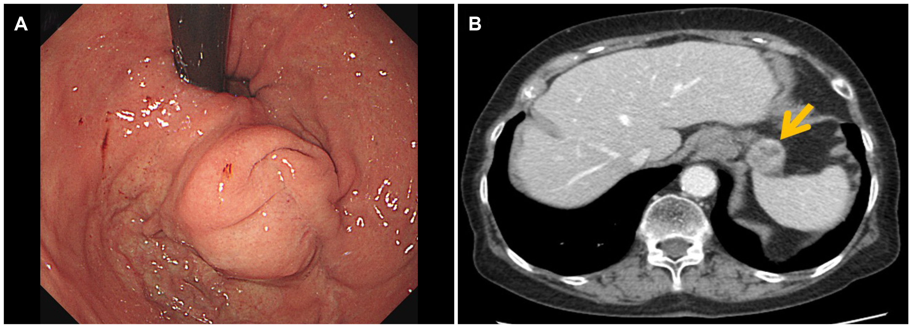
Fig. 3 Endoscopic images of the recurred gastric lesion and abdominal computed tomography findings at the first recurrence. (A) A 2-cm sized spherical shaped subepithelial mass lying on the anastomosis site. (B) An exophytic, heterogeneously enhancing mass at the anastomosis site of the stomach, measuring 2.6 cm, without signs of extragastric metastasis (arrow).
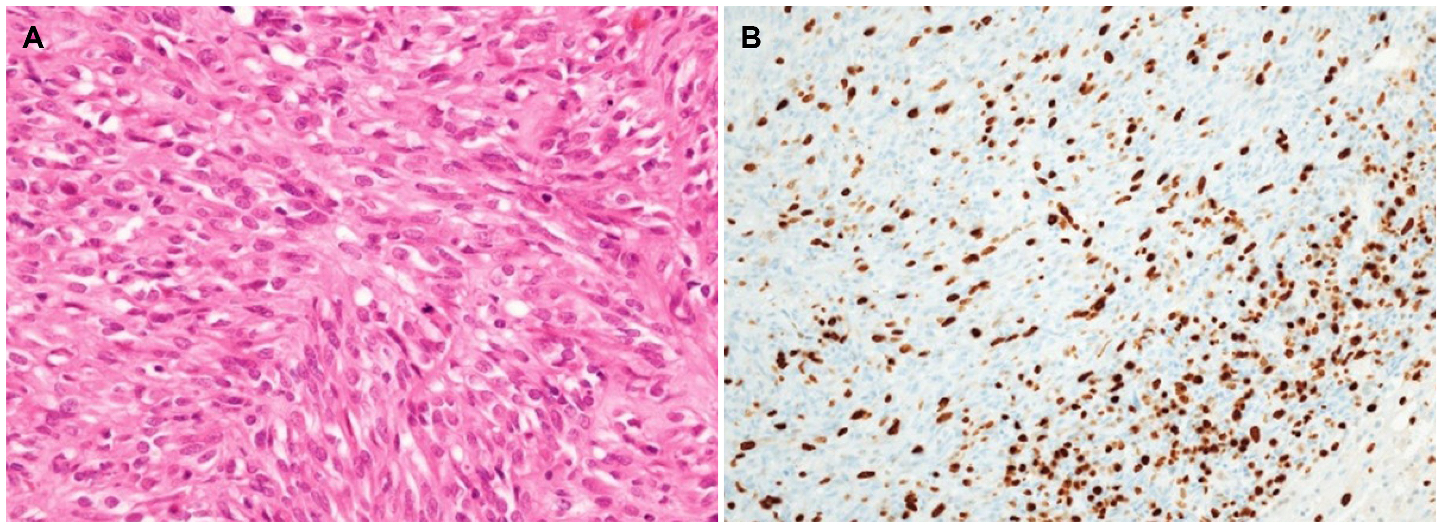
Fig. 4 Histologic features of a gastric gastrointestinal stromal tumor at the second surgery. (A) Histopathological evaluation revealed spindle cell proliferation with frequent mitoses up to 159/50 high-power fields (H&E, ×40) (B) Ki-67 proliferation index is 30% (Immunohistochemical staining, ×20).
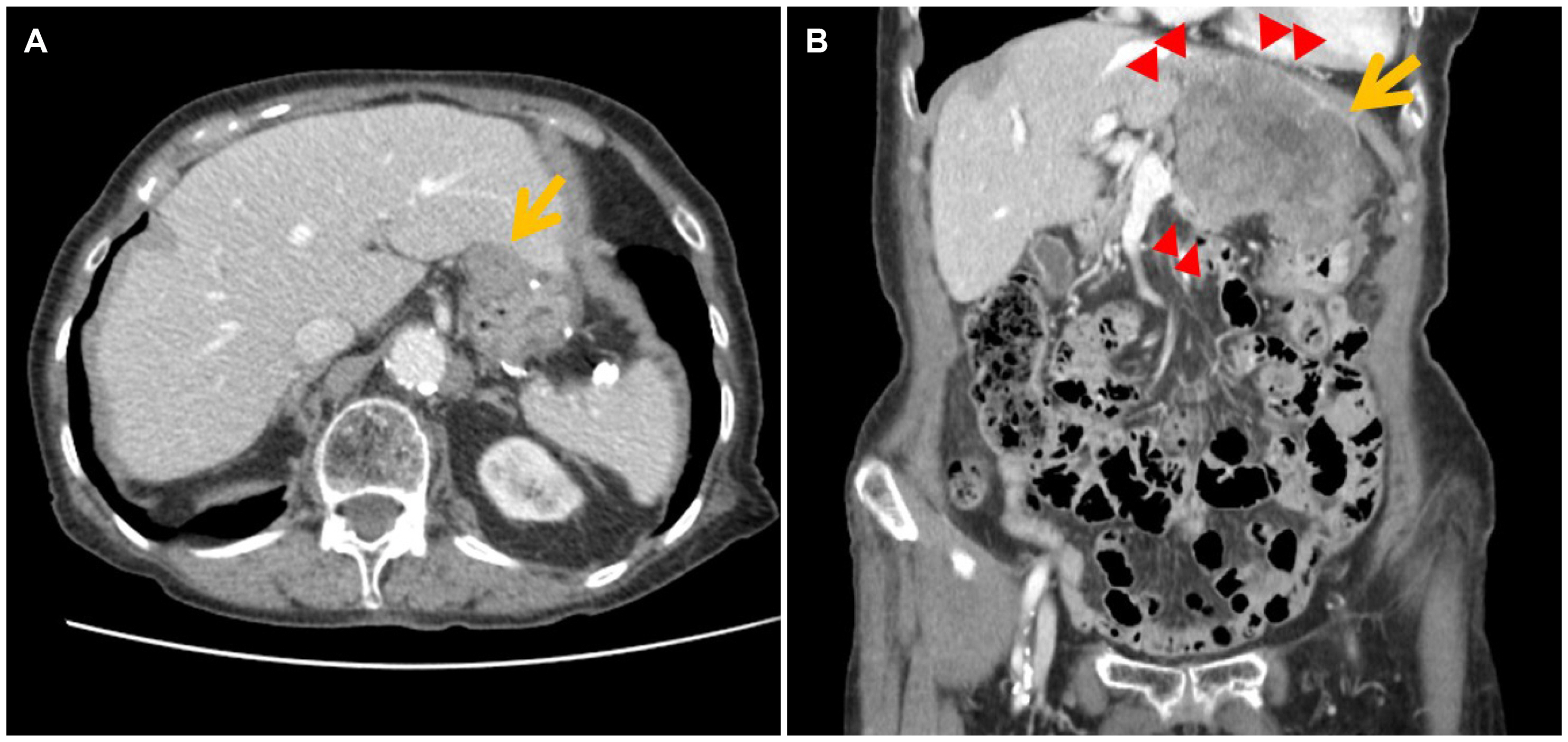
Fig. 5 Follow-up abdominal computed tomography findings after the second surgery. (A) A newly appeared 2.6-cm sized mass was found at the gastric anastomosis site (arrow), 1.5 years after the second surgery. (B) After 6 months of observation, the recurrent gastric lesion increased in size (2.6→9.4 cm, arrow) with invasion to the adjacent liver, pancreas, diaphragm, and pericardial fat (arrowheads).
Reference
-
1. Sorour MA, Kassem MI, Ghazal Ael-H, El-Riwini MT, Abu Nasr A. 2014; Gastrointestinal stromal tumors (GIST) related emergencies. Int J Surg. 12:269–280. DOI: 10.1016/j.ijsu.2014.02.004. PMID: 24530605.
Article2. Deshaies I, Cherenfant J, Gusani NJ, et al. 2010; Gastrointestinal stromal tumor (GIST) recurrence following surgery: review of the clin-ical utility of imatinib treatment. Ther Clin Risk Manag. 6:453–458. DOI: 10.2147/TCRM.S5634. PMID: 20957137. PMCID: PMC2952484.3. Zhao R, Wang Y, Huang Y, et al. 2017; Adjuvant imatinib for patients with high-risk gastrointestinal stromal tumors: a retrospective cohort study. Sci Rep. 7:16834. DOI: 10.1038/s41598-017-17266-5. PMID: 29203825. PMCID: PMC5715066.
Article4. Raut CP, Espat NJ, Maki RG, et al. 2018; Efficacy and tolerability of 5-year adjuvant imatinib treatment for patients with resected intermediate- or high-risk primary gastrointestinal stromal tumor: the PERSIST-5 clinical trial. JAMA Oncol. 4:e184060. DOI: 10.1001/jamaoncol.2018.4060. PMID: 30383140. PMCID: PMC6440723.5. Joensuu H, Eriksson M, Sundby Hall K, et al. 2012; One vs three years of adjuvant imatinib for operable gastrointestinal stromal tumor: a randomized trial. JAMA. 307:1265–1272. DOI: 10.1001/jama.2012.347. PMID: 22453568.6. Judson I, Bulusu R, Seddon B, Dangoor A, Wong N, Mudan S. 2017; UK clinical practice guidelines for the management of gastrointestinal stromal tumours (GIST). Clin Sarcoma Res. 7:6. DOI: 10.1186/s13569-017-0072-8. PMID: 28465823. PMCID: PMC5408425.
Article7. Demetri GD, von Mehren M, Blanke CD, et al. 2002; Efficacy and safety of imatinib mesylate in advanced gastrointestinal stromal tumors. N Engl J Med. 347:472–480. DOI: 10.1056/NEJMoa020461. PMID: 12181401.8. Blanke CD, Rankin C, Demetri GD, et al. 2008; Phase III randomized, intergroup trial assessing imatinib mesylate at two dose levels in patients with unresectable or metastatic gastrointestinal stromal tumors expressing the kit receptor tyrosine kinase: S0033. J Clin Oncol. 26:626–632. DOI: 10.1200/JCO.2007.13.4452. PMID: 18235122.
Article9. Latagliata R, Ferrero D, Iurlo A, et al. 2013; Imatinib in very elderly patients with chronic myeloid leukemia in chronic phase: a retrospective study. Drugs Aging. 30:629–637. DOI: 10.1007/s40266-013-0088-6. PMID: 23681399.
Article10. Ogata K, Kimura A, Nakazawa N, et al. 2018; Long-term imatinib treatment for patients with unresectable or recurrent gastrointestinal stromal tumors. Digestion. 97:20–25. DOI: 10.1159/000484102. PMID: 29393163.
Article
- Full Text Links
-
- Actions
-
Cited
- CITED
-
- Close
- Share
-
- Similar articles
-
- A Case of Epithelioid Type Gastric Gastrointestinal Stromal Tumor with Gastrointestinal Bleeding
- Association between Recurrence and Survival Rates According to the Location of Gastric Gastrointestinal Stromal Tumor
- Systemic Treatment of the Gastrointestinal Stromal Tumor (GIST)
- Extragastrointestinal Stromal Tumor Mimicking Gastric Subepithelial Tumor
- Tumor Size is Associated with Long-term Outcomes after Resection of Gastric Gastrointestinal Stromal Tumors
