Anat Cell Biol.
2020 Sep;53(3):279-283. 10.5115/acb.20.048.
Variation in the vertebral levels of the origins of the abdominal aorta branches: a retrospective imaging study
- Affiliations
-
- 1Department of Internal and Family Medicine, Faculty of Medicine, The Hashemite University, Zarqa, Jordan
- 2School of Medicine, The University of Jordan, Amman, Jordan
- 3Faculty of Medicine, Al-Balqa’ Applied University, Al-Salt, Jordan
- 4Department of Anatomy and Histology, School of Medicine, The University of Jordan, Amman, Jordan
- 5Department of General Surgery and Urology, Faculty of Medicine, Jordan University of Science and Technology, Irbid, Jordan
- 6Prince Hamzah Hospital, Amman, Jordan
- KMID: 2507640
- DOI: http://doi.org/10.5115/acb.20.048
Abstract
- Our aim was to investigate the variation in the vertebral levels of the origins of the celiac artery, superior and inferior mesenteric arteries, paired renal arteries, and common iliac arteries. We conducted a retrospective imaging study in a large public secondary hospital on a nonrandom sample of 227 participants. We consecutively included adult patients who had undergone computed tomography angiography of the abdomen and excluded patients with a history of any vertebral abnormality or whose images revealed evidence of a vertebral abnormality or a congenital anomaly of any of the branches of the abdominal aorta. The primary outcome was the frequency distribution of the vertebral levels of the landmarks. The secondary outcomes were the intercorrelations of the vertebral levels of the landmarks and their relationships with age, sex, weight, height, and body mass index. The celiac artery originated at T11/T12–L1/L2, followed by the superior mesenteric artery at T12–L2, the paired renal arteries at T12/L1–L2/L3, the inferior mesenteric artery at L2–L4, and the common iliac arteries at L3–L5. The vertebral levels of the landmarks were positively intercorrelated and stronger between proximate pairs. In addition, the vertebral levels of the landmarks were related to age, but not sex, weight, height, or body mass index. The intercorrelations suggest that a considerable proportion of the variation is accounted for by ‘trickle-down’ variation; variation in the vertebral level of a proximal landmark results in variation in the vertebral level of the immediate distal landmark. The overarching parameter remains unidentified.
Keyword
Figure
-
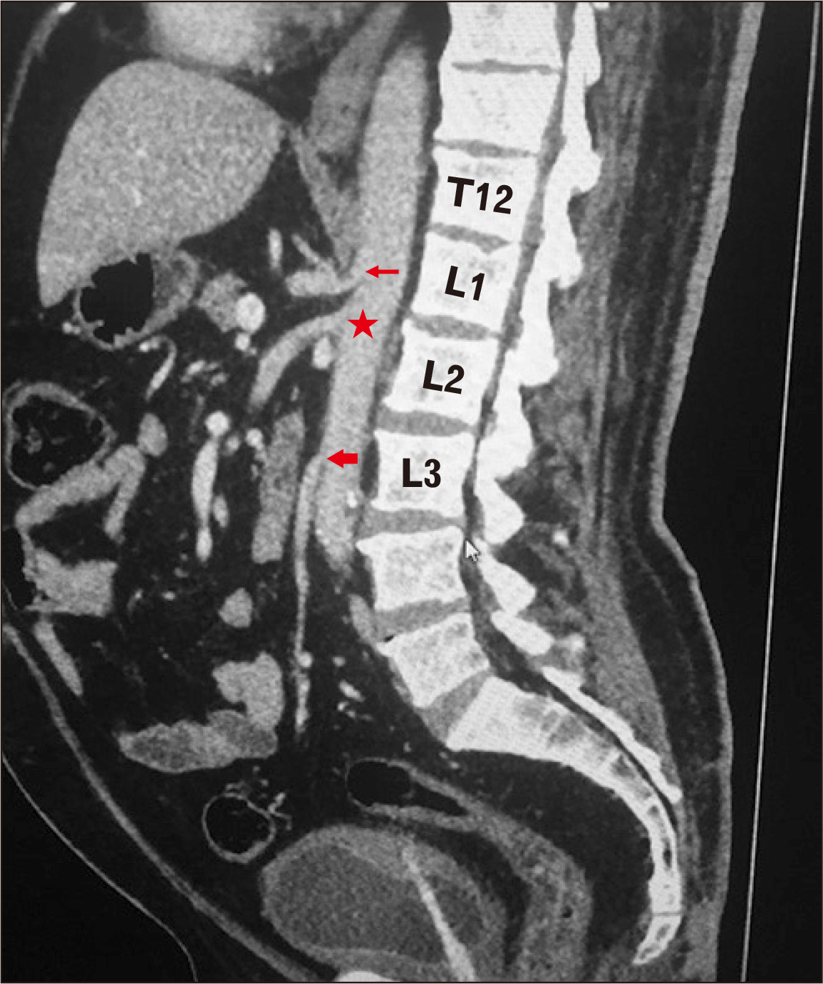
Fig. 1 Computed tomography angiography image showing the celiac (narrow red arrow), superior mesenteric (red star), and inferior mesenteric (wide red arrow) arteries.
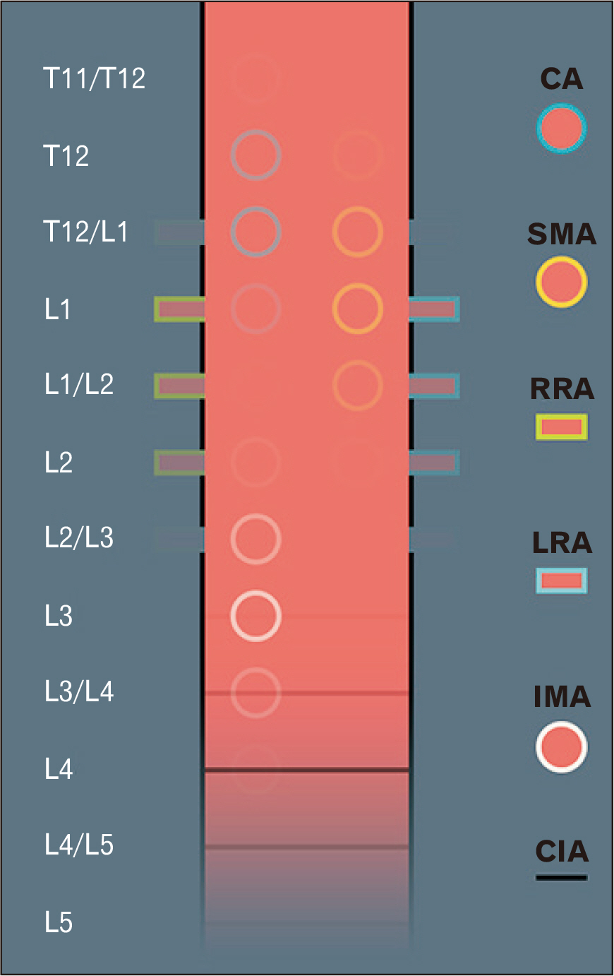
Fig. 2 The vertebral levels of the origins of the abdominal aorta branches (n=227 participants). We used the proportions of incidence in our study to determine the degree of opacity of the visual representation of the variants. CA, celiac artery; CIA, common iliac arteries; IMA, inferior mesenteric artery; LRA, left renal artery; RRA, right renal artery; SMA, superior mesenteric artery.
Reference
-
References
1. Drake RL, Vogl AW, Mitchell AWM. Gray's anatomy for students. Churchill Livingstone;Edinburgh:2. Goyal R, Aggarwal A, Gupta T, Gulati A, Jaggi S, Mirjalili SA, Sahni D. 2020; Reappraisal of the classical abdominal anatomical landmarks using in vivo computerized tomography imaging. Surg Radiol Anat. 42:417–28. DOI: 10.1007/s00276-019-02326-4. PMID: 31511961.3. Mirjalili SA, McFadden SL, Buckenham T, Stringer MD. 2012; A reappraisal of adult abdominal surface anatomy. Clin Anat. 25:844–50. DOI: 10.1002/ca.22119. PMID: 22744875.4. Shen XH, Su BY, Liu JJ, Zhang GM, Xue HD, Jin ZY, Mirjalili SA, Ma C. 2016; A reappraisal of adult thoracic and abdominal surface anatomy via CT scan in Chinese population. Clin Anat. 29:165–74. DOI: 10.1002/ca.22556. PMID: 26032163.
Article5. Campbell I. 2007; Chi-squared and Fisher-Irwin tests of two-by-two tables with small sample recommendations. Stat Med. 26:3661–75. DOI: 10.1002/sim.2832. PMID: 17315184.
Article6. Cole TJ. 2015; Too many digits: the presentation of numerical data. Arch Dis Child. 100:608–9. DOI: 10.1136/archdischild-2014-307149. PMID: 25877157. PMCID: PMC4483789.
Article7. George R. 1935; Topography of the unpaired visceral branches of the abdominal aorta. J Anat. 69(Pt 2):196–205. PMID: 17104532. PMCID: PMC1249104.8. Panagouli E, Lolis E, Venieratos D. 2011; A morphometric study concerning the branching points of the main arteries in humans: relationships and correlations. Ann Anat. 193:86–99. DOI: 10.1016/j.aanat.2010.10.009. PMID: 21169000.
Article9. Komutrattananont P, Mahakkanukrauh P, Das S. 2019; Morphology of the human aorta and age-related changes: anatomical facts. Anat Cell Biol. 52:109–14. DOI: 10.5115/acb.2019.52.2.109. PMID: 31338225. PMCID: PMC6624342.
Article
- Full Text Links
-
- Actions
-
Cited
- CITED
-
- Close
- Share
-
- Similar articles
-
- MR Findings of Thoracic and Abdominal Aortic Aneurysms: Comparison with Anglographic and Surgical Findings
- A radiological study on normal variations of abdominal aorta and its major branches
- Anomalous Origins of the Bilateral Vertebral Arteries Arising from the Aortic Arch: A Case Report
- Cord-like Atresia of the Abdominal Aorta Due to Takayasu Arteritis in Middle Aged Woman: A case Report
- Variations in the branching pattern of the celiac trunk and its clinical significance
