Korean J Ophthalmol.
2020 Apr;34(2):176-177. 10.3341/kjo.2019.0101.
Leptomeningeal Seeding in Choroidal Melanoma after Enucleation Surgery
- Affiliations
-
- 1Department of Ophthalmology, Institute of Vision Research, Severance Hospital, Yonsei University College of Medicine, Seoul, Korea.
- KMID: 2507420
- DOI: http://doi.org/10.3341/kjo.2019.0101
Figure
-
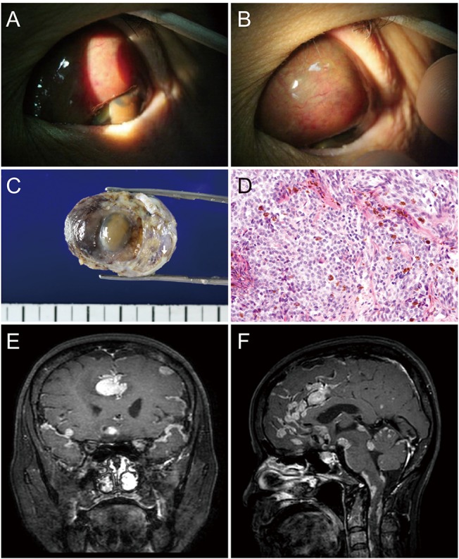
Fig. 1 (A,B) Slit-lamp photograph of recurred choroidal melanoma with abrupt rupture through the sclera and extraocular extension into the superior subconjunctival space. (C) Gross photograph of specimen after enucleation. The length between two bold lines is 1 cm. (D) Histologic findings of epitheloid cell type choroidal melanoma (hematoxylin and eosin stain, ×200). (E,F) Magnetic resonance imaging findings of extensive leptomeningeal seeding in the brain.
Reference
-
1. Borkar SA, Satyarthee GD, Das P, Suri V. Isolated brain metastasis from malignant melanoma of choroid seven years following enucleation. Neurol India. 2009; 57:92–94. PMID: 19305092.
Article2. Margo CE. The Collaborative Ocular Melanoma Study: an overview. Cancer Control. 2004; 11:304–309. PMID: 15377989.
Article3. Bansal AS, Bianciotto CG, Maguire JI, et al. Safety of pars plana vitrectomy in eyes with plaque-irradiated posterior uveal melanoma. Arch Ophthalmol. 2012; 130:1285–1290. PMID: 23044941.
Article
- Full Text Links
-
- Actions
-
Cited
- CITED
-
- Close
- Share
-
- Similar articles
-
- A Case of Malignant Melanoma of the Choroid
- Pigmented Choroidal Metastases Masquerading as Choroidal Melanoma
- A Choroidal Schwannoma Confirmed by Surgical Excision
- Surgical Resection of Metastatic Choroidal Melanoma in the Rib and Bronchus: A case report
- Fluorescein and Indocyanine Green Angiography of Choroidal Tumors
