Importance of Head Position after Gas Tamponade in Bilateral Descemet's Membrane Detachment Following Cataract Surgery
- Affiliations
-
- 1Department of Ophthalmology, HanGil Eye Hospital, Incheon, Korea.
- 2Department of Ophthalmology, HanGil Eye Hospital, Catholic Kwandong University College of Medicine, Incheon, Korea.
- KMID: 2507419
- DOI: http://doi.org/10.3341/kjo.2019.0119
Figure
-
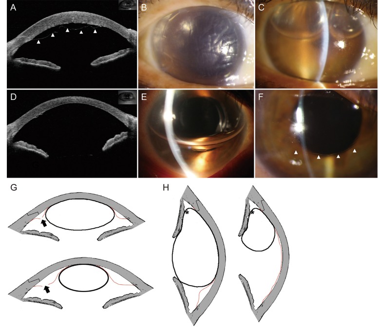
Fig. 1 A comparison of supine and lateral decubitus position after gas tamponade. (A) Anterior optical coherence tomography revealed total Descemet's membrane detachment (DMD) (arrowheads). (B,C) Slit lamp photographs depicted extensive DMD, and DMD remained with gas bubbles. (D,E) DMD recovered dramatically the day after the left-decubitus position was adopted. (F) Only a small amount of DMD (arrowhead) was detected. (G) In the supine position, the bubble in the anterior chamber could not seal off the incision site and fluids could flow into the incision site (arrows). (H) In a decubitus position, the bubble sealed off the incision site more effectively, even when the bubble became smaller (stellates). The red demarcation line represents Descemet's membrane.
Reference
- Full Text Links
-
- Actions
-
Cited
- CITED
-
- Close
- Share
-
- Similar articles
-
- A Case of Total Descemet's Nembrane Detachment Treated by Non-expansible SF6 Gas Iinfusion.
- Case Series of Descemet Membrane Detachment Associated with Cataract Surgery
- Three Cases of Descemet's Membrane Detachment after Cataract Surgery
- Spontaneous Reattachment of Descemet's Membrane Detachment at Postoperative Two Months, Which Occurred During Cataract Surgery
- A Case of Surgical Repair of Extensive Descemet's Membrane Detachment during Cataract Surgery
