Korean J Ophthalmol.
2020 Apr;34(2):106-112. 10.3341/kjo.2019.0113.
Macular Microvasculature in High Myopia without Pathologic Changes: An Optical Coherence Tomography Angiography Study
- Affiliations
-
- 1Department of Ophthalmology, Asan Medical Center, University of Ulsan College of Medicine, Seoul, Korea.
- 2College of Medicine, King Saud University, Riyadh, Saudi Arabia.
- KMID: 2507399
- DOI: http://doi.org/10.3341/kjo.2019.0113
Abstract
- Purpose
To investigate macular microvasculature changes using optical coherence tomography angiography (OCTA) and analyze their correlation with the structural parameters in highly myopic eyes.
Methods
We measured the area of the foveal avascular zone (FAZ) and the parafoveal vessel density in the superficial and deep retinal plexuses using OCTA. The magnification effect of the FAZ area was corrected using Bennett's formula. Retinal thickness measured at each corresponding area of the OCTA parameters, subfoveal choroidal thickness, and ocular characteristics were reviewed, and the relationships between the microvasculature measurements and the ocular structural characteristics were explored.
Results
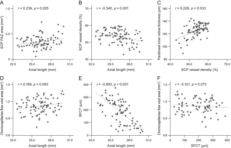
Fifty-two eyes with high myopia and 52 normal sex- and age-matched controls were included in the analysis. The FAZ area was significantly larger in the myopic eyes (p = 0.023). The superficial parafoveal vascular density was significantly decreased (p= 0.007) in the myopic eyes compared with the normal eyes, whereas there was no significant difference in the deep parafoveal vascular density (p = 0.226). Regarding the retinal thickness, only the parafoveal inner retinal thickness was significantly smaller in the myopic eyes than in the normal eyes (p = 0.023). The FAZ and subfoveal choroidal thickness were significantly correlated with the axial length, and the parafoveal inner retinal thickness was significantly correlated with the superficial parafoveal vascular density (all p < 0.05).
Conclusions
The FAZ was enlarged and the parafoveal vascular density was reduced in the highly myopic eyes. The decrease was prominent in the superficial capillary plexuses and well-correlated with the retinal thickness profiles. The macular microvascular network alteration may be attributed to the ocular axial elongation that occurs with myopia.
Keyword
Figure
Reference
-
1. Fan H, Chen HY, Ma HJ, et al. Reduced macular vascular density in myopic eyes. Chin Med J (Engl). 2017; 130:445–451. PMID: 28218219.
Article2. Gupta P, Saw SM, Cheung CY, et al. Choroidal thickness and high myopia: a case-control study of young Chinese men in Singapore. Acta Ophthalmol. 2015; 93:e585–e592. PMID: 25529388.
Article3. Kumar A, Chawla R, Kumawat D, Pillay G. Insight into high myopia and the macula. Indian J Ophthalmol. 2017; 65:85–91. PMID: 28345561.
Article4. Lam DS, Leung KS, Mohamed S, et al. Regional variations in the relationship between macular thickness measurements and myopia. Invest Ophthalmol Vis Sci. 2007; 48:376–382. PMID: 17197557.
Article5. Li M, Yang Y, Jiang H, et al. Retinal microvascular network and microcirculation assessments in high myopia. Am J Ophthalmol. 2017; 174:56–67. PMID: 27818204.
Article6. Milani P, Montesano G, Rossetti L, et al. Vessel density, retinal thickness, and choriocapillaris vascular flow in myopic eyes on OCT angiography. Graefes Arch Clin Exp Ophthalmol. 2018; 256:1419–1427. PMID: 29876731.
Article7. Ng DS, Cheung CY, Luk FO, et al. Advances of optical coherence tomography in myopia and pathologic myopia. Eye (Lond). 2016; 30:901–916. PMID: 27055674.
Article8. Sayanagi K, Ikuno Y, Uematsu S, Nishida K. Features of the choriocapillaris in myopic maculopathy identified by optical coherence tomography angiography. Br J Ophthalmol. 2017; 101:1524–1529. PMID: 28314832.
Article9. Wang NK, Lai CC, Chou CL, et al. Choroidal thickness and biometric markers for the screening of lacquer cracks in patients with high myopia. PLoS One. 2013; 8:e53660. PMID: 23349728.
Article10. Yang Y, Wang J, Jiang H, et al. Retinal microvasculature alteration in high myopia. Invest Ophthalmol Vis Sci. 2016; 57:6020–6030. PMID: 27820633.
Article11. Lim MC, Hoh ST, Foster PJ, et al. Use of optical coherence tomography to assess variations in macular retinal thickness in myopia. Invest Ophthalmol Vis Sci. 2005; 46:974–978. PMID: 15728555.
Article12. Luo HD, Gazzard G, Fong A, et al. Myopia, axial length, and OCT characteristics of the macula in Singaporean children. Invest Ophthalmol Vis Sci. 2006; 47:2773–2781. PMID: 16799013.
Article13. Zhao M, Wu Q, Hu P, Jia L. Macular thickness assessed with optical coherence tomography in young Chinese myopic patients. J Ophthalmol. 2015; 2015:715798. PMID: 26609427.
Article14. Zhao Z, Zhou X, Jiang C, Sun X. Effects of myopia on different areas and layers of the macula: a Fourier-domain optical coherence tomography study of a Chinese cohort. BMC Ophthalmol. 2015; 15:90. PMID: 26231216.
Article15. Al-Sheikh M, Phasukkijwatana N, Dolz-Marco R, et al. Quantitative OCT angiography of the retinal microvasculature and the choriocapillaris in myopic eyes. Invest Ophthalmol Vis Sci. 2017; 58:2063–2069. PMID: 28388703.
Article16. Samuel NE, Krishnagopal S. Foveal and macular thickness evaluation by spectral OCT SLO and its relation with axial length in various degree of myopia. J Clin Diagn Res. 2015; 9:NC01–NC04.
Article17. de Carlo TE, Romano A, Waheed NK, Duker JS. A review of optical coherence tomography angiography (OCTA). Int J Retina Vitreous. 2015; 1:5. PMID: 27847598.
Article18. Hassan M, Sadiq MA, Halim MS, et al. Evaluation of macular and peripapillary vessel flow density in eyes with no known pathology using optical coherence tomography angiography. Int J Retina Vitreous. 2017; 3:27. PMID: 28781889.
Article19. Wang Q, Chan S, Yang JY, et al. Vascular density in retina and choriocapillaris as measured by optical coherence tomography angiography. Am J Ophthalmol. 2016; 168:95–109. PMID: 27183862.
Article20. Kim YJ, Jo J, Lee JY, et al. Macular capillary plexuses after macular hole surgery: an optical coherence tomography angiography study. Br J Ophthalmol. 2018; 102:966–970. PMID: 28982954.
Article21. Iafe NA, Phasukkijwatana N, Chen X, Sarraf D. Retinal capillary density and foveal avascular zone area are age-dependent: quantitative analysis using optical coherence tomography angiography. Invest Ophthalmol Vis Sci. 2016; 57:5780–5787. PMID: 27792812.
Article22. Huynh SC, Wang XY, Rochtchina E, Mitchell P. Distribution of macular thickness by optical coherence tomography: findings from a population-based study of 6-year-old children. Invest Ophthalmol Vis Sci. 2006; 47:2351–2357. PMID: 16723444.
Article23. Sato A, Fukui E, Ohta K. Retinal thickness of myopic eyes determined by spectralis optical coherence tomography. Br J Ophthalmol. 2010; 94:1624–1628. PMID: 20494914.
Article24. Wu PC, Chen YJ, Chen CH, et al. Assessment of macular retinal thickness and volume in normal eyes and highly myopic eyes with third-generation optical coherence tomography. Eye (Lond). 2008; 22:551–555. PMID: 17464309.
Article25. Agawa T, Miura M, Ikuno Y, et al. Choroidal thickness measurement in healthy Japanese subjects by three-dimensional high-penetration optical coherence tomography. Graefes Arch Clin Exp Ophthalmol. 2011; 249:1485–1492. PMID: 21556938.
Article26. Flores-Moreno I, Lugo F, Duker JS, Ruiz-Moreno JM. The relationship between axial length and choroidal thickness in eyes with high myopia. Am J Ophthalmol. 2013; 155:314–319. PMID: 23036569.
Article27. Wei WB, Xu L, Jonas JB, et al. Subfoveal choroidal thickness: the Beijing Eye Study. Ophthalmology. 2013; 120:175–180. PMID: 23009895.
Article28. Mo J, Duan A, Chan S, et al. Vascular flow density in pathological myopia: an optical coherence tomography angiography study. BMJ Open. 2017; 7:e013571.
Article
- Full Text Links
- Actions
-
Cited
- CITED
-
- Close
- Share
- Similar articles
-
- Optical Coherence Tomographic Finding in a Case of Macular Coloboma
- Macular Microvasculature Features of the Eyes of Healthy Korean Children Using Optical Coherence Tomography Angiography
- Availability of Optical Coherence Tomography in Diagnosis and Classification of Choroidal Neovascularization
- Effects of Vitrectomy on the Treatment of a Lamellar Macular Hole in Highly Myopic Patients
- Photodynamic Therapy of Subfoveal Choroidal Neovascularization in Pathologic Myopia


